The
n e w e ng l a n d j o u r na l
of
m e dic i n e
clinical practice
Superior Vena Cava Syndrome with Malignant Causes Lynn D. Wilson, M.D., M.P.H., Frank C. Detterbeck, M.D., and Joachim Yahalom, M.D. This Journal feature begins with a case vignette highlighting a common clinical problem. Evidence supporting various strategies is then presented, followed by a review of formal guidelines, when they exist. The article ends with the authors’ clinical recommendations.
A 58-year-old man presents with a 2-week history of progressive dyspnea on exertion, neck swelling, decreased appetite, and fatigue. There is no history of syncope or dysphagia. He smoked cigarettes until 5 years ago. The physical examination reveals a heart rate of 105 beats per minute, a respiratory rate of 20 breaths per minute, and superficial vascular distention over the neck, chest, and upper abdomen. Stridor is not present. How should his case be evaluated and managed?
The Cl inic a l Probl e m From the Departments of Therapeutic Radiology (L.D.W.) and Surgery (F.C.D.), Yale University School of Medicine; and the Yale Comprehensive Cancer Center (L.D.W., F.C.D.) — both in New Haven, CT; and the Department of Radiation Oncology, Memorial Sloan-Kettering Cancer Center, and Weill Medical College of Cornell University, New York (J.Y.). Address reprint requests to Dr. Wilson at the Department of Therapeutic Radiology, Yale University School of Medicine, HRT 132, 333 Cedar St., New Haven, CT 06520, or at
[email protected]. N Engl J Med 2007;356:1862-9.
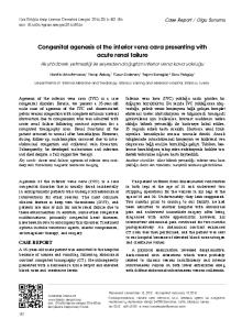
The superior vena cava syndrome, which occurs in approximately 15,000 persons in the United States each year, encompasses a constellation of symptoms and signs resulting from obstruction of the superior vena cava. The increased venous pressure in the upper body results in edema of the head, neck, and arms, often with cyanosis, plethora, and distended subcutaneous vessels (Fig. 1A). Edema may cause functional compromise of the larynx or pharynx, manifested as cough, hoarseness, dyspnea, stridor, and dysphagia. Cerebral edema may lead to headache, confusion, and coma. The decreased venous return may result in hemodynamic compromise; this complication may be a consequence of obstruction of the superior vena cava (intrinsic or due to extrinsic compression), compression of the heart by a large mass in the chest, or both. Symptoms develop over a period of 2 weeks in approximately a third of patients, and over longer periods in other cases.1-5
Copyright © 2007 Massachusetts Medical Society.
Anatomy and Physiology
The superior vena cava carries blood from the head, arms, and upper torso to the heart; it carries approximately one third of the venous return to the heart. Compression of the superior vena cava may result from the presence of a mass in the middle or anterior mediastinum (generally to the right of midline), consisting of enlarged right paratracheal lymph nodes, lymphoma, thymoma, an inflammatory process, or an aortic aneurysm, for example. Thrombosis of the superior vena cava without extrinsic compression can also occur (Fig. 1B). When the superior vena cava is obstructed, blood flows through a collateral vascular network to the lower body and the inferior vena cava or the azygos vein. It generally takes several weeks for the venous collaterals to dilate sufficiently to accommodate the blood flow of the superior vena cava.6,7 In humans with obstruction of the superior vena cava, the cervical venous pressure is usually increased to 20 to 40 mm Hg (normal range, 2 to 8 mm Hg).8-10 The severity of the symptoms depends on the degree of narrowing of the superior vena cava and the speed of the onset of the narrowing. 1862
n engl j med 356;18 www.nejm.org may 3, 2007
The New England Journal of Medicine Downloaded from nejm.org at THE OHIO STATE UNIV on June 29, 2011. For personal use only. No other uses without permission. Copyright © 2007 Massachusetts Medical Society. All rights reserved.
clinical pr actice
Figure 1. The Superior Vena Cava Syndrome. Clinical findings in a patient with the superior vena cava syndrome, including facial edema, plethora, jugular venous distention, and prominent superficial vascularity of neck and upper chest, are shown in Panel A. The vascular anatomy of the upper chest, including the heart, superior vena cava, inferior vena cava, and subclavian vessels, is shown in Panel B. The tumor is shown compressing the superior vena cava.
Edema in the upper body as a result of obstruction of the superior vena cava is visually striking but often of little consequence. However, cerebral edema, although rare, can be serious or fatal. The upper respiratory tract may become narrowed by nasal and laryngeal edema. Serious effects of obstruction of the superior vena cava are rare; among 1986 patients with obstruction of the superior vena cava, only one death was documented.11 In case reports of neurologic or laryngeal compromise, it is unclear whether other contributing factors such as brain metastases or tracheal compression were present.10,11
Etiologic Factors
Infectious causes (especially syphilitic aortic aneurysm and tuberculosis) accounted for the majority of cases of obstruction of the superior vena cava until about 50 years ago. These causes became rare, and malignant conditions accounted for more than 90% of cases approximately 25 years ago.1,12,13 Currently, obstruction of the superior vena cava caused by thrombosis or nonmalignant conditions accounts for approximately 35% of cases, reflecting the increased use of intravascular devices such as catheters and pacemakers.14 The most common malignant causes are non–small-cell lung cancer
n engl j med 356;18 www.nejm.org may 3, 2007
The New England Journal of Medicine Downloaded from nejm.org at THE OHIO STATE UNIV on June 29, 2011. For personal use only. No other uses without permission. Copyright © 2007 Massachusetts Medical Society. All rights reserved.
1863
The
n e w e ng l a n d j o u r na l
(approximately 50% of patients), small-cell lung cancer (approximately 25% of patients), lymphoma, and metastatic lesions (each approximately 10% of patients); the clinical features that may suggest these diagnoses are summarized in Table 1.1,4,5,13,15-17 Recognition of a nonmalignant cause of the superior vena cava syndrome is typically straightforward, particularly when the syndrome is associated with the use of an implanted intravascular device. An aortic aneurysm is easily recognized on computed tomography (CT). The diagnosis of fibrosing mediastinitis, although a rare cause, requires a biopsy.
S t r ategie s a nd E v idence Clinical Evaluation
Clinical diagnosis of obstruction of the superior vena cava is made on the basis of signs and symptoms (Table 2).1,4,5,13,15,18 The history taking should attend to the duration of symptoms, previous diagnoses of malignant conditions, or previous intravascular procedures. In most cases, symptoms are progressive over several weeks, and in some cases they may improve as collateral circulation develops. The severity of the symptoms is important in determining the urgency of intervention. Imaging
The most useful imaging study is CT of the chest after the administration of contrast material (which
of
m e dic i n e
is needed to evaluate the superior vena cava). Complications, including excessive bleeding from the venipuncture sites and reactions to contrast medium, are uncommon.11,14,19 Venography is generally warranted only when an intervention (placement of a stent or surgery) is planned.20 Magnetic resonance imaging may be useful for patients who cannot tolerate the contrast medium. Positronemission tomography (PET) is sometimes useful, because it may influence the design of the radiotherapy field (Fig. 2).21 The clinical history combined with CT imaging will generally differentiate between vena caval thrombosis and extrinsic compression. A tissue diagnosis is necessary to confirm the presence of malignant conditions. Clinical assessment is warranted to determine whether a peripheral biopsy site (e.g., a palpable supraclavicular lymph node) might be accessible before proceeding to an invasive procedure such as mediastinoscopy for tissue diagnosis. Cytologic examination of the sputum may result in diagnosis in patients who have endobronchial cancer. Pleural effusion is common (affecting about two thirds of patients with the superior vena cava syndrome); thoracentesis and cytologic analysis should be strongly considered because they are simple to perform and expedient, although they yield a diagnosis in only about 50% of such patients.15 Bronchoscopy has a diagnostic yield of 50 to 70% and transthoracic needle-aspiration biopsy has a yield of approximately 75%, whereas mediastinoscopy or mediastinotomy has
Table 1. Malignant Causes of the Superior Vena Cava Syndrome.* Tumor Type
Proportion
Suggestive Clinical Features
% (range) Non–small-cell lung cancer
50 (43–59)
History of smoking; often age >50 yr
Small-cell lung cancer
22 (7–39)
History of smoking; often age >50 yr
Lymphoma
12 (1–25)
Adenopathy outside the chest; often age