Summary of five case studies on the treatment of venous leg ulcers with a new two layer compression system in a community setting
by
� ylvie Hampton MA BSc (Hons) DpSN RGN, S Andy Kerr RN DipHe, Mike Crossley RN RM Tissue Viability Consultancy Services Eastbourne, UK
Summary of five case studies on the treatment of venous leg ulcers with a new two layer compression system in a community setting � ylvie Hampton MA BSc (Hons) DpSN RGN, S Andy Kerr RN DipHe, Mike Crossley RN RM Tissue Viability Consultancy Services, Eastbourne, UK
Overview Ulcers of the lower leg are common debilitating wounds of the elderly. In the U.S., venous ulcers have an incidence of 1.42% in women over 65 years of age.1 Broadly, these ulcers can be split into two main types with those originating from poor arterial perfusion being in the minority and those occurring due to venous problems accounting for approximately 70% of those presenting. The ulcer etiology is identified by measuring the ankle/ brachial pressure index (ABPI). Ulcers having an ABPI >0.8 are regarded as being suitable for treatment with compression therapy. Compression therapy has been long recognized as the standard treatment for venous leg ulcers.2, 3, 4 A new two layer bandage system, 3MTM CobanTM 2 Layer Compression System, hereafter called Coban 2 Layer System, has been developed to improve ease of application and patient comfort while providing sustained compression. The first layer (comfort layer) is composed of a foam laminated to a latex-free cohesive bandage. This is wrapped upwards around the foot and leg of the patient with a minimal overlap. The second layer (compression layer) is wrapped over the first layer with a 50% overlap, using full stretch to provide effective compression. Upon application, the two layers cohere to form one thin, conforming compression bandage. In this paper we report on a series of case studies in a community setting, evaluating clinical acceptability and product performance (slippage and wear time) of the new Coban 2 Layer System. All patients gave informed consent, were over 18 years of age, had one or more venous leg ulcer(s) and an ABPI >0.8, making them suitable for compression therapy.
�
The circumference of the ankle and calf was measured at each assessment. To assess slippage, the bandage height was measured (both at time of application and removal) from floor to top of the bandage with the patient stance standardized. Digital photographs recorded the condition of the patient’s leg, wound and bandage at times of application and removal. Exudate levels were assessed by experienced clinicians with a background in tissue viability, using subjective terms such as minimal / moderate / heavy.
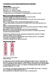
Patient 1 This case concerns a lady, MH, of 80 years, living at home and acting as a primary caregiver for her husband. She is fully active but has a limited social life due to caring for her husband. Doppler assessment provided an ABPI of 0.97. The shape of the leg was normal and the ulcer on the left leg had been present for seven months and situated over the medial malleolus. The wound bed was sloughy and there was significant erythema to the surrounding tissue with a smaller adjacent wound (Figure 1). The ulcer had shown little progress for the previous six months.
Figure 1
The patient’s medical history includes vein stripping of the ulcerated leg and she has hypertension. Her previous ulcer treatment was a silver dressing and short-stretch compression bandages. At first assessment, the wound was 1.5 cm x 1.5 cm and was square in shape with atrophy blanche at the wound margins. Although, exudate levels were minimal the peri-wound area was macerated and had an area of erythema previously treated with topical hydrocortisone. The tissue surrounding the erythema was healthy. The ankle measured 22.5 cm at the first assessment and after four weeks treatment the ankle had reduced to 20.5 cm. The calf measurement had not changed appreciably. The bandage was easy to apply, conformed to the leg without effort and no additional reshaping of the leg was required. The appearance of the applied bandage was aesthetically pleasing. This reassured both clinician and patient that the system had been applied correctly. Strikethrough was minimal and only occurred after the first seven days’ wear time. After two weeks, the erythema was reduced at the wound margins and the smaller wound was fully healed (Figure 2). At the end of the first month, the wound bed had reduced slough. Granulation tissue could be seen at the wound margins and under the slough. The smaller wound remained healed.
several years of hypersensitivity to a variety of dressings. The patient’s ulcers were of two years’ duration and static in nature. The wound bed was partially sloughy with some granulation tissue and generally did not appear to be making progress. There was maceration to the surrounding tissue and the ulcers were deep with callus formation to the periwound area. The larger ulcerated area covered 8.5 cm x 6.5 cm and the smaller ulcer measured 2 cm x 2 cm (Figure 3). While the exudate levels of the ulcers were moderate, the surrounding skin of the leg was dry and scaly.
Figure 3
The larger ulcer had bridges of vulnerable tissue which were protected with an alcohol-free, no sting barrier film.* After 17 days of treatment with the new compression system, the ulcers had not changed in size; however, the wound bed showed increased granulation and was without slough (Figure 4). The peri-wound area had an appearance of being slightly macerated, when in fact, the callus had rehydrated and along with the surrounding tissue, had improved.
Figure 2
Patient 2 MT is a 70-year-old gentleman who has a right leg amputation due to trauma. He lives with his wife and is fully independent with a full active social life and drives long journeys to see his relatives. Doppler assessment showed an ABPI of 1.2 signifying calcification of the arteries. The arterial sounds were bi-phasic, turbulent and dampened. Past treatment had been short-stretch compression therapy with a non-adherent iodine dressing. This was the dressing the patient insisted on using after
Figure 4
In spite of this patient’s hypersensitivity to many dressings, he did not react to this new compression system.
*3M™ Cavilon™ No Sting Barrier Film
�
This gentleman was committed to his previous treatment, even though that treatment was considered inappropriate. Nevertheless, he showed a high level of adherance to this new compression treatment.
Patient 3 PK is a 61-year-old lady with osteoporosis and osteoarthritis in both her spine and right knee. She lives in a flat, on her own, and one of her pleasures is taking her dog for a walk. Doppler assessment showed an ABPI of 1.16 and the arterial sounds were elastic and tri-phasic.
The patient’s view of this bandage system, in terms of wound improvement and comfort levels, is very positive and has requested its continued use after completion of the case study.
Patient 4 This 83-year-old gentleman, MG, has an ulcer on the right leg, the treatment of which has been complicated by a leg deformity due to a road traffic accident at the age of 18 (Figure 7).
The patient has an eight year history of ulceration. The most recent treatment was shortstretch compression therapy and a foam dressing containing silver. The ulcer (6 cm x 2 cm) was situated on the medial aspect of the lower gaiter region (Figure 5). At the first assessment, there was no granulation tissue apparent, with pale patches within the wound bed. The exudate was green, suggestive of the presence of Pseudomonas, and the wound margins were scaly in appearance.
Figure 7
Figure 8
The ulcer was of two years’ duration. Previous compression therapy had been unsuccessful, due to the cylindrical shape of the patient’s leg resulting in bandage slippage and, thus, increased risk of pressure damage related to bandage wrinkling. Figure 5
After three weeks of treatment, the wound had improved, showing signs of granulation and epithelialization (Figure 6).
It was important for this gentleman’s therapy that the compression system was adaptable to his individual limb requirements. A modification of the standard application technique was to apply an additional comfort layer around the calf region. This enabled accommodation of the irregular contours of the leg. The cohesive characteristics of the new compression system reduce the potential for slippage, thus enhancing patient comfort and prolonging effective wear time.
Figure 6 �
Patient 5
Overall Conclusions
This 92-year-old lady, VW, is in general good health with a previous history of mild heart failure not requiring drug therapy. Her mobility is excellent, her outlook on life is positive with a good social life, including daily walks to the Day Club and trips with her son on weekends.
The new 3M™ Coban™ 2 Layer Compression System was applied by clinicians experienced in the application of compression bandage systems. Compared to other multilayer compression systems, the application technique is easy to learn and the potential for inappropriate application is diminished. This two layer bandage system could easily be removed, in one piece, by cutting with bandage scissors.
She presented with a traumatic wound that had not healed after three months of self care. Her leg was edematous, with a pale appearance. The wound size was 1 cm x 3.8 cm and crescent shaped (Figure 9). Doppler assessment showed an ABPI of 0.9, making her suitable for compression therapy.
Figure 9
On application of the new compression system, no re-shaping of the leg was required. As the primary dressing, a hydrogel sheet was used, followed after two weeks by a foam dressing. At no time did the bandage slip during wear. The maximum system wear time was seven days. The edema at the ankle was reduced by 1.8 cm in the first week with a final reduction of 3.2 cm in the fourth week. The wound healed within four weeks, undoubtedly due to edema reduction and enhanced venous return through effective compression (Figure 10).
Figure 10
5
The Coban 2 Layer System successfully provided compression therapy to patients with a variety of leg shapes. The bandage maintained its integrity and shape. Any reduction in bandage height during wear was due to a slight rolling at the top end of the bandage rather than slippage. On two occasions, the bandage was changed due to strikethrough as a direct result of moderate to high exudate levels. One patient had two occasions when the bandage was changed earlier than seven days, as changes had to be arranged around her social requirements. In summary, a full seven day wear time was achieved on 15 out of the 19 wear time episodes. The minimum wear time was four days. In no instances were bandages changed due to slippage or loss of compression. In general, wound condition improved in all cases presented. All five patients found the Coban 2 Layer System comfortable and this could be associated with improved adherence levels and requests for continuing therapy in this system. In the authors’ experience, the Coban 2 Layer System is an improvement on existing compression bandages and deserves further assessment.
References 1. �Margolis DJ, Bilker W, Santanna J, and Baumgarten. Venous leg ulcer: Incidence and prevalence in the elderly. J Am Acad Dermatol 2002; 46: 381–6.
3. �Rojas Al, Phillips TJ. Venous ulcers and their management. In: Falanga V, editor. Cutaneous wound healing. Martin Dunitz Ltd; 2001. p. 263–86.
2. �Kantor J, Margolis DJ. Management of leg ulcers. Seminars in Cutaneous Medicine and Surgery 2003; 22(3): 212–221.
4. �Cullum N, Nelson EA, Fletcher AW, Sheldon, TA. Compression for venous leg ulcers (Review) The Cochrane Database of Systematic Reviews 2001; 1–34. This study was funded by 3M Deutschland GmbH, Neuss, Germany
3 Health Care 3M Center, Building 275-4W-02 St. Paul, MN 55144-1000 USA 1 800 228-3957 3M.com/healthcare
3M Canada Post Office Box 5757 London, Ontario N6A 4T1 Canada 1 800 563-2921
3M, Cavilon and Coban are trademarks of 3M.
Please Recycle Recycle, Neg
Printed in U.S.A. © 3M 2006 All Rights Reserved 70-2009-7387-6