J Vet Intern Med 2014;28:1498–1503
Association between Aortoseptal Angle in Golden Retriever Puppies and Subaortic Stenosis in Adulthood M.C. Belanger, E. C^ ot�e, and G. Beauchamp Background: Predicting subaortic stenosis (SAS) in adult Golden Retriever dogs (GRs) by evaluating them as puppies is hampered by the progressive expression of the SAS phenotype in youth. In some children who develop SAS as adults, an abnormal aortoseptal angle (AoSA) precedes development of stenosis. Objectives: To determine the normal AoSA in young adult GRs using echocardiography; to assess the value of AoSA in GR puppies for predicting development of the SAS phenotype. Animals: Forty-eight 2- to 6-month-old GR puppies. Methods: Prospective study. Puppies were recruited from clients and breeders. Puppies were evaluated with a physical examination and an echocardiogram, and this evaluation was repeated when they were 12–18-month-old adults. Puppies were classified as unaffected (WNL) or affected (SAS) retroactively, based on their results as adults. Results: In WNL young adult GRs, mean � SD AoSA was 152.3 � 6.5°. Mean � SD AoSA in SAS puppies (144.9 � 8.6°) was significantly different from mean AoSA in WNL puppies (155.7 � 8.8°, P < .01). No puppy with AoSA >160° had the SAS phenotype as a young adult; 93% (75.7–99.1%) of puppies with AoSA 160 mmHg); overt signs of illness; lack of identifiable tattoo or microchip, or inconsistency in identification from the first to the second evaluations; noncooperation during evaluation; failure to return for the second evaluation; or any combination of these factors.
Echocardiography Echocardiographic examinations (2D, M-mode, and Doppler) were performed as previously described, with right-sided longaxis LVOT and subcostal views used for calculation of AoSA15,20 and LVOT velocity, respectively.21–23 Echocardiograms were performed with dogs in lateral recumbency using ultrasound units equipped with either 1.5–4.0 MHz or 3.5–8.0 MHz phased-array transducersb,c and simultaneous electrocardiographic display. No animal was sedated; to encourage cooperation through postprandial relaxation, owners were asked to feed each puppy in the 15 minutes that preceded the echocardiogram. Measurement of AoSA on all echocardiograms was performed posthoc by a single evaluator (EC). Before blinding, the evaluator identified a brief (4- to 20 heartbeats) echocardiographic cine loop for each animal in each age group (puppy or young adult) that accurately demonstrated the LVOT and IVS, as previously described.21 Then, the primary investigator (MCB) removed all clinical information (ie, name, date, and all other identifying
A
1499
B
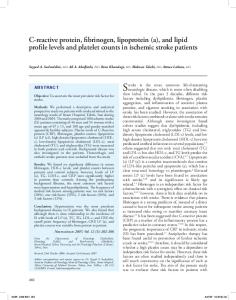
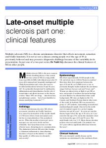
Fig 1. (A) Aortoseptal angle measurement in a Golden Retriever puppy that developed SAS as a young adult. The angle is formed by the long axis of the ascending aorta and the plane of the IVS. The midline axis of the aortic root is constructed by bisecting the aortic root at the level of the annulus and above the sinotubular junction (line a). The midline axis of the IVS is constructed by bisecting the septum at the level of the mitral leaflet tips and 2 cm apically from that point (line b). AoSA = 142°. Note the rightward deviation of the aortic axis (right side = top of image) causing this lower/steep AoSA. This 2-month-old puppy had an LVOT Vmax of 1.5 m/s on this echocardiogram, but 2.8 m/s on the echocardiogram performed at age 14 months. (B) Aortoseptal angle measurement in a normal Golden Retriever puppy. AoSA = 158°. AoSA, aortoseptal angle; IVS, interventricular septum; LVOT Vmax, peak left ventricular outflow tract velocity; SAS, subaortic stenosis.
1500
Belanger et al
the auscultatory and Doppler echocardiographic criteria for SAS later, as a young adult. The SAS phenotype was defined as the presence of a basilar systolic ejection murmur and a Doppler echocardiographic subcostal LVOT peak velocity (LVOT Vmax) ≥2.3 m/s on the second evaluation.24 This diagnosis was not based on any other imaging criteria. To attempt to separate SAS GRs from WNL GRs with greater accuracy, a secondary analysis was planned a priori. In this additional analysis, an equivocal group was empirically defined as GRs with Vmax = 2.0–2.5 m/s at the second evaluation.3,17 For this secondary analysis, GRs in the equivocal group were excluded; GRs with an LVOT Vmax 2.5 m/s were categorized as having the SAS phenotype (SAS-UNEQ).
Statistical Analyses Target sample size was calculated using Lehr’s formula (sample size = 16/[standard difference/standard deviation]2), where the standard difference is the smallest difference of interest, and the probabilities of type I and II errors are 0.05 and 0.2, respectively.25 The standard deviation (SD) was extrapolated from similar work on Boxers dogs (SD = 4.17°).15 Using a smallest difference of interest of 2° in AoSA, a sample size of 16/(2/ 4.17)2 = 69 individuals was projected to generate results that met these statistical criteria. The reliability of AoSA measurements was assessed by determining an intraclass correlation coefficient obtained from a linear mixed model of the triplicate measurements for adults and puppies. A 2-way repeated measures ANOVA with group as between-subject factor and age as within-subject factor was used for determining the effect of group and age and their interaction on angle and LVOT Vmax values. The model for LVOT Vmax also took into account the unequal variances between the 2 groups. Pearson’s correlation coefficient was used for examining the relationship between the angle and LVOT Vmax values for each group separately. Receiver-operator characteristic (ROC) curve analysis was used for evaluating sensitivity and specificity with respect to disease status at different thresholds of AoSA or LVOT Vmax values for puppies and adults separately. Ninetyfive percent confidence intervals (95% CI) were calculated for the various estimates of sensitivity and specificity. A P value 2.3 m/s as puppies had the SAS phenotype as young adults. However, many puppies with LVOT Vmax 2.3 m/s as young adults, which illustrates the variability in this measurement as dogs grow. Furthermore, adult dogs with LVOT Vmax >2.3 m/s can have such high velocities in the absence of detectable lesions of the LVOT, aortic valve, and ascending aorta, as demonstrated in other breeds.17,26 Therefore, both the sensitivity and specificity observed in this study must be considered in light of the extensive limitation imposed by a diagnosis of SAS reached only through auscultation and Doppler echocardiography. Conversely, the findings of this study suggest that the static nature of AoSA during a dog’s growth is different from the changes that occur with Doppler-assessed LVOT velocities: AoSA did not change significantly when puppies were re-examined as young adults, whereas LVOT Vmax was significantly higher in adult dogs compared to when the same dogs were puppies. This observation is consistent with the notion that LVOT Vmax is not a specific marker of SAS unless it is very high.26,31 AoSA might therefore allow for an early, and more accurate, diagnosis of SAS in growing GRs. The study results also showed that LVOT Vmax could be within normal limits in some GR puppies and then be abnormally high in the same dogs as young adults, and that in such cases, an abnormal AoSA suggested the SAS phenotype when the GRs were still puppies (n = 3/10; 30%). Moreover, no dogs in Group SAS had a normal AoSA and an abnormal LVOT Vmax as puppies and then developed an abnormal AoSA (and retained an abnormal LVOT Vmax) as young adults. Specifically, in this study population, finding a normal AoSA (>155°) in a GR puppy strongly suggested that the animal would likely not develop the SAS phenotype as a young adult. Similarly, abnormal AoSA (