This free educational material is provided by
333 N. Commercial Street, Suite 100, Neenah, WI 54956
Stroke, Seizures, Multiple Sclerosis, and Dementia Donald L. Renfrew, MD This chapter discusses the diagnosis and imaging of TIA/Stroke, seizures, and dementia. The five main points of this chapter are:
1. Neurologic symptoms need to be placed into one of several broad categories to plan imaging. 2. Both transient ischemic attacks and stroke require aggressive, timely management and work-up. 3. Patients with suspected epilepsy should be sent to a specialist for work-up, and MR should be performed. 4. Patients with possible multiple sclerosis should undergo MRI. 5. Patients with dementia should undergo MRI.
NEUROLOGIC SYMPTOMS NEED TO BE PLACED INTO ONE OF SEVERAL BROAD CATEGORIES TO PLAN IMAGING Primary care practitioners see many patients who have a neurologic abnormality. Symptoms from these abnormalities may indicate an obvious specific diagnosis, such as the acutely hemiparetic patient from a stroke or the patient who has just suffered a
loss of consciousness and tonic-clonic movements from epilepsy. Such patients belong in the emergency room or in the hospital, with imaging as outlined below. Other clear-cut symptoms include patients with likely abnormalities of the cranial nerves or the cranial nerves’ associated central nervous system structures (e.g. anosmia, double vision, hearing loss/tinnitus/dizziness), discussed in Chapter 5. Syncope, an abrupt transient loss of consciousness followed by complete recovery, most frequently represents a vasovagal attack. Other causes include cardiac disease (especially bradyarrhythmia or tachyarrhythmia). Often, syncope remains unexplained and neurologic disease accounts for very few cases1. Evaluation of patients with syncope uses primarily (non-imaging) cardiologic tests with neurologic testing of low yield unless there are suspicious neurologic findings (Figure 1)2. Patients with syncope following exertion or with accompanying angina should be seen urgently in the emergency room and/or by a cardiologist to exclude a cardiac cause, whereas patients with syncope and dyspnea should undergo urgent computed tomographic angiography of the chest to exclude pulmonary embolism3 (see page 149).
For additional free educational material regarding symptoms and imaging, please visit www.symptombasedradiology.com
Page 42
Strokes, Seizures, Multiple Sclerosis, and Dementia
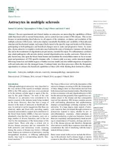
Figure 1. Temporal lobe astrocytoma in a 77 year old woman who experienced syncope. A. Nonenhanced axial CT study shows no abnormality. B. Axial FLAIR MR image demonstrates increased signal intensity along the left temporal lobe (arrow). Astrocytoma was found on biopsy. Negative neurologic symptoms include loss of Categorizing neurologic symptoms with more vision, hearing, cutaneous sensation, or the ability to subtle or fleeting neurologic findings presents a move a body part, and indicate a loss of neurologic challenge. Such symptoms often arise from one of a function. Negative symptoms favor a TIA (if limited number of diseases including transient transient) or stroke (if fixed), and transient sensory ischemic attack (TIA)/stroke, seizures, migraine deficits are the most common presentation of auras (see pages 27-28), and multiple sclerosis. patients with multiple sclerosis (MS)5. Migraine Historical features which help indicate a specific auras, which often start with positive symptoms, disease include whether the apparent neurologic may progress to negative symptoms in the same symptoms are positive or negative, the progression and modality: for example, paresthesias may precede course of the symptoms, and the duration of the cutaneous numbness4. symptoms4.
Progression and course of symptoms Positive or negative symptoms Examples of positive neurologic symptoms include seeing bright lines or shapes, hearing noises, having a burning sensation or paresthesias, or experiencing jerking or repetitive rhythmic movements. Such symptoms indicate active discharge of central nervous system neurons4, as will be encountered with seizures or migraine auras.
The positive neurologic symptoms in seizure typically progress rapidly within a single modality, as do the negative neurologic symptoms of TIA/stroke and MS. The initially positive, then negative symptoms of migraine aura typically slowly progress and may switch from one modality (seeing bright lights) to another (paresthesias). Symptoms of MS characteristically come and go (the
Chapter 4
Stroke, Seizures, Multiple Sclerosis, and Dementia
catch-phrase describing the disease is “multiple lesions in time and space”).
Duration Duration of neurologic symptoms ranges from mere seconds to permanent. Seizures represent the shortest duration process with neurologic symptoms lasting from a few seconds up to a few minutes; many TIAs last shorter than five minutes; migraine auras often last 20 to 30 minutes4; multiple sclerosis attacks by definition last more than 24 hours5, and strokes produce long-term and often permanent neurologic symptoms.
BOTH TRANSIENT ISCHEMIC ATTACKS AND STROKE REQUIRE AGGRESSIVE, TIMELY MANAGEMENT AND WORK-UP The term “transient ischemic attack” (TIA) was originally defined as symptoms or signs of brain ischemia lasting less than 24 hours. The definition has been modified6, recognizing that the original supposition that neurologic symptoms lasting less than 24 hours were not associated with brain infarction is false. In fact, ischemic symptoms lasting between one and twenty-four hours are often associated with brain infarction7. TIAs are currently defined as a transient episode of neurologic dysfunction caused by ischemia without acute infarction6. Even though they are defined as not being associated with infarction, TIAs may still represent the harbinger of a subsequent stroke8, much like a sentinel headache may precede a severe or even fatal aneurysmal subarachnoid hemorrhage (see pages 32-34). For this reason, TIAs require urgent work-up and management, either within the hospital or on a very closely monitored outpatient basis8.
Page 43
Clinical risk assessment for impending stroke after TIA Johnston et al9 used a scoring system based on: age (>60 years = 1 point); blood pressure elevation when first assessed after TIA (systolic > 140 mmHg or diastolic > 90 mmHg = 1 point); clinical features (unilateral weakness = 2 points; isolated speech disturbance = 1 point); duration of TIA (> 60 minutes = 2 points, 10-59 minutes = 1 point; < 10 minutes = 0 points); and diabetes (present = 1 point) to stratify risk, with the estimated 2-day risk of stroke at 8.1% for scores of 6 or 7, 4.1% for scores of 4 or 5; and 1.0% for scores of 3 or less.
Imaging of TIA The goals of imaging in a patient with an apparent TIA include: to exclude intracranial hemorrhage (more typically accompanied by a fixed neurological defect, headache, or both – see page 34 for further discussion of intracranial hemorrhage associated with headache); to evaluate for a possible alternative explanation of the neurologic symptoms such as brain tumor (Figure 2); to document any actual infarct accompanying the apparent TIA (which would, by definition, indicate that the transient symptoms do not, in fact, represent a TIA) (Figure 3); and to evaluate a (usually vascular) source of the TIA, including disease of the carotid bifurcations, intracranial vasculature, and heart (Figure 4). While CT may be used in the emergent setting to exclude hemorrhage, TIA patients by definition have no ongoing symptoms and should therefore be capable of undergoing MRI examination, which should be performed both without and with contrast, and which should include diffusion weighted imaging (DWI) sequences (Figure 3). DWI sequences will typically demonstrate cerebral infarction within minutes of onset, and are typically positive hours before T1 and T2 weighted sequences10. MRI examination performed with gradient echo sequences is also capable of detecting intracranial hemorrhage.
Page 44
Strokes, Seizures, Multiple Sclerosis, and Dementia
Figure 2. Glioblastoma multiforme in a 57 year old woman with transient verbal difficulty initially thought to be ischemic. A. Axial unenhanced T1 weighted image demonstrates a mass in the left temporal lobe with increased signal intensity along the anteromedial margin compatible with recent hemorrhage (arrow). B. Axial T1 postcontrast image demonstrates intense contrast enhancement of the lesion. C. Axial FLAIR image demonstrates the lesion and marked adjacent white matter vasogenic edema (arrow). D. Axial T2 image demonstrates focal areas of decreased signal intensity (arrow) compatible with intratumoral hemorrhage.
Chapter 4
Stroke, Seizures, Multiple Sclerosis, and Dementia
Page 45
Figure 3. Stroke in an 85 year old woman with transient weakness six days prior to MR study. A. Axial unenhanced CT study shows no abnormality. B. Axial T1 MR weighted image also shows no abnormality. C. Axial FLAIR MR image demonstrates several focal areas of increased signal intensity, including one in the right cerebral hemisphere white matter (arrow). D. Axial diffusion weighted MR image demonstrates increased signal intensity indicating diffusion restriction (arrow) at the location of the lesion noted on image C., whereas the other white matter lesions seen on image C. show no diffusion restriction. These imaging findings indicate an acute infarction, and even through the patient’s symptoms completely resolved she did not, by definition, have a TIA. The other areas of increased signal in C. likely represent chronic microvascular ischemic changes.
Page 46
Strokes, Seizures, Multiple Sclerosis, and Dementia
Figure 4. Extensive vascular disease in a 74 year old man with hypertension and a 2 minute episode of dizziness and sweating. Brain MR study (not shown) was normal. A. Arch and carotid MR angiogram shows multiple irregularities in the vascular tree including an approximately 75% stenosis at the origin of the right internal carotid artery (arrow). B. Axial magnetic resonance angiogram source images from the circle of Willis study demonstrates nearly complete occlusion of the right intracranial internal carotid artery (arrow). C. MR angiogram maximum intensity projection of the circle of Willis also demonstrates severe stenosis of the intracranial internal carotid artery (arrow).
Chapter 4
Stroke, Seizures, Multiple Sclerosis, and Dementia
Magnetic resonance imaging following TIA may include a magnetic resonance angiogram (MRA) for evaluation of the arch and carotid arteries (typically performed with contrast material, and using the same bolus of contrast as the contrast-enhanced brain MRI), and imaging of the circle of Willis(also known as MRA-COW which may be performed either without or with contrast material) (Figure 4). For patients who cannot undergo MRI (because of aneurysm clips, pacers, retained metallic foreign bodies within the orbit, etc.), CT of the brain without and with contrast, and CT angiography of the arch, carotids, and circle of Willis may be performed. Ultrasound examination of the carotid arteries may also be used to screen for carotid stenosis, ulceration, dissection, hematoma, and aneurysms. For further discussion of vascular evaluation of the arch, carotid arteries, and intracranial vasculature, see page 170. Transthoracic or transesophageal echocardiography (with greater sensitivity) evaluates for cardiac sources of emboli causing TIAs.
Imaging of Stroke Patients with obvious stroke belong in an emergency room or in the hospital. The critical decision regarding treatment of these patients is whether to administer fibrinolytic therapy as soon as possible11, at present limited to recombinant tissuetype plasminogen activator or tPA (alteplase). ER physicians and neurologists in stroke centers usually make this decision on the basis of multiple criteria including: duration of symptoms of less than 4.5 hours; historical exclusion criteria (stroke or head trauma in the previous 3 months, previous intracranial hemorrhage, major surgery in the prior 14 days, etc.); clinical exclusion criteria (spontaneously clearing stroke symptoms, minor or isolated neurologic signs, persistent blood pressure elevation, etc.); laboratory exclusion criteria (platelets < 100,000 cc3, serum glucose < 50 mg/dl, INR > 1.7, etc.); and CT exclusion criteria (evidence of hemorrhage or evidence of multilobar infarction with hypodensity involving greater than 33% of the cerebral hemisphere (Figure 5)). Given the multiple exclusion criteria, few patients are truly eligible for
Page 47
alteplase. For those who are eligible, the increased chances of a complete recovery (38% versus 21% with placebo) must be weighed against the approximately 10-fold increase in symptomatic intracerebral hemorrhage12. Even more controversial than the intravenous administration of alteplase within 4.5 hours is the administration of IV alteplase after this time-limited therapeutic window (based on imaging studies) or intra-arterial administration of the same drug (also based on imaging studies). In explanation: both contrast-enhanced CT and contrast-enhanced MR are capable of creating perfusion maps of the brain following stroke. These maps differentiate infarcted brain (incapable of recovery) from stunned but not infarcted brain, which is at least theoretically capable of recovery. Preventing infarction of stunned brain is the purpose of administering alteplase and some centers use either CT or MR perfusion imaging to supplement the usual exclusion rules and time from the acute event to make decisions regarding intravenous alteplase10. In addition, both CTA and MRA may be performed acutely to delineate intraarterial thrombus, which (particularly if accompanied by stunned but not yet infarcted brain) forms the target of intra-arterial alteplase. Perfusion imaging studies to evaluate patients for possible alteplase therapy (either intravenous or intra-arterial) are typically done emergently within the setting of a stroke center. In patients who are not candidates for alteplase, CT still needs to be performed to evaluate for intracranial hemorrhage. In addition, the same MR imaging considerations listed for patients with TIAs above apply to patients with stroke: MR imaging of the brain without and with contrast should be performed to document the stroke (Figure 6) and exclude alternative diagnoses (Figure 7); vascular imaging should be performed, including either MRA of the arch and carotids and MRA-COW (Figure 6) or CTA of the arch and carotids and COW or ultrasound of the carotids; and echocardiography should be performed to exclude a cardiac source of embolism.
Page 48
Strokes, Seizures, Multiple Sclerosis, and Dementia
Figure 5. Huge acute cerebral stroke in an 88 year old woman who woke with left hemiparesis. A. Unenhanced axial CT at the level of the suprasellar cistern demonstrates hypodensity and effaced sulci in the right temporal lobe (arrow). Note the calcified vascular tree. B. Unenhanced axial CT superior to A. also demonstrates hypodensity and effaced sulci (arrow). C. Unenhanced axial CT at the level of the basal ganglia demonstrates a large area of the right cerebral hemisphere (basically the entire distribution of the right middle cerebral artery) with effaced sulci and hypodensity (arrows). D. Unenhanced axial CT through the mid lateral ventricles shows extension of the large, acute infarct through the cerebral convexity. Such a large acute stroke, involving more than 33% of the hemisphere, contraindicates fibrinolytic therapy for stroke.
Chapter 4
Stroke, Seizures, Multiple Sclerosis, and Dementia
Page 49
Figure 6. Acute left thalamic stroke in a 76 year old man with acute dizziness. A Unenhanced axial CT demonstrates a subtle hypodensity in the left thalamus (arrow). B. Axial T2 weighted MR image showing focal increased signal intensity in the left thalamus (arrow) C. Axial diffusion weighted MR image demonstrates increased signal indicating restricted diffusion (arrow). D. Circle of Willis magnetic resonance angiogram maximum intensity projection shows severe stenosis of the contralateral right middle cerebral artery, documenting severe vascular disease at a location other than that causing the patient’s acute stroke.
Page 50
Strokes, Seizures, Multiple Sclerosis, and Dementia
Figure 7. Viral cerebrellitis in a 48 year old woman with unsteady gait, dizziness, and blurred vision. A Sagittal magnetic resonance FLAIR image through the right cerebellar hemisphere shows abnormal signal (arrow). B. Sagittal magnetic resonance FLAIR image through the contralateral cerebellar hemisphere shows much less abnormal signal (arrow). The patient had viral meningitis six years before the onset of symptoms. .
PATIENTS WITH SUSPECTED EPILEPSY SHOULD BE SENT TO A SPECIALIST FOR WORK-UP, AND MR SHOULD BE PERFORMED Seizures may be the result of a reversible medical disorder or epilepsy. Multiple medical disorders may provoke seizure, including: hypoglycemia; nonketotic hyperglycemia; rapid falls in serum sodium concentration; hypocalcemia; renal failure/uremia; hyperthyroidism; acute intermittent porphyria; cerebral anoxia (e.g. carbon monoxide poisoning, drowning), and drug toxicity or withdrawal13. Initial testing should be directed toward excluding such medical disorders. In the acute setting following the first seizure, a head CT will usually be obtained mainly to exclude
intracranial hemorrhage, brain abscess, and tumor (Figure 8)14. Assuming that medical disorders have been largely or completely excluded and epilepsy remains the likely diagnosis in a patient with seizure, most primary care practitioners refer the patient to specialists or subspecialists for further evaluation13, since epilepsy is relatively rare, the diagnosis has such a significant impact, and treatment is often lifelong. With respect to imaging epilepsy, the goal is to find a structural cause of the epilepsy, particularly in those cases where surgery is contemplated secondary to failed medical therapy. Structural causes include hippocampal sclerosis, brain tumor, dysplasia, and vascular malformations15. Knake et al16 showed that imaging performed on a high field strength magnet (3.0T rather than 1.5T) depicted causative lesions with much greater sensitivity and
Chapter 4
Stroke, Seizures, Multiple Sclerosis, and Dementia
Page 51
Figure 8. Subdural hematoma in a 60 year old anticoagulated woman with new seizures. A. Axial unenhanced T1 weighted MR image shows a crescent of increased signal along the left cerebral hemisphere (arrow), typical of a subdural hematoma. B. Axial FLAIR image also demonstrates a crescent of increased signal along the left cerebral hemisphere (arrow). accuracy: in a subgroup of 23 patients with a normal interpretation at 1.5T, new lesions were detected on a 3.0T study in 15 (63%). Phal et al 17 found that epilepsy imaging performed on a 3.0T MRI showed increased imaging quality, detected more structural lesions, and improved characterization of lesions compared to 1.5T. While imaging at 1.5T may be diagnostic in some cases, it is not possible to predict which cases will be falsely negative or equivocal. Given the necessity to re-image with a 3.0T MR in negative or equivocal cases, direct referral for initial imaging at 3.0T performed at an epilepsy center may be preferred when feasible.
PATIENTS WITH POSSIBLE MULTIPLE SCLEROSIS SHOULD UNDERGO MR While the hallmark of multiple sclerosis (MS) is multiple lesions in time and space (neurologic location), most patients (approximately 85%) will
initially present with a clinically isolated syndrome18, usually one of the following: 1. 2.
3. 4.
Transient sensory or motor deficits, affecting 40-50% of patients5. Monocular visual loss or visual field loss from optic neuritis, affecting 15-20% of patients19. Diplopia, affecting about 7% of patients. Balance problems and/or vertigo, affecting about 5% of patients.
Patients who present with a clinically isolated syndrome should undergo contrast-enhanced MR imaging20. While diagnosis of MS was at one time confirmed using the Poser criteria, which require at least two clinical episodes, waiting for a second
Page 52
Strokes, Seizures, Multiple Sclerosis, and Dementia
Figure 9. Multiple sclerosis in a 52 year old man with new onset poor coordination of the lower extremities. A Axial FLAIR brain MR shows multiple foci increased signal intensity (arrows). B. Axial FLAIR brain MR at a slightly lower location again shows multiple foci of increased signal intensity (arrows). C. Axial T1 weighted image shows decreased signal intensity at the location of the increased signal on B. D. Sagittal FLAIR brain MR shows multiple lesions of the corpus callosum (arrows), typical of multiple sclerosis.
Chapter 4
Stroke, Seizures, Multiple Sclerosis, and Dementia
episode is no longer acceptable since there is disease-modifying therapy5. Therefore, the McDonald criteria are used instead of the Poser criteria. The McDonald criteria allow a diagnosis of multiple sclerosis with one clinical attack and either abnormal cerebrospinal fluid protein with oligoclonal bands or an abnormal MRI, with three out of the following four MRI abnormalities: one gadolinium enhancing or nine T2-hyperintense lesions (if no gadolinium enhancing lesion is seen); one or more infratentorial lesions; one or more juxtacortical lesions; and three or more periventricular lesions21. As indicated in the McDonald criteria, MR abnormalities associated with multiple sclerosis include areas of increased signal on T2 weighted images (Figure 9), which sometimes show matched decreased signal on T1 weighted images and contrast enhancement. These are, of course, not specific findings and similar imaging features may be seen in acute disseminated encephalomyelitis, vasculitis, Lyme disease, and migraine headache. Systemic lupus erythematosis may not only demonstrate similar lesions at MR, but occasionally presents with recurrent neurologic symptoms prior to the systemic manifestations of the disease, complicating diagnosis20. Patients with multiple sclerosis may be followed with serial MR examinations to document progression or regression of lesions in response to therapy. Enhancing lesions indicate breakdown of the blood-brain barrier, which is generally taken as a proxy for “inflammation” and disease activity22. Several maneuvers may document additional contrast-enhancing lesions compared to standard technique (including magnetization transfer pulse sequences, triple-dose contrast, and delayed imaging), but such techniques are not routinely required and are not incorporated into MS diagnostic criteria22.
PATIENTS WITH DEMENTIA SHOULD UNDERGO MR The fourth edition of the American Psychiatric Association Diagnostic and Statistical Manual (also known as “DSM-IV”) defines dementia as a disorder characterized by impairment of memory and at least
Page 53
one other cognitive domain (aphasia, apraxia, agnosia, or executive function) which represents a decline from a prior level of function severe enough to interfere with daily function and independence 23. Self-reported memory loss does not appear to correlate with the development of dementia, while spouse (or other informant) reported memory loss is a much better indicator of the presence (or future development) of dementia24. The diagnosis typically rests on a clinical history, supplemented by cognitive tests such as the Mini-Mental State Examination25, Clinical Dementia Rating26, the “mini-cog” test27 or formal neuropsychologic testing24. Major dementia syndromes include Alzheimer’s disease (accounting for 60 to 80% of the total), vascular dementia, dementia with Lewy bodies, dementia with Parkinson’s disease, and frontotemporal dementia28. Normal pressure hydrocephalus (NPH) may also be associated with dementia (along with gait disturbance and urinary incontinence), and is characterized by pathologically enlarged ventricles with normal opening pressure on lumbar puncture29. This diagnosis may be confirmed by the patient’s clinical response to removal of 30 – 50 mL of cerebrospinal fluid, although there is little consensus regarding the diagnosis of NPH or the selection of patients with possible NPH for therapeutic shunt placement29. The American Association of Neurology recommends imaging with either CT or MRI in the routine initial evaluation of all patients with dementia30. MR imaging (preferred to CT) can accomplish the following in patients with newly diagnosed dementia: 1. 2. 3.
4.
5.
Exclude subdural hematoma. Exclude cerebral neoplasm. Evaluate for disproportionate distention of the lateral ventricles relative to the sulci, suggestive of normal pressure hydrocephalus (NPH) (Figure 10). Evaluate for disproportionate frontal lobe atrophy, suggesting frontotemporal dementia. Evaluate for multiple prior strokes suggesting vascular dementia.
Page 54
Strokes, Seizures, Multiple Sclerosis, and Dementia
Most MR studies will demonstrate nonspecific generalized atrophy, since this is the most common finding in Alzheimer’s disease, and Alzheimer’s disease accounts for the majority of dementias. While MR may also allow hippocampal volume measurement in Alzheimer’s disease, it is not clear that this finding adds to the clinical diagnosis31. FDG-PET may be useful in distinguishing Alzheimer’s disease from frontotemporal dementia, but typically there is little therapeutic imperative to make this distinction31.
SUMMARY
Figure 10. Normal pressure hydrocephalus in a 78 year old with dementia, incontinence, and gait abnormality. Axial unenhanced CT study shows bilaterally enlarged ventricles with disproportionate distension compared to the sulci.
Neurologic symptoms may indicate migraine aura, TIA/stroke, seizure, or multiple sclerosis. TIA and stroke require urgent work-up, seizures require specialist assessment and MR imaging (preferably at 3.0T if possible), and multiple sclerosis requires MR scanning. Dementia patients should undergo MR imaging upon initial diagnosis.
Chapter 4
Stroke, Seizures, Multiple Sclerosis, and Dementia
REFERENCES Alboni P, Brignole M, Menozzi C et al. Diagnostic value of history in patients with syncope with or without heart disease. J Am Coll Cardiol 2001;37:1921 – 1928. 2 Pires LA, Ganji JR, Jarandila R, Steele R. Diagnostic patterns and temporal trends in the evaluation of adult patients hospitalized with syncope. Arch Intern Med 2001;161:1889 – 1895. 3 Olshansky B. Evaluation of syncope in adults. UpToDate, accessed 10/21/09. 4 Caplan LR. Differential diagnosis of brain ischemia. UpToDate, accessed 10/19/09. 5 Pruit A. Management of multiple sclerosis. Chapter 17 of Goroll AH, Mulley AG (editors): Primary Care Medicine: Office Evaluation and Management of the Adult Patient. Wolters Kluwer/Lippincott Williams & Wilkins, Philadelphia, 2009. 6 Kistler JP, Furie KL, Ay H. Definition of transient ischemic attack. UpToDate, accessed 10/26/09. 7 Caplan LR. Overview of the evaluation of stroke. UpToDate, accesed 10/10/09. 8 Kistler JP, Furie KL, Ay H. Initial evaluation and management of transient ischemic attack and minor stroke. UpToDate, accessed 10/10/09. 9 Johnston SC, Rothwell PM, Nguyen-Huynh MN et al. Validation and refinement of scores predict very early stroke risk after transient ischaemic attack. Lancet 2007;366:29-292. 10 Oliveira-Filho J, Koroschetz WJ. Neuroimaging of acute ischemic stroke. 11 Oliveira-Filho J, Samuels OB. Fibrinolytic (thrombolytic) therapy for acute ischemic stroke. 12 Tissue plasminogen activator for acute stroke. The National nstitute of Neurological Disorders and Stroke rt-PA Stroke Study Group. N Engl J Med 1995;333:1581-1587. 13 Schachter SC. Evaluation of the first seizure in adults. UpToDate, accessed 10/10/09. 1
Page 55
Harden CL, Huff JS, Schwartz TH et al. Reassessment: neuroimaging in the emergency patients presenting with seizure (an evidence-based review): report of the Therapeutics and Technology Assessment Subcommittee of the American Academy of Neurology. Neurology 2007;69:17721780. 15 Hirsch LJ, Arif H. Neuroimaging in the evaluation of seizures and epilepsy. UpToDate accessed 10/21/09. 16 Knake S, Triantafyllou C, Wald LL et al. 3T phased array MRI improves the presurgical evaluation in focal epilepsies: a prospective study. Neurology 2005;65:1026-1031. 17 Phal PM, Usamanov A, Nesbit GM et al. Qualitative comparison of 3-T and 1.5-T MRI in the evaluation of epilepsy. Am J Roentgen 2008: 191:890-895. 18 Stern SDC, Cifu AS, Altkorn D. I have a patient with dizziness. How do I determine the cause? Chapter 13 in Symptoms to Diagnosis: an EvidenceBased Guide. McGraw Hill Medical, New York, 2010. 19 Olek MJ. Diagnosis of multiple sclerosis in adults. UpToDate, accessed 10/26/09. 20 Olek MJ. Clinically isolated syndromes suggestive of multiple sclerosis. UpToDate, accessed 10/26/09. 21 McDonald WI, Compston A, Edan G et al. Recommended diagnostic criteria for multiple sclerosis: guidelines from International Panel on the diagnosis of multiple sclerosis. Ann Neurol; 2001:50:121-127. 22 Simon JH. Update on multiple sclerosis. Radiol Clin N Am 2006;44:79-100. 23 American Psychiatric Association Diagnostic and Statistical Manual, 4th Edition, APA Press, Washington DC, 1994. 24 Shadlen MF, Larson EB. Evaluation of cognitive impairment and dementia. UpToDate, accessed 10/10/09. 25 Tangalos EG, Smith GE, Ivnik RJ et al. The MiniMental State Examination in general medical practice: clinical utility and acceptance. Mayo Clin Proc 1996;71:829-837. 14
Page 56
Strokes, Seizures, Multiple Sclerosis, and Dementia
Morris JC. The Clinical Dementia Rating (CDR): current version and scoring rules. Neurology 1993;43:2412-2414. 27 Borson S, Scanlan J, Brush M et al. The mini-cog: a cognitive “vital signs” measure for dementia screening in multi-lingual elderly. Int J Geriatr Psychiatry 2000;15:1021-1027. 28 Shadlen MF, Larson EB. Dementia syndromes. UpToDate, accessed 10/21/09. 29 Graff-Radford NR. Normal pressure hydrocephalus. UpToDate, accessed 10/24/09. 30 Knopman DS, DeKosky ST, Cummings JL et al. Practice parameter: diagnosis of dementia (an evidence-based review). Report of the Quality Standards Subcommittee of the American Academy of Neurology. Neurology 2001;56:1143-1153. 31 Grabowski TJ. Clinical manifestations and diagnosis of Alzheimer’s disease. UpToDate accessed 10/21/09. 26