Downloaded from www.ajronline.org by 37.44.207.151 on 01/15/17 from IP address 37.44.207.151. Copyright ARRS. For personal use only; all rights reserved
Lowe et al. Neonatal Spine Sonography
Pe d i a t r i c I m a g i n g • P i c t o r i a l E s s a y
Sonography of the Neonatal Spine: Part 2, Spinal Disorders Lisa H. Lowe1,2 Andrew J. Johanek1,3 Charlotte W. Moore1,2 Lowe LH, Johanek AJ, Moore CW
OBJECTIVE. The objective of part 2 of this article is to focus on key imaging features of common skin-covered spinal anomalies (spina bifida occulta) and to distinguish them from normal variants (previously discussed in part 1). CONCLUSION. Modern imaging technology allows accurate neonatal spinal sonographic screening and the characterization of spinal abnormalities within the first few days of life. It is useful to determine the type of lesion present and to guide the type and timing of therapy. ongenital spinal anomalies are the result of three basic abnormal embryologic processes (see part 1 of this article under Embryology). First, premature separation of the skin ectoderm from the neural tube can lead to entrapment of mesodermal elements, such as fat. Second, failed neurulation leads to dysraphisms, such as myelomeningocele. Last, anomalies of the filum terminale, such as fibrolipomas and caudal regression syndrome, are caused by disembryogenesis of the caudal cell mass [1–3].
C
Keywords: neonatal imaging, neuroradiology, pediatric radiology, sonography, spine DOI:10.2214/AJR.05.2160 Received December 16, 2005; accepted after revision February 28, 2006. Awarded Bronze Medal poster exhibit at the 2005 annual meeting of the American Roentgen Ray Society, New Orleans, LA. 1Department
of Radiology, The University of Missouri–Kansas City, Kansas City, MO.
2Department
of Radiology, Children’s Mercy Hospital and Clinics, 2401 Gillham Rd., Kansas City, MO 64108. Address correspondence to L. H. Howe (
[email protected]).
3Department of Radiology, St. Luke’s Hospital, Kansas City,
MO. CME This article is available for CME credit. See www.arrs.org for more information. AJR 2007; 188:739–744 0361–803X/07/1883–739 © American Roentgen Ray Society
AJR:188, March 2007
Classification Congenital spinal dysraphisms can be classified on the basis of the presence or absence of a soft-tissue mass and skin covering [2]. Those without a mass include tethered cord, diastematomyelia, anterior sacral meningocele, and spinal lipoma. Those with a skincovered soft-tissue mass include lipomyelomeningocele and myelocystocele. Those with a back mass but without skin covering include myelomeningocele and myelocele. Several common disorders will be discussed in this article, including tethered cord, spinal lipoma, lipomyelomeningocele, fatty filum and filar fibrolipoma, and caudal regression syndrome. Finally, the usefulness of sonography in failed lumbar puncture with subdural hematoma will be discussed. Tethered Cord Tethered cord, or low-lying conus medullaris, is caused by incomplete regressive differentiation and failed involution of the ter-
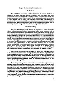
minal cord. Symptoms occur because of traction on the abnormally anchored filum terminale and adjacent nerve roots. Children with tethered cord may present at any age with difficulty ambulating, weakness, stiffness, abnormal reflexes, bladder dysfunction, and, less often, bowel dysfunction [2]. Sonographically, tethered cord is diagnosed in neonates by the presence of a low-lying conus (below the L2–L3 disk space) and lack of normal nerve root motion during realtime sonography [4, 5] (Fig. 1). However, in older patients, clinical correlation is required because the conus may be normally positioned but still be tethered (tight filum syndrome) [6]. In this situation, assessment of normal nerve root motion, whenever possible, at real-time imaging is more important [1, 6]. Other associated spinal findings include a thickened filum terminale, fibrolipoma, spinal dysraphism, syringomyelia, scoliosis, congenital spinal masses (lipomas, dermoids), cysts (myelocele), and sinus tracts that contain fluid (Fig. 2). Other nonneurologic anomalies are common as well, including tracheoesophageal fistula, congenital heart disease, and renal anomalies (VATER syndrome). Treatment centers on surgical release of the filum and preservation of nerve root function. Early recognition and treatment of tethered cord is important to preserve normal function, which may be irreversibly lost if treatment is delayed [6]. The prognosis varies with the severity of anomalies present. Retethering is common with normal growth and may require re-release surgery.
739
Downloaded from www.ajronline.org by 37.44.207.151 on 01/15/17 from IP address 37.44.207.151. Copyright ARRS. For personal use only; all rights reserved
Lowe et al. Spinal Lipoma Spinal lipomas are caused by premature disjunction (embryologic separation of neural ectoderm from cutaneous tissue elements) that allows mesenchyma to be trapped between the neural folds and remain in contact with the neural canal [1, 2]. Spinal lipomas are composed of normal fat, may grow significantly during the first year of life, and may change in size with weight. They may be intradural, extradural, or a combination of both. In addition to fat, 84% of lipomas also contain neural tissue or meninges [2, 3]. Associations include tethered cord, dysraphism (4%), fatty filum or lipoma of filum (12%), and vertebral anomalies [1–3] (Fig. 3). On MRI, the mass follows fat signal. The prognosis of these lesions is varied depending on their ability to be resected and the presence and type of various associated anomalies. Treatment consists of resection, when possible, or debulking. Spina Bifida Occulta with Lipomyelomeningocele Spina bifida occulta is defined as any skincovered osseous defect of posterior elements through which various combinations of neural elements (neural placode), meninges, CSF, and adipose tissue protrude [1, 2] (Fig. 4). The cause is defective disjunction and neurulation with entrapped mesenchyma in contact with the incompletely closed neural tube. The presentation is usually at an age younger than 6 months; the disease rarely presents in adulthood. Lipomyelomeningoceles, with an incidence of two in 1,000, encompass 20% of skin-covered lumbosacral masses and 20–50% of occult dysraphic spinal lesions [2, 4]. Treatment and prognosis depend on the specific anomalies present.
740
Fatty Filum and Filar Fibrolipoma Fatty filum and filar lipomas are due to a minor anomaly of canalization and retrogressive differentiation with persistent or dedifferentiated fatty tissue [2]. Minimal fat in filum is often asymptomatic and has been seen in 6% of normal spines at autopsy [1] (Fig. 5). It is considered a normal variant when it is an isolated finding in a normalsize filum (< 1–2 mm) [1, 4]. When the fatty tissue forms a mass, a filar lipoma is diagnosed. Associated anomalies include myelomeningocele and tethered cord. The treatment and prognosis vary depending on the clinical symptoms and specific anomalies present. Caudal Regression Syndrome Caudal regression syndrome, which is thought to be due to abnormal mesodermal formation of the caudal cell mass (possibly from hyperglycemia), affects one in 7,500 children [3]. It occurs most often in children of diabetic mothers (Fig. 6) and is also associated with various other genitourinary, anal, vertebral, and limb anomalies [1, 3]. The presentation and imaging appearance vary with the degree of deformity, ranging from minimal to severe regression of the coccyx, sacrum, and lumbar spine. Progressive absence of bone structures occurs in a caudal to cranial direction [1] (Fig. 6). Subdural Hematoma Subdural hematoma is uncommon in neonates; it may be iatrogenic after failed attempts at neonatal lumbar puncture [7]. Sonography is useful to determine whether the thecal sac is compressed by a hematoma.
If it is not, sonography can be used to determine the best timing and level for a potential reattempt at lumbar puncture [7] (Fig. 7). Conclusion Modern sonography technology allows accurate screening and characterization of spinal abnormalities during the first few days of life. It is useful for determining the type of lesion present in order to guide the type and timing of intervention.
References 1. Barkovich AJ. Normal development of the neonatal and infant brain, skull, and spine. In: Barkovich, AJ. Pediatric neuroimaging, 4th ed. Philadelphia, PA: Lippincott Williams & Wilkins, 2005:710, 723, 732, 735 2. Dick EA, Patel K, Owens CM, De Bruyn R. Spinal ultrasound in infants. Br J Radiol 2002; 75:384–392 3. Unsinn KM, Geley T, Freund MC, Gassner I. US of the spinal cord in newborns: spectrum of normal findings, variants, congenital anomalies, and acquired diseases. RadioGraphics 2000; 20:923–938 4. Byrd SE, Darling CF, McLone DG. Developmental disorders of the pediatric spine. Radiol Clin North Am 1991; 29:711–752 5. Hill CA, Gibson PJ. Ultrasound determination of the normal location of the conus medullaris in neonates. Am J Neuroradiol 1995; 16:469–472 6. Selcuki M, Vatansever S, Inan S, Erdemli E, Bagdatoglu C, Polat A. Is a filum terminale with a normal appearance really normal? Childs Nerv Syst 2003; 19:3–10 7. Coley BD, Shiels WE 2nd, Hogan MJ. Diagnostic and interventional ultrasonography in neonatal and infant lumbar puncture. Pediatr Radiol 2001; 31:399–402
AJR:188, March 2007
Downloaded from www.ajronline.org by 37.44.207.151 on 01/15/17 from IP address 37.44.207.151. Copyright ARRS. For personal use only; all rights reserved
Neonatal Spine Sonography
A
Fig. 1—Tethered cord in 2-day-old boy with multiple congenital anomalies. A, Longitudinal sonogram shows low-lying conus (arrowhead) at L5 vertebra and thickened, echogenic fatty filum (arrow). B, Sagittal T1-weighted MR image confirms thick, fatty filum (arrow) overlapping tethered cord from L4 to S1 level.
B
A
Fig. 2—Syrinx and tethered cord in 1-week-old girl with imperforate anus and scoliosis. A, Longitudinal sonogram reveals low-lying conus at L4 vertebra with hypoechoic cystic space (arrow) expanding lumbar spinal cord. B, Sagittal T2-weighted MR image confirms conus is tethered at S1 level (arrowhead) and lumbar spinal cord contains large, hyperintense, fusiform syrinx (arrow).
B
AJR:188, March 2007
741
Downloaded from www.ajronline.org by 37.44.207.151 on 01/15/17 from IP address 37.44.207.151. Copyright ARRS. For personal use only; all rights reserved
Lowe et al.
A
B
Fig. 3—Intradural lipoma and tethered cord in 2-week-old girl with hairy patch on lower back. A, Longitudinal sonogram reveals typical features of hyperechoic lipoma (calipers) attached to dorsal aspect of thoracolumbar spinal cord. Conus is tethered to mass at L3–L4 disk space (arrow). B, Transverse sonogram at L3 vertebra shows conus (arrow) tethered to dorsal lipoma (arrowhead).
A
C Fig. 4—Lipomyelomeningocele in 1-day-old girl with soft-tissue swelling on lower back. A and B, Longitudinal (A) and transverse (B) sonograms show lumbosacral dysraphism through which spinal cord (straight arrow), hyperechoic fatty tissue (curved arrow), and hypoechoic CSF (arrowhead, B) pass. C, T1-weighted sagittal MR image confirms lumbosacral dysraphism with intra- and extradural adipose tissue (arrows), neural tissue (arrowhead), and tethered cord.
B
742
AJR:188, March 2007
Downloaded from www.ajronline.org by 37.44.207.151 on 01/15/17 from IP address 37.44.207.151. Copyright ARRS. For personal use only; all rights reserved
Neonatal Spine Sonography
A
B
Fig. 5—Fatty filum in 23-week-old boy with sacral dimple who is otherwise developmentally normal. A, Longitudinal sonogram shows focus of segmental increased echogenicity within filum (arrowhead) posterior to L4 vertebral body. B, Axial T1-weighted MR image confirms fat in filum as localized area of increased signal intensity (arrowhead).
Fig. 6—Caudal regression syndrome in 3-day-old girl of diabetic mother. A, Longitudinal sonogram shows blunted distal cord (arrow), typical of caudal regression syndrome. B, Sagittal T1-weighted MR image confirms blunted conus medullaris and associated fat in filum (arrow) as well as absence of sacrum and coccyx (arrowhead).
A
AJR:188, March 2007
B
743
Downloaded from www.ajronline.org by 37.44.207.151 on 01/15/17 from IP address 37.44.207.151. Copyright ARRS. For personal use only; all rights reserved
Lowe et al.
A
B
Fig. 7—Subdural hematoma in febrile 2-month-old boy after multiple attempts at lumbar puncture. A, Longitudinal sonogram identifies hemorrhage as circumferential, echogenic material in subdural space (straight arrow) that displaces dura (curved arrows) from posterior elements (arrowhead) and collapses normal CSF-containing thecal sac. B, Transverse sonogram also reveals circumferential echogenic subdural blood (arrows) obliterating normal CSF, which contains thecal sac.
F O R YO U R I N F O R M AT I O N
The reader’s attention is directed to part 1 accompanying this article, titled “Sonography of the Neonatal Spine: Part 1, Normal Anatomy, Imaging Pitfalls, and Variations That May Simulate Disorders,” which begins on page 733.
F O R YO U R I N F O R M AT I O N
This article is available for CME credit. See www.arrs.org for more information.
744
AJR:188, March 2007