ORIGINAL CONTRIBUTION
First Unaffected Pregnancy Using Preimplantation Genetic Diagnosis for Sickle Cell Anemia Kangpu Xu, PhD Zhong Ming Shi, MD Lucinda L. Veeck, MLT, DSc Mark R. Hughes, MD, PhD Zev Rosenwaks, MD
S
ICKLE CELL ANEMIA IS ONE OF THE
most common human autosomal recessive disorders. It is caused by a mutation substituting thymine for adenine in the sixth codon (GAG to GTG) of the gene for the b-globin chain on chromosome 11p, thereby encoding valine instead of glutamic acid in the sixth position of the globin chain. The frequency of sickle cell trait (carrier status) among the African American population at birth is about 8%, and the incidence of sickle cell anemia at birth is 0.16%, or 1 per 625 births.1 Furthermore, the widespread presence of the sickle gene in other ethnic groups has also been confirmed.2 For example, in urban centers in the United States, nearly 10% of patients with various sickling disorders identify themselves as non–African American.3 Children affected with sickle cell anemia experience recurrent episodes of pain (during sickle cell crises) and increased susceptibility to potentially lifethreatening conditions, including bacterial infections, cerebrovascular accidents, and organ failure. According to US statistics collected between 1981 and 1992, there were 6.8 deaths per 1000 AfSee also Patient Page.
Context Sickle cell anemia is a common autosomal recessive disorder. However, preimplantation genetic diagnosis (PGD) for this severe genetic disorder previously has not been successful. Objective To achieve pregnancy with an unaffected embryo using in vitro fertilization (IVF) and PGD. Design Laboratory analysis of DNA from single cells obtained by biopsy from embryos in 2 IVF attempts, 1 in 1996 and 1 in 1997, to determine the genetic status of each embryo before intrauterine transfer. Setting University hospital in a large metropolitan area. Patients A couple, both carriers of the recessive mutation for sickle cell disease. Interventions Standard IVF treatment, intracytoplasmic sperm injection, embryo biopsy, single-cell polymerase chain reaction and DNA analyses, embryo transfer to uterus, pregnancy confirmation, and prenatal diagnosis by amniocentesis at 16.5 weeks’ gestation. Main Outcome Measure DNA analysis of single blastomeres indicating whether embryos carried the sickle cell mutation, allowing only unaffected or carrier embryos to be transferred. Results The first IVF attempt failed to produce a pregnancy. Of the 7 embryos analyzed in the second attempt, PGD indicated that 4 were normal and 2 were carriers; diagnosis was not possible in 1. Three embryos were transferred to the uterus on the fourth day after oocyte retrieval. A twin pregnancy was confirmed by ultrasonography, and subsequent amniocentesis revealed that both fetuses were unaffected and were not carriers of the sickle cell mutation. The patient delivered healthy twins at 39 weeks’ gestation. Conclusion This first unaffected pregnancy resulting from PGD for sickle cell anemia demonstrates that the technique can be a powerful diagnostic tool for carrier couples who desire a healthy child but wish to avoid the difficult decision of whether to abort an affected fetus. JAMA. 1999;281:1701-1706
rican American children aged 1 to 4 years due to sickle cell disease.4 Life expectancy for persons with sickle cell disease varies and there is an age-related pattern in mortality rates: a peak in patients younger than 5 years, with a gradual increase starting in late adolescence.5 Progress in the treatment of sickle cell disease has been slow.6 At present,
©1999 American Medical Association. All rights reserved.
Downloaded From: http://jama.jamanetwork.com/pdfaccess.ashx?url=/data/journals/jama/4633/ on 01/27/2017
www.jama.com Author Affiliations: The Center for Reproductive Medicine and Infertility and the Department of Obstetrics and Gynecology, Weill Medical College of Cornell University, New York, NY (Drs Xu, Shi, Veeck, and Rosenwaks); and the Department of Reproductive Genetics, Wayne State University, Detroit Medical Center, Detroit, Mich (Dr Hughes). Corresponding Author and Reprints: Zev Rosenwaks, MD, The Center for Reproductive Medicine and Infertility, Weill Medical College of Cornell University, 505 E 70th St, Room HT326a, New York, NY 10021 (e-mail:
[email protected]). JAMA, May 12, 1999—Vol 281, No. 18
1701
PREIMPLANTATION DIAGNOSIS FOR SICKLE CELL ANEMIA
there is no satisfactory treatment for the sickling condition, although blood transfusion may reduce the risk of a first stroke in children7 and gene therapy holds promise for a curative approach.8 Early prenatal diagnosis of the disease is critical because it allows a couple to consider pregnancy termination as an option. The first DNA diagnostic procedure for prenatal purposes was reported 20 years ago.9 Subsequently, it was recognized that the mutation itself affected the cleavage site of a restriction enzyme, DdeI, that could recognize the DNA sequence of CTNAG (N = A, T, C, or G). While DNA from a normal allele (CTGAG) would be digested by the enzyme, DNA from an affected allele in which A is substituted by T (CTGTG) would not.10,11 The resulting differences between DNA fragment sizes can then be recognized by electrophoresis, thus forming the basis for diagnosis. With the advent of polymerase chain reaction (PCR), rapid DNA analysis methods have become available, and these techniques are now widely used for prenatal diagnosis.12-16 An alternative and powerful diagnostic tool for identifying sickle cell status in embryos is preimplantation genetic diagnosis (PGD), which became possible nearly a decade ago.17 Preimplantation genetic diagnosis takes advantage of assisted reproductive techniques in conjunction with modern molecular methods. With PGD, the genetic status of an embryo can be determined before transfer into the uterus after in vitro fertilization (IVF), thus eliminating the risks of bearing a child with the disease. While PGD for sickle cell anemia has been performed in the mouse model,18 routine clinical application in the human previously has not been successful. In this article, we describe our experience using PGD to determine the precise genetic status of embryos generated by assisted reproduction for a couple who are heterozygous carriers of the sickle cell mutation. METHODS A 34-year-old female patient had undergone 2 previous induced abortions 1702
JAMA, May 12, 1999—Vol 281, No. 18
because she was carrying fetuses affected with sickle cell anemia. Genetic diagnosis indicated that both female and male partners were carriers of the sickle cell mutation. After extensive counseling, the couple gave informed consent and elected to undergo preimplantation genetic diagnosis. The study was approved by the Weill Medical College of Cornell University (New York, NY) Institutional Review Board. To confirm the genetic status of the couple and establish a protocol for single-copy gene amplification from single cells, blood was collected from both partners. Lymphocytes were isolated using Ficoll-Paque density gradient separation (Pharmacia Biotech Inc, Piscataway, NJ) with the protocol provided by the manufacturer. Single lymphocytes were loaded into 0.5-mL tubes containing 5 µL of lysis buffer19 and stored at −20°C before trial testing. On the day of preliminary trial testing, sample tubes were removed from the freezer and heated to 65°C for 10 minutes before they were placed back on ice. Five microliters of neutralization buffer was added to each tube. A nested PCR approach was used for the amplification of the region surrounding the sickle cell mutation. The primers used have been described previously.17,19 Polymerase chain reaction was performed after adding a standard mixture of all components, including 2.5 mmol of Mg2+, 0.2 mmol of dNTPs (containing dATP, dCTP, dGTP, and dTTP, Perkin Elmer, Foster City, Calif), 100 ng of primers, and 2 U of Taq polymerase (AmpliTaq, Perkin-Elmer). A hot start at 95°C was applied for 3 minutes to ensure complete denaturation of the template. For the PCR profile, the following parameters were used: 93°C denaturation for 30 seconds; 50°C for 40 seconds for annealing; and 72°C for 45 seconds for extension. A total of 20 amplification cycles were applied for outer primers. For the inner primer set, identical parameters were used, except that the annealing temperature was raised to 55°C and a total of 40 cycles were used. Following the nested PCR amplification, 18 µL of amplified product was
digested with the restriction enzyme DdeI (GIBCO/BRL, Rockville, Md) for 3 hours. Subsequently, 10 µL of digested product was run on a 10% acrylamide gel. On completion of electrophoresis, the gel was stained with ethidium bromide and photographed by UV transillumination. As predicted, unaffected DNA showed 3 bands (201, 90, and 74 base pairs [bp]), carrier DNA showed 4 bands (291, 201, 90, and 74 bp), and an affected homozygous sample showed only 2 bands (291 and 74 bp). Testing of single lymphocytes from the male and female subjects showed the same predicted patterns (4 bands of predicted sizes), together with an unaffected DNA control (3 bands) (FIGURE 1). The IVF procedure has been described previously.20 Briefly, to ensure that several embryos would become available for DNA analysis, multiple ovarian follicular development was initiated with gonadotropin therapy. After pituitary desensitization with gonadotropin hormone–releasing hormone agonist (leuprolide acetate, TAP Pharmaceutical, Chicago, Ill), ovarian stimulation was begun on day 3 of the ensuing menstrual cycle using intramuscular administration of a combination of urofollitropin (75 U of pure follicle-stimulating hormone) and menotropins (150 U of folliclestimulating hormone and luteinizing hormone) (Serono Laboratories, Norwell, Mass). Follicular growth was monitored by daily serum estradiol levels and pelvic ultrasonograms. To induce final oocyte maturation, 3300 IU of human chorionic gonadotropin was administered when 2 follicles of 18 mm in average diameter were observed on ultrasonogram. Transvaginal oocyte retrieval was performed 35 hours later. To avoid sperm contamination and possible amplification of sperm DNA, intracytoplasmic sperm injection was used. After 16 hours of incubation, fertilization was confirmed by the identification of 2 pronuclei. Normally fertilized concepti were then transferred to droplets of human tubal fluid (made on site) supplemented with 15% ma-
©1999 American Medical Association. All rights reserved.
Downloaded From: http://jama.jamanetwork.com/pdfaccess.ashx?url=/data/journals/jama/4633/ on 01/27/2017
PREIMPLANTATION DIAGNOSIS FOR SICKLE CELL ANEMIA
ternal serum under mineral oil (ER Squibb & Sons Inc, Princeton, NJ). Biopsy was performed on the morning of the third day after harvest. All embryos were maintained at 37°C in an atmosphere of 5% carbon dioxide. Cleavage rate and morphologic appearance were assessed daily. Blastomere biopsy was carried out in the early morning, approximately 65 hours after oocyte collection. Briefly, a holding pipette was used to stabilize the embryo (at the 9 o’clock position). A hole was made at the 3 o’clock position by expelling a small amount of acidified Tyrode solution (pH, 2.35) onto the zona pellucida through a smallbore pipette. Reverse suction was applied as soon as a hole of appropriate size was created to reduce possible damage caused by exposing the blastomeres to the acidic solution. A large inner-diameter biopsy pipette replaced the pipette containing the acidified Tyrode solution. Subsequently, 1 or 2 cells were aspirated, depending on the total cell number of the embryo. Blastomeres were rinsed in biopsy medium 3 times before loading into a PCR tube containing 5 µL of lysis buffer. Tubes were processed and PCR amplification and restriction enzyme analysis were performed as described herein. Micromanipulated embryos were further cultured in medium droplets overnight. Embryo transfer was performed in the afternoon of day 4. Pregnancy was determined by serum b–human chorionic gonadotropin measurement on cycle days 28 and 35, followed by ultrasonographic assessment at 7 weeks’ gestation. The genetic status of the fetuses was confirmed after amniocentesis by an independent laboratory. Cultured amniocytes were also sent to our laboratory for follow-up PCR analysis. RESULTS Polymerase chain reaction and restriction enzyme analysis of single lymphocytes from both the female and male partners clearly indicated that each carried the sickle cell mutation. Forty-six of 48 single lymphocytes, 24 from the
ase chain reaction amplification was successful in 7 of 9 blastomeres (5 of 6 embryos). Amplification failed in 1 cell from embryo 5 and in 1 cell from embryo 6. Restriction enzyme digestion demonstrated that 4 were homozygous unaffected, 2 were carriers (FIGURE 2), and 1 was of unknown status due to PCR amplification failure. On the afternoon of day 4, all concepti that underwent biopsy demonstrated further cleavage. Selection of embryos for transfer was based on PGD diagnosis, growth rate, and morphology. Two unaffected embryos were of poor quality, displaying slow cleavage and multinucleation (embryo 2) or high fragmentation (embryo 4) and therefore were not suitable for transfer (TABLE). Because there were only 3 highquality transferable embryos—2 unaffected (embryo 1 with 15 cells; embryo 6 with 8 cells) and 1 carrier (embryo 8 with 10 cells) (FIGURE 3 and the Table)—and because the patient was willing to accept a fetus of carrier status, all 3 embryos were transferred. A twin pregnancy was confirmed by ultrasonography at 7 weeks. Amnio-
female and 24 from the male, were successfully amplified. As predicted, 4 bands of correct size were obtained from both partners. An example of the gel is shown in Figure 1. During the first IVF attempt in November 1996, 18 oocytes were retrieved. Four of 6 mature oocytes were normally fertilized after single-sperm injection by intracytoplasmic sperm injection, yielding 4 embryos. Embryo biopsy was performed on all 4 embryos on day 3 by removing a single blastomere from each conceptus. Polymerase chain reaction and restriction enzyme analysis revealed that 1 was homozygous unaffected, 2 were carriers, and 1 was homozygous affected. Transfer of 1 unaffected embryo on day 4 failed to result in a pregnancy. The second IVF attempt was initiated in August 1997, during which 16 oocytes were retrieved. Of those, 8 were mature and underwent intracytoplasmic sperm injection. Seven concepti cleaved at least once by the following day. On the morning of the third day, 2 cells were removed from 2 embryos and 1 cell from 5 embryos. Polymer-
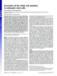
Figure 1. Establishment of Single-Cell PCR and Confirmation of the Carrier Status of Female and Male by Restriction Enzyme Analysis M
1
2
3
4
5
6
7
8
9
10
364 bp 291 bp
201 bp
90 bp 74 bp
βA/βS
βA/βA
βS/βS
Female
Male
PCR indicates polymerase chain reaction; bp, base pair; and M, DNA size marker (100-bp ladder). Lanes 1, 3, 5, 7, and 9 are PCR-amplified but undigested DNA (364 bp), and lanes 2, 4, 6, 8, and 10 are DdeI-digested DNA. Lanes 1 and 2 are from a known carrier DNA (bA/bs, 4 bands); lanes 3 and 4 are from a known unaffected DNA (bA/bA, 3 bands); lanes 5 and 6 are from known affected DNA (bs/bs, 2 bands); lanes 7 and 8 are from the female partner; and lanes 9 and 10 are from the male partner.
©1999 American Medical Association. All rights reserved.
Downloaded From: http://jama.jamanetwork.com/pdfaccess.ashx?url=/data/journals/jama/4633/ on 01/27/2017
JAMA, May 12, 1999—Vol 281, No. 18
1703
PREIMPLANTATION DIAGNOSIS FOR SICKLE CELL ANEMIA
centesis performed by an independent laboratory at 16.5 weeks revealed that neither fetus harbored the sickle cell mutation. DNA analysis of amniocytes shipped from the prenatal diagnostic laboratory to our own laboratory also showed that both fetuses were unaffected (FIGURE 4). The patient delivered healthy, unaffected fraternal twin girls at 39 weeks’ gestation. COMMENT After natural conception, couples who carry autosomal recessive mutations risk a 25% chance of delivering an af-
fected child, and half of the offspring may carry the mutation. Although prenatal testing is currently available, some couples have strong personal objections to aborting affected fetuses. For these couples, PGD provides a realistic alternative to prenatal testing. Although the first pregnancy achieved by PGD for sex determination to avoid the transmission of a sexlinked disorder occurred nearly a decade ago,17 PGD for single-gene defects is still in the experimental stage because of its complexity and technical difficulties. At present, PGD is prima-
Figure 2. Restriction Analysis (DdeI) of PCR-Amplified DNA From Each Blastomere From 6 Embryos M
1
2
3
4
5
6
7
8
M
1
2
3
4
5
6
7
8
364 bp 291 bp 201 bp
90 bp 74 bp βA/βA 1
βA/βA 2
βA/βS 3
βA/βA Control
βA/βA 1
βA/βA 2
βA/βS 3
βA/βS Sperm
PCR indicates polymerase chain reaction; bp, base pair; and M, DNA size marker (100-bp ladder). For both panels, lanes 1, 3, 5, and 7 are PCR amplified but undigested DNA (364 bp) and lanes 2, 4, 6, and 8 are DdeIdigested DNA. Left, Lanes 1 and 2 (products from the blastomere of embryo 1) = bA/bA; lanes 3 and 4 (embryo 2) = bA/bA; lanes 5 and 6 (embryo 3) = bA/bs; and lanes 7 and 8 (unaffected DNA control) = bA/bA. Right, Lanes 1 and 2 (products from the blastomere of embryo 4) = bA/bA; lanes 3 and 4 (embryo 6) = bA/bA; lanes 5 and 6 (embryo 8) = bA/bs; and lanes 7 and 8 (products from spermatozoa of the male partner) = bA/bs. Note that in lane 8 on the left and lanes 2 and 4 on the right, a faint band (