Chiumello and Brioni Critical Care (2016) 20:132 DOI 10.1186/s13054-016-1304-7
REVIEW
Open Access
Severe hypoxemia: which strategy to choose Davide Chiumello1,2* and Matteo Brioni2
Abstract Background: Acute respiratory distress syndrome (ARDS) is characterized by a noncardiogenic pulmonary edema with bilateral chest X-ray opacities and reduction in lung compliance, and the hallmark of the syndrome is hypoxemia refractory to oxygen therapy. Severe hypoxemia (PaO2/FiO2 < 100 mmHg), which defines severe ARDS, can be found in 20–30 % of the patients and is associated with the highest mortality rate. Although the standard supportive treatment remains mechanical ventilation (noninvasive and invasive), possible adjuvant therapies can be considered. We performed an up-to-date clinical review of the possible available strategies for ARDS patients with severe hypoxemia. Main results: In summary, in moderate-to-severe ARDS or in the presence of other organ failure, noninvasive ventilatory support presents a high risk of failure: in those cases the risk/benefit of delayed mechanical ventilation should be evaluated carefully. Tailoring mechanical ventilation to the individual patient is fundamental to reduce the risk of ventilation-induced lung injury (VILI): it is mandatory to apply a low tidal volume, while the optimal level of positive end-expiratory pressure should be selected after a stratification of the severity of the disease, also taking into account lung recruitability; monitoring transpulmonary pressure or airway driving pressure can help to avoid lung overstress. Targeting oxygenation of 88–92 % and tolerating a moderate level of hypercapnia are a safe choice. Neuromuscular blocking agents (NMBAs) are useful to maintain patient–ventilation synchrony in the first hours; prone positioning improves oxygenation in most cases and promotes a more homogeneous distribution of ventilation, reducing the risk of VILI; both treatments, also in combination, are associated with an improvement in outcome if applied in the acute phase in the most severe cases. The use of extracorporeal membrane oxygenation (ECMO) in severe ARDS is increasing worldwide, but because of a lack of randomized trials is still considered a rescue therapy. Conclusion: Severe ARDS patients should receive a holistic framework of respiratory and hemodynamic support aimed to ensure adequate gas exchange while minimizing the risk of VILI, by promoting lung recruitment and setting protective mechanical ventilation. In the most severe cases, NMBAs, prone positioning, and ECMO should be considered.
Background Since the first description, acute respiratory distress syndrome (ARDS) has been redefined several times in order to ameliorate the accuracy of the clinical diagnosis [1–3]. However, independently from the different proposed definitions, the hallmark of ARDS is the arterial hypoxemia refractory to the oxygen therapy, * Correspondence:
[email protected] 1 Dipartimento di Anestesia, Rianimazione ed Emergenza-Urgenza, Fondazione IRCCS Ca’ Granda—Ospedale Maggiore Policlinico, Via F. Sforza 35, Milan, Italy 2 Dipartimento di Fisiopatologia Medico-Chirurgica e dei Trapianti, Università degli Studi di Milano, Milan, Italy
due to pulmonary shunt. Two thresholds for severe hypoxemia (PaO2/FiO2 < 150 or 100 mmHg) have been proposed; both of these are associated with the highest mortality (up to 45 %), duration of mechanical ventilation, and risk of ventilation-induced lung injury (VILI) [3–5]. According to the recent Berlin definition, ARDS is characterized by an inflammatory lung edema of recent onset, causing severe respiratory failure which requires invasive ventilation or noninvasive ventilation (NIV) [3]. Classically, the increases in lung edema (i.e., lung weight) and in pleural pressure, raising the hydrostatic
© 2016 Chiumello and Brioni. Open Access This article is distributed under the terms of the Creative Commons Attribution 4.0 International License (http://creativecommons.org/licenses/by/4.0/), which permits unrestricted use, distribution, and reproduction in any medium, provided you give appropriate credit to the original author(s) and the source, provide a link to the Creative Commons license, and indicate if changes were made. The Creative Commons Public Domain Dedication waiver (http://creativecommons.org/publicdomain/zero/1.0/) applies to the data made available in this article, unless otherwise stated.
Chiumello and Brioni Critical Care (2016) 20:132
pressure transmitted throughout the lung, reduce the lung gas volume and promote the development of nonaerated regions (consolidated or atelectatic), mainly in the more dependent lung regions [6]. The increasing knowledge of ARDS pathophysiology through the years has led to suggestions for the application of a lung-protective ventilatory strategy, which in addition to ensuring adequate oxygenation (PaO2 between 60 and 80 mmHg) should minimize VILI [3]. Unfortunately, completely “safe” lung-protective ventilation does not exist, and the ventilatory support should be individualized according to the best compromise among respiratory mechanics, recruitability, gas exchange, and hemodynamics. In this clinical review we will present expert opinion on the different lung support and adjuvant therapies which have been proposed within the framework of the clinical management of ARDS with severe hypoxemia (i.e., severe ARDS, with PaO2/FiO2 < 100 mmHg). Noninvasive support
The possible use of NIV in patients with ARDS, although it could reduce the intrapulmonary shunt and decrease the work of breathing, is still controversial because of the high risk of failure and the possible risks associated with a delay in starting invasive mechanical ventilation. The last consensus conference on NIV pointed out that “larger controlled studies are required to determine the potential benefit of adding NIV to standard medical treatment in the avoidance of endotracheal intubation” [7]. In a recent meta-analysis, which included 13 studies with a total of 540 patients mainly treated with bilevel positive airway pressure, the intubation rate ranged between 30 and 86 % and the mortality rate from 15 to 71 % [8]. Unfortunately, the majority of these studies were not randomized, the studies presented great heterogeneity, and none of them compared NIV with invasive ventilation; consequently, it is not possible to make firm conclusions. Because of the high risk of failure, NIV should be reserved for ARDS patients without extralung organ failures, and should be provided in the ICU where strict monitoring and prompt intubation is always possible without delay. If after the first few hours there is no significant improvement in gas exchange or the respiratory rate, NIV should be stopped and invasive mechanical ventilation should be started. A possible alternative to NIV could be application of the high-flow nasal cannula (HFNC) system, which can deliver a very high, heated, and humidified oxygen flow through the nose [9]. HFNCs are able to increase the end-expiratory lung volume, reduce the work of breathing, and improve CO2 clearance and oxygenation. In addition to these beneficial effects, and contrary to NIV, HFNCs do not require any nasal or mask interface,
Page 2 of 9
which significantly improves long-term tolerance and use. HFNCs, originally developed for neonatal and pediatric settings, have been evaluated recently in adult patients. In an observational study in ARDS patients (33 % and 29 % with severe and moderate ARDS respectively), HFNCs failed in 40 % of the patients, who were subsequently intubated [10]. The main reasons for intubation included worsening of hypoxemia and hemodynamic or neurologic failure [10]. This rate of intubation was similar to that found by Antonelli et al. (46 %), who tested NIV in ARDS patients [11]. Presently, only one randomized study in patients with acute respiratory failure without cardiogenic edema has evaluated HFNCs (with gas flow rate of 50 l/minute) compared with NIV (set with a pressure support to ensure a tidal volume between 7 and 10 ml/kg and positive end-expiratory pressure (PEEP) between 2 and 10 cmH2O) and oxygen therapy [12]. The intubation rate was not different between the three groups (from 38 to 50 %), but the intensive care mortality was significantly lower in the HFNC group. Presently the indications and the standards of monitoring for HFNCs in ARDS patients are similar to those of NIV. PEEP and lung recruitment
Although PEEP and lung recruitment are usually considered separately in the lung-protective ventilation protocols, they are strictly related. Accordingly to a physical model, in order to recruit the lung (i.e., to inflate the collapsed lung regions) and to keep these regions open, we have to overcome the superimposed pressure generated by the lung mass and by the chest wall [13]. To recruit the lung, several types of recruitment maneuvers (RMs) have been proposed: the sigh, in which higher tidal volumes are intermittently delivered during ventilation; the sustained inflation, induced by a static increase in airway pressure applied for 20–40 seconds; and the extended sigh, in which a stepwise increase of PEEP is applied [14]. Independently of the applied RMs, the main goal is to reinflate the “closed” pulmonary units by applying a high transpulmonary pressure for an adequate period of time. In the majority of patients, a RM is able to improve oxygenation for a certain period of time without major side effects [14]; however, the RMs alone were not associated with a reduction in mortality [15]. During the decades, the “philosophy” of PEEP has changed significantly. From a simple tool used to increase oxygenation at the beginning of the history of mechanical ventilation, PEEP has in recent years gained a primary role in the framework of the lung-protective strategy, avoiding intratidal opening and closing and decreasing lung inhomogeneities [4, 16–18]. Owing to the different amounts of lung edema, the total lung recruitability (estimated by lung computed tomography (CT)
Chiumello and Brioni Critical Care (2016) 20:132
Page 3 of 9
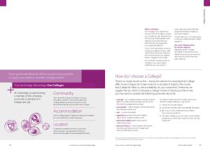
scan) was found to range from 0 to 70 % of the total lung weight [19] (Fig. 1). Presently, although the lung CT scan requires the transport of patients outside the ICU and the use of X-ray radiation, it remains the gold standard to compute lung recruitability [20, 21]. The use of a visual scale to estimate lung recruitment and the application of a low-dose protocol for CT scan acquisition have shown promising results [22, 23]. In addition, a recent observational study showed that the CT scan, independent from estimation of lung recruitment, contributed to diagnosis in 53 % of patients and induced a therapeutic change in 54 % of cases [24]. As an alternative, lung ultrasound showed a reliable accuracy in estimating lung recruitability, but further studies are necessary to confirm its use [25]. Although several experimental and observational studies found a beneficial effect for the use of higher PEEP in ARDS [19, 26, 27], the three most recent randomized trials (ALVEOLI, ExPress, and LOV) did not show any difference in outcome between a low and a high PEEP ventilator strategy [28–30]. However, when combining these data considering only the subgroup of the most severe patients (PaO2/FiO2 < 200 mmHg), the use of higher PEEP level significantly decreased mortality [31, 32]. This suggests that the greater the severity (and the higher the amount of lung edema), the higher the positive effect of PEEP in reducing VILI. This has also been confirmed in an observational study, in which higher PEEP levels significantly reduced the opening and closing effects only in patients with higher recruitability [16].
However, the relationship between lung edema/mass and recruitability has been questioned by Cressoni et al. [13], who found that the PEEP levels necessary to keep the lung open are independent from total lung recruitability. These results suggest that recruitability depends also on the nature of edema, time of onset, and distribution of the disease within the lung parenchyma. Several approaches have been proposed to tailor PEEP for the individual patient. The most common approach is to titrate PEEP according to an oxygenation/saturation target based on a PEEP/FiO2 table [30]. An alternative method, based on respiratory mechanics, is to increase PEEP by maintaining a constant tidal volume, not overcoming a safe limit of airway pressure (26–28 cmH2O) [28], or, after a RM, to decrease PEEP until a reduction of compliance appears [33, 34]. Despite the possible uncertainties regarding the end-expiratory absolute esophageal pressure as a reliable estimation of the pleural pressure [35], Talmor et al. [36] showed a better oxygenation and compliance when PEEP was set according to an end-expiratory transpulmonary pressure between 0 and 10 cmH2O (absolute method). Alternative to the absolute value, the changes in esophageal pressure due to PEEP and tidal volume (elastance method) have been used to compute the total end-inspiratory transpulmonary pressure, as a better marker of lung stress compared with airway inspiratory pressure in the presence of alteration in chest wall elastance [37]. By computing the endinspiratory transpulmonary pressure vs airway pressure, Grasso et al. [38] showed that it was possible to increase
High lung recruitability
5 cmH2O
45 cmH2O
Low lung recruitability Fig. 1 Example of lung CT scan of patients with high (upper panel) or low (lower panel) potential of lung recruitment
Chiumello and Brioni Critical Care (2016) 20:132
PEEP, improving oxygenation and avoiding extracorporeal membrane oxygenation (ECMO) support without overcoming the lung stress. However, when these two methods (absolute and elastance) were compared, the resulting PEEP levels were significantly discordant and furthermore the recommended PEEP changes were in the opposite direction in up to 30 % of the patients [39]. Recently our group compared the previous published methods for selecting PEEP (based on gas exchange, respiratory mechanics, and transpulmonary pressure) with lung recruitability and severity of the disease [40]. The method based on gas exchange (i.e., FiO2/PEEP table of the LOV study [30]) was the only one which provided PEEP levels according to the severity of the disease; on the contrary, the other methods suggested similar levels of PEEP that were not related to the severity or to lung recruitability [40]. Interestingly, obese patients with ARDS presented a significantly lower lung gas volume but similar lung recruitability and chest wall elastance compared with normal body weight patients [41]. Based on the available data, it is clear that “perfect PEEP”—which can simultaneously provide the best oxygenation, compliance, and reduction of VILI—does not exist. Therefore, we suggest performing, in the acute phase, a stratification of patients according to the severity of ARDS before any PEEP selection. This can be done easily by ventilating the patient with pure oxygen at PEEP 5 cmH2O [5]. In the case of severe ARDS, the lung recruitability should be computed by lung CT scan or lung ultrasound, and high PEEP levels (i.e., >15 cmH2O) following the PEEP/FiO2 table of the LOV study [30] should be applied. On the contrary, in mild to moderate ARDS, low PEEP levels (