original article
J Bras Patol Med Lab, v. 49, n. 2, p. 84-90, abril 2013
Immunological parameters of macrophages infected by methicillin resistant/sensitive Staphylococcus aureus Parâmetros imunológicos de macrófagos frente à infecção por Staphylococcus aureus meticilina sensível/resistente Natália Gomes de Morais1; Thacianna Barreto da Costa1; Thays Miranda de Almeida2; Maiara Santos Severo3; Célia Maria Machado Barbosa de Castro4
abstract Introduction: Could changes in Staphylococcus aureus cellular walls, which are commonly associated with multidrug resistance phenotype, influence their immune evasion mechanisms? Objective: To evaluate the microbicide response and survival of alveolar macrophages after in vitro infection with methicillin-sensitive and methicillin-resistant Staphylococcus aureus. Material and methods: We used 20 adult, male, albino, Wistar rats. The alveolar macrophage samples were obtained after tracheostomy and bronchoalveolar lavage. The alveolar macrophages were cultured in the proportion of 1:1 cells/ml Roswell Park Memorial Institute (RPMI) medium/well and isolated by plate adhesion. For the assessment of immunological parameters, four systems were established: negative control, positive control, methicillin sensitive S. aureus (MSSA) and methicillin resistant S. aureus (MRSA). Results: When comparing MRSA and MSSA systems, there was no significant difference as to adhesion and phagocytosis rates, superoxide anion production and macrophage viability. By analyzing the kinetics of nitric oxide production, after 4 to 10 hours of cellular culture incubation, there was lower average production of this radical in the MRSA system when compared to MSSA. However, after 12 hours, no differences were detected between both systems. Conclusion: It is claimed that methicillin resistance may be a factor that influences the bacteria’s ability to escape from macrophage microbicide response. Although the results of some immunological parameters were similar in the surveyed systems, the oscillations occurred during the production of nitric oxide may contribute significantly to the survival of Staphylococcus aureus. Key words: macrophages; Staphylococcus aureus; methicillin.
Introduction The organism invasion by pathogens may cause deregulation of microbicide response in immune system cells(23). This mechanism is triggered by several microorganisms, since it enables the development of the infection process and it also optimizes hostage survival(11). In order to spread infection, Staphylococcus aureus has developed several mechanisms to resist to the immunological system, which involve countless virulence factors that are coordinately expressed during the different stages of infection. This wide range of factors, either occurring on the surface or secreted in the external medium, may reduce phagocytic microbicide response, induce cellular death
and/or inhibit antibiotic action(8, 23). Despite its scientific relevance, there are hardly any studies that evaluate the activation and survival of macrophages after infection by multi-resistant bacterial strains. Since the 1960’s, infections by methicillin resistant S. aureus (MRSA) have become a major public health problem worldwide, inasmuch as it is linked with higher mortality rates(5). It is believed that MRSA strains have distinctive virulence mechanisms, which may be more exacerbated in comparison with MSSA strains(8). Several clinical studies based on morbidity and mortality rates show that MRSA is more virulent than MSSA. Nevertheless, laboratory studies that assess the presence and magnitude of pathogenic
First submission on 28/06/12; last submission on 29/12/12; accepted for publication on 05/01/13; published on 20/04/13
1. Master’s degree in Tropical Medicine; attending doctorate in Tropical Medicine at Universidade Federal de Pernambuco (UFPE). 2. Biomedical scientist; attending master in Public Health at UFPE. 3. Master’s degree in Tropical Medicine, attending doctorate in Entomology at University of California Riverside. 4. Post-doctorate in Immunology at Universidad de Salamanca-Spain; associate professor at UFPE.
84
Natália Gomes de Morais; Thacianna Barreto da Costa; Thays Miranda de Almeida; Maiara Santos Severo; Célia Maria Machado Barbosa de Castro
mechanisms and virulence factors of MSSA and MRSA strains have yielded conflicting results(12, 20). Thus, the present investigation had the objective to evaluate immunological parameters of alveolar macrophages of rats after in vitro cellular infection with MSSA and MRSA strains.
Material and methods
BAL cells were counted with the aid of a Newbauer camera. The concentration 106 cells/ml of RPMI was standardized. Subsequently, the cells were transferred to 35 mm diameter culture dishes (6 wells, Falcon®). After an hour at 37ºC and 5% CO2 , the supernatant with non-adherent cells was discarded and 1 ml of RPMI medium was added.
Systems
The study protocol was approved by the Ethics Committee in Animal Experiments from Universidade Federal de Pernambuco – Centre of Biological Sciences (Protocol 239/09 – 24/11/2009).
Two systems were applied for the assessment of phagocytic index: • MSSA, MA and 100 µl of bacterial inoculum of sensitive strain (ATCC 29213); • MSSA, MA and 100 µl of bacterial inoculum of resistant strain (ATCC 33591).
Animals We applied 20 male, albino, Wistar rats (90-120 days) from the vivarium of UFPE-Nutrition Department. The animals were kept in propylene cages with galvanized wire lid, containing 3 animals per cage at most, with free access to water and food. The room temperature was controlled at 22 ± 1ºC, and the animals were exposed to 12:12h photoperiodic light-dark cycles.
Staphylococcus aureus strains We used methicillin resistant Staphylococcus aureus (The American Type Culture Collection [ATCC] 33591) and methicillin sensitive S. aureus (ATCC 29213) due to its distinctive resistance profile and its relevance concerning public health. The bacteria were maintained in Tryptic Soy Broth (TSB) with 20% glycerol at -20ºC. 24 hours prior to each experiment, strains were seeded in blood agar dishes (agar with 5% lamb blood) and incubated at 37ºC. Some colonies were transferred to tubes containing sterile PBS in order to achieve approximately 0.15 nm turbidity by spectrophotometer at 570 nm. According to Lu and McEwan(12), this absorbance corresponds to 106 UFC/ml of PBS.
Bronchoalveolar lavage (BAL) The animals were anesthetized with chloralose-urethane (0.5 and 12.5%, respectively) at the proportion of 8 ml/kg administered intraperitoneally. The BAL was collected with a saline injection (0.9%) through a plastic cannula inserted in the trachea. Several aliquots of 3 ml were injected and collected in 50 ml conical polypropylene tubes (Falcon, Sigma). We obtained approximately 30 ml of BAL per animal.
Alveolar macrophage culture The BAL was centrifuged at 1,500 RPM for 15 minutes. The precipitate was resuspended in RPMI 1640 (Gibco-Invitrogen Corporation®) with 3% fetal bovine serum (Gibco-Invitrogen Corporation®) and antibiotics (penicillin 100 U/ml and streptomycin 100 μg/ml). After dilution in Trypan blue (1:10), the
85
Three systems were established for the adhesion index (AI) and superoxide anion kinetics: negative control (NC), containing exclusively alveolar macrophages, MSSA and MRSA, as described previously. The positive control (PC) was included for the assessment of nitric oxide kinetics and for the viability of alveolar macrophages, which contained MA and 10 µl of LPS (Escherichia coli serotype; 055:B5, Sigma®). The plates were incubated at 37ºC in humid atmosphere and 5% of CO2.
Adhesion index After incubation for an hour, 10 µl of culture supernatant was collected and added to Trypan blue at 1:10 dilution. The counting was performed with Newbauer camera. The AI was calculated through the formula described by Fuente et al.(2): IA = 100-non-adherent cells/ml divided by the initial number of cells/ml × 100. The results were expressed in percentage of non-adherent cells.
Phagocytic index (FI) In this research, two systems (MSSA and MRSA) were established in test tubes. The contents were homogenized and uniformly distributed onto slides for optic microscopy. The slides were incubated at 37ºC for an hour. Subsequently, they were washed for the removal of non-adherent cells, stained and assessed through optic microscopy by a skilled observer, who was unaware of the systems surveyed herein. The results were expressed in percentage of phagocytic cells after total counting of 100 cells.
Kinetics of anion superoxide production (O2-) O2- was detected by its ability to reduce iron-cytochrome C chemically (30 mg/ml in Hank’s Balanced Salt Solution [HBSS], 2.4 × 10⁻³ M, firon-cytochrome C of horse mitochondrion, type VI, SIGMA Chemical Company, St. Louis, MO). After adding phorbol miristate acetate/ PMA (Sigma®) to Hank’s solution (HBSS, Gibco-Invitrogen Corporation®), 3 systems of discontinuous analysis (NC, MSSA and MRSA) were prepared at 2 μg/ml with assessment between the first and the second hour of incubation. The reading was performed
Immunological parameters of macrophages infected by methicillin resistant/sensitive Staphylococcus aureus
through immunosorbent assay linked with enzyme (ELISA- BIORAD, Model 680) with a 55 nm filter. The quantification of O2- was expressed in nM/ml.
MR SA
MS SA
NC
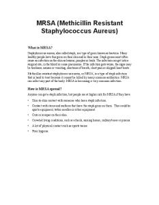
Figure 1 – AI of alveolar macrophages from adult Wistar rats for the following systems: NC, MSSA and MRSA. Values expressed as mean ± standard error (n = 20) AI: adhesion index; NC: negative control; MSSA: methicillin sensitive S. aureus; MRSA: methicillin resistant S. aureus.
Phagocytic index As to PI, there was no difference between macrophages infected in vitro with MRSA (12.1 ± 3.0 %) and MSSA (10.4 ± 3.2 %), p > 0.05 (Figure 2). 20
15
10
5
0
SA
The analysis of variance (ANOVA) was applied for the comparison of multiple means. When ANOVA results revealed significant difference, Tukey test was performed in order to identify inconsistency in results. In the analysis of only two systems, which were used in the assessment of superoxide kinetics and FI, test t Student was employed for parametric data and Mann-Whitney test for non-parametric data. The results were expressed by mean ± standard error. The significance was defined by p < 0.05. The statistical program employed in the analysis was Sigma Stat®.
0
MR
Statistical analysis
50
SA
The cellular viability was assessed by mitochondrial reduction of 3-[4.5-dimethylthiazol-2-il]-2.5- diphenyl tetrazolium bromide (MTT) to formazan, as described by Mosmann(15). After a 24-hour incubation period, the cell cultures were washed with PBS (1×) at room temperature. Subsequently, they were incubated with 550 µl of PBS and 55 µl of MTT solution for two hours under light protection. After this period, 200 µl of PBS and 200 µl of dimethylsulfoxide (DMSO) were added and, afterwards, the cellular monolayer was scraped. The quantification of formazan solution was performed with ELISA reader (BIORAD, Model 680) with a 570 nm filter. The results were expressed in percentage of viable cells, applying NC results as reference.
100
MS
Viability of alveolar macrophages
Adherence index (%)
The NO production was determined through the concentration of supernatant nitric in culture cells(10). 100 µl of supernatant cells was collected every two hours totaling 24 hours of incubation. 50 µl of Griess reagent (1.5% of sulfanilamide in 5% of H3PO4, 0.1% in H2O of naphthyl ethylenediamine dihydrochloride) was added to the cell culture supernants for nitric quantification. After 15 minutes at rest and at room temperature, the reading was performed by Elisa (BIORAD, Model 680) with a 550 nm filter. The nitric concentration was calculated through mean values of NaNO2 standard curve and the collected data were expressed in µM of nitrite/ml.
Phagocytic index (%)
Kinetics of nitric oxide production (NO)
150
Figure 2 – PI of alveolar macrophages from adult Wistar rats for MSSA and MRSA systems. Values expressed as mean ± standard error (n = 20) PI: phagocytosis index; MSSA: methicillin sensitive S. aureus; MRSA: methicillin resistant S. aureus.
Kinetics of O2- production
Results Adhesion index The analysis of non-adherent cells revealed no difference among the systems: NC (87.5 ± 3.0 %), MSSA (90.3 ± 2.6 %) and MRSA (93.3 ± 3.1 %), p > 0.05 (Figure 1).
86
The kinetics of O2- production in the supernatant of macrophage cultures was higher in all systems in the second hour of incubation: NC (1h: 1.8 ± 0.1/2h: 3.7± 0.2 nM/ml, p < 0.001); MSSA (1h: 1.9 ± 0.1/2h: 3.8 ± 0.2 nM/ml, p < 0.001); MRSA (1h: 2.2 ± 0.1/2h: 4.1 ± 0.2 nM/ml, p = 0.009). However, regarding comparison within systems, there was no difference in O2- production when analyzed in the first two hours of incubation, p > 0.05 (Figure 3).
Natália Gomes de Morais; Thacianna Barreto da Costa; Thays Miranda de Almeida; Maiara Santos Severo; Célia Maria Machado Barbosa de Castro
MRSA MSSA NC
5
NC 15
PC MSSA MRSA
10
3
NO (µM/ml)
O2- (nM/ml)
4
2 1
5
2h Incubation time
Viability of AM There was a reduction in the percentage of viable cells infected with MSSA and MRSA strains in comparison with PC ((LPS: 69.2 ± 0.8%; MSSA: 18.5 ± 0.22%, p < 0.001; MRSA: 20.7 ± 0.2%, p < 0.001) (Figure 5).
Discussion In the present investigation, the coincubation of alveolar macrophages with Staphylococcus aureus strains did not induce any change in the adhesion index of these cells. This finding may be justified by the capacity of Staphylococcus aureus to evade immune system recognition by means of polysaccharide capsules or biofilm
87
24 h
22 h
18 h 20 h
16 h
14 h
12 h
10 h
8h
6h
80
60
40
20
SA MR
SA
0
MS
When comparing NC and PC, there was a higher production of nitrogen reactive species in PC after an 8h incubation period, remaining steady until the end of the analysis (p < 0.001). In the MSSA and MRSA systems, there were differences in the production of NO in the following periods: 4h, 6h, 8h and 10h. Nevertheless, after 12h the nitrite concentration within the systems was similar to NC. During the kinetic period, the mean nitrite production was lower in MRSA in contrast with MSSA, p < 0.05 (Figure 4).
Figure 4 – Kinetics of NO production by alveolar macrophages from adult Wistar rats for NC, PC, MSSA and MRSA systems. Values expressed as mean ± standard error (n = 20) *: p < 0.05 between NC and PC; **: p < 0.05 between NC and MSSA; #: p < 0.05 between NC and MRSA. NO: nitric oxide; NC: negative control; PC: positive control; MSSA: methicillin sensitive S. aureus; MRSA: methicillin resistant S. aureus.
PC
The kinetics of NO revealed an increase in the production of NO when compared with other incubation periods (p < 0.001). Similar results were detected in PC, MSSA and MRSA, with p < 0.001. The maximum liberation peak of this free radical occurred after a 22h incubation period in PC (CP-22h: 7.55 ± 0.295 µM/ml), 4h in the MSSA system (MSSA-4h: 11.778 ± 0.413 µM/ml) and 8h in the MRSA system (MRSA-8h: 11.663 ± 0.16 µM/ml).
Incubation time
Cellular viability (%)
Kinetics of NO production
4h
0
Figure 3 – Kinetics of O production by alveolar macrophages from adult Wistar rats for NC, MSSA and MRSA systems after one and two-hour incubation periods. Values expressed as mean ± standard error (n = 20) *: p < 0.05 between 1h and 2h for NC, MSSA and MRSA. NC: negative control; MSSA: methicillin sensitive S. aureus; MRSA: methicillin resistant S. aureus. 2-
2h
1h
0
Figure 5 – Viability of alveolar macrophages from adult Wistar rats for PC, MSSA and MRSA systems. Values expressed as mean ± standard error (n = 20) *: p < 0.05 among PC, MSSA and MRSA.
and the fact that they produce or secrete specific molecules that block phagocyte receptors(3, 4). Some studies suggest that both MSSA and MRSA present the same magnitude in terms of binding to human epithelial cells(4) and feline corneocytes(26). Furthermore, there was no difference as to phagocytic index when comparing MSSA and MRSA strains. Similar results were reported by Salgado et al.(21), who compared phagocytic susceptibility to MSSA and MRSA. Mekontso-Dessap et al.(14) also observed no difference neither in the phagocytic function of Wistar rat neutrophils nor in the survival rate of bacteria inside the cell when compared with methicillin sensitive and resistant strains.
Immunological parameters of macrophages infected by methicillin resistant/sensitive Staphylococcus aureus
The phagocytosis of microorganisms activate oxidase dependant on adenine dinucleotide phosphatese (NADPH), which induces the production of high levels of superoxide, a process commonly denominated respiratory burst. Studies on the release of this anion describe that there is increasing production of O2- during the first 6h after antigenic stimulus with phorbol myristate acetate(17). Herein, the generation of O2- in supernatant of alveolar macrophage culture increased in the first to the second hour of the incubation period in the control systems as well as in MSSA and MRSA strain systems. According to Mandell(13), regardless of the resistance antimicrobial profile, S. aureus seems to neutralize oxygen intermediates formed after its internalization through the release of catalase and staphyloxanthin enzymes. As to NO production, there was an increase after stimulation with LPS in all analyzed periods. Ferreira-Silva et al.(24) yielded similar results evaluating the production of NO in Wistar rat macrophages after contact with LPS, which corroborates our investigation. According to Hwang et al.(9), LPS is regarded as an efficient macrophage stimulator, insofar as it induces the production of NO, hence being a positive control for the activation of these cells. Pumerantz et al.(16) claimed that No produced by alveolar macrophages play a major microbicide role against S. aureus. According to Sasaki et al.(22), the expression of induced nitric oxyde synthase is initiated 3h after S. aureus infection. During the kinetics, a maximum peak of NO release was observed after 4h in the MSSA system and 8h in the MRSA system. These results evinced that the resistant strain activates a more delayed microbicide response of macrophages in comparison with sensitive strains. Thus, this may enable the multiplication of resistant bacteria inside the phagocytes, inasmuch as they do not present antimicrobial mechanisms until the macrophagic activation occurs. After No production peaks, a reduction in the concentration of nitrite was detected in the supernatant of MSSA and MRSA systems, and after a 12 hour incubation period, it was similar to basal levels of NC. According to Richardson et al.(18), S. aureus may evade multiple components of the innate immune response, including the microbicide action of NO. These researchers proved that S. aureus is able to adapt metabolically to nitrosative stress, inasmuch as it expresses inducible dehydrogenase (NO-L-lactato). Its production allows S. aureus to maintain homeostasis during nitrosative redox stress. The kinetics of NO production in MSSA and MRSA strains showed that there was oscillation during 4h, 6h, 8h and 10h of incubation. The mean concentration of nitrite during this period was lower in MRSA in comparison with MSSA. This result implies that macrophages may have been activated with lower intensity during the infection by MRSA or that this bacterium may have distinctive mechanisms of evasion to microbicide response. Vaudaux e Waldvogel(25) analyzed PI cytosis of polimorphonuclear leucocytes (PMN) through an in vitro assay. After 30 and 60 minutes of incubation, MRSA strains showed a better survival to phagocytic action of PMN in comparison with MSSA strains.
88
According to Cutler(1), methicillin resistant staphyloccocus present thicker cellular walls and different lipidic content in contrast with those sensitive to this antibiotic. Accordingly, it is possible to claim that the morphology of resistant strains enables a better protection against destruction by lysosomal enzymes or by the oxidative metabolism of activated phagocytes. In 2011, Ferreira et al.(6) affirmed that MRSA is a major pathogen in nosocomial infections and it is associated with high rates of mortality opposed to infections by MSSA. As to the viability rate of Wistar rat alveolar macrophages, there was a reduction after a 24h incubation period with MSSA and MRSA in comparison with PC. This finding may explain the low levels of nitrite in culture supernatant. According to Deleo et al.(3), there are two possible interactions between bacterium-phagocyte. The bacterial phagocytosis triggers the production of oxygen and nitrogen reactive species as well as lysosome degranulation. This process occurs along with apoptosis induction, hence combating the infection. Alternatively, some bacterial pathogens may cause lysis of phagocytes or induce apoptosis, enabling the bacteria to remain in the medium or the development of the disease. In order to escape cytoplasmatic confinement, S. aureus induces phagocyte death by triggering pyroptosis or by releasing leukocidins(11). The main characteristic of pyroptosis is the rupture of the plasmatic membrane with the release of pro-inflammatory intracellular content via caspase-1 (cytoplasmatic protease)(7). The action mechanism of leukodicins causes leukocyte destruction due to the formation of pores in the cellular membrane. Kubica et al.(11) confirmed that after phagocytosis by macrophages, S. aureus, regardless of its antimicrobial resistance profile, survives intracellularly in vacuoles for 3-4 days prior to escaping into the cytoplasm and ultimate cellular lysis. Besides remaining several days inside macrophages, the bacterium was able to escape intracellular confinement and to proliferate in the hostage organism. Different bacterial strains may express a higher or lower number of virulence factors such as adhesins and toxins or differ in their ability to produce biofilm and resist to phagocytosis. According to Rossney et al.(19), the expression regulation of virulence factors plays a pivotal role in the pathogenicity of S. aureus. According to Gordon e Lowy(7), MRSA strains may contain genetic factors that maximize virulence, which allow these bacteria to cause particular clinical syndromes. Further studies with molecular biology techniques could elucidate the differences in macrophage immune response to methicillin resistant and sensitive strains.
Conclusions There are some evidences that methicillin resistance constitutes a factor that influences the ability to evade microbicide response in alveolar macrophages from adult Wistar rats. Although the adhesion capacity, PI, O2- production and viability of infected phagocytes were not altered, there were oscillations in the kinetics of ON production, which may contribute significantly to its pathogenicity and bacterial survival.
Natália Gomes de Morais; Thacianna Barreto da Costa; Thays Miranda de Almeida; Maiara Santos Severo; Célia Maria Machado Barbosa de Castro
resumo Introdução: Modificações nas paredes celulares das cepas de Staphylococcus aureus relacionadas com o fenótipo de multirresistência poderiam influenciar seus mecanismos de evasão frente à resposta imune? Objetivo: Avaliar a resposta microbicida e a sobrevivência de macrófagos alveolares após infecção in vitro com Staphylococcus aureus meticilina sensível/resistente. Material e métodos: Utilizaram-se 20 ratos adultos, machos, albinos e da linhagem Wistar. Os macrófagos alveolares foram obtidos após procedimento cirúrgico de traqueostomia, por meios da coleta do lavado broncoalveolar. Os macrófagos alveolares foram cultivados na proporção de 1:1 células/ml de Roswell Park Memorial Institute (RPMI)/poço e isolados pela capacidade de adesão à placa. Para avaliação de parâmetros imunológicos, foram estabelecidos quatro sistemas: controle negativo, controle positivo, S. aureus sensível a meticilina (MSSA) e S. aureus resistente à meticilina (MRSA). Resultados: Ao comparar os sistemas de MSSA e MRSA, não foi observada diferença no índice de aderência, na taxa de fagocitose, na produção do ânion superóxido e na viabilidade dos macrófagos. Ao analisar a cinética de óxido nítrico, houve menor produção média desse radical para o MRSA quando comparado com o MSSA, no período de 4 a 10 horas de incubação das culturas celulares. Entretanto, após 12 horas, não foi detectada divergências entre esses sistemas. Conclusão: Sugere-se que a resistência à meticilina poderá ser um fator que influenciará a capacidade de evasão da bactéria à resposta microbicida dos macrófagos. Apesar dos resultados de alguns parâmetros imunológicos terem sido similares entre os sistemas analisados, as oscilações ocorridas durante a produção do óxido nítrico poderão contribuir de forma importante para a sobrevivência da bactéria Staphylococcus aureus. Unitermos: macrófagos; Staphylococcus aureus; meticilina.
References 1. CUTLER, R. R. Relationship between antibiotic resistance, the production of virulence factors, and virulence for experimental animals in Staphylococcus aureus. J Med Microbiol, v. 12, p. 55-62, 1979. 2. DE LA FUENTE, M.; DEL RIO, M.; FERRANDEZ, M. D.; HERNANZ, A. Modulation of phagocytic function in murine peritoneal macrophages by bombesin, gastrin-releasing peptide and neuromedin C. Immunol, v. 73, p. 205-11, 1991.
11. KUBICA, M. et al. Potential new pathway for Staphylococcus aureus dissemination: the silent survival of S. aureus phagocytosed by human monocyte-derived macrophages. PLoS ONE, v. 3, n. 1, p. 1409-35, 2008. 12. LU, Y. F.; MCEWAN, N. A. Staphylococcal and micrococcal adherence to canine and feline corneocytes: quantification using a simple adhesion assay. Vet Dermatol, v. 18, p. 29-35, 2007. 13. MANDELL, G. L. Uptake, transport, delivery and intracellular activity of antimicrobial agents. Pharmacotherapy, v. 25, n. 12, p. 130S-133S, 2005.
3. DELEO, F. R.; DIEP, B.; OTTO, M. Host defense and pathogenesis in Staphylococcus aureus infections. J Immun, v. 180, p. 500-9, 2009.
14. MEKONTSO-DESSAP, A. et al. Blood neutrophil bactericidal activity against methicillin-resistant and methicillin-sensitive Staphylococcus aureus during cardiac surgery. Shock Aug, n. 24, v. 2, p. 109-13, 2005.
4. DUCKWORTH, G. J.; JORDENS, J. Z. Adherence and survival properties of an epidemic methicillin-resistant strain of Staphylococcus aureus compared with those of methicillin-sensitive strains. J Med Microbiol, v. 32, p. 195-200, 1990.
15. MOSMANN, T. Rapid colorimetric assay for cellular growth and survival: application to proliferation and cytotoxicity assays. J Immun Meth, v. 65, n. 1/2, p. 55-63, 1983.
5. DURAN, N.; OZER, B.; DURAN, G. G.; ONLEN, Y.; DEMIR, C. Antibiotic resistance genes and susceptibility patterns in Staphylococci. Indian J Med Res, v. 135, p. 389-96, 2012.
16. PUMERANTZ, A. et al. Preparation of liposomal vancomycin and intracellular killing of methicillin-resistant Staphylococcus aureus (MRSA). Int J Antimicrob Agents, v. 23, p. 1-5, 2010.
6. FERREIRA, J. P.; ABREU, M. A.; RODRIGUES, P.; CARVALHO, L.; CORREIA, J. A. Staphylococcus aureus resistente à meticilina e abcesso hepático. Análise retrospectiva de 117 casos. Acta Med Port, v. 24, p. 399-406, 2011.
17. QUINN, M. T.; AMMONS, M. C.; DELEO, F. R. The expanding role of NADPH oxidases in health and disease: no longer just agents of death and destruction. Clin Sci, v. 111, p. 1-20, 2006.
7. GORDON, R. J.; LOWY, F. D. Pathogenesis of methicillin-resistant Staphylococcus aureus infection. Clin Infect Dis, v. 16, p. 209-13, 2008.
18. RICHARDSON, A. R.; LIBBY, S. J.; FANG, F. C. A nitric oxide-inducible lactate dehydrogenase enables Staphylococcus aureus to resist innate immunity. Science, v. 319, p. 1-21, 2008.
8. HASTE, N. M.; FARNAES, L.; PERERA, V. R.; FENICAL, W.; NIZET, V.; HENSLER, M. E. Bactericidal kinetics of marine-derived napyradiomycins against contemporary methicillin-resistant Staphylococcus aureus. Marine Drugs, n. 9, p. 680-9, 2011. 9. HWANG, T. L. et al. YC-1 potentiates cAMP-induced CREB activation and nitric oxide production in alveolar macrophages original research article. Toxicology and Applied Pharmacology, v. 260, n. 2, p. 193-200, 2012. 10. IMANISHI, N. et al. Induction of inducible nitric oxide (NO) synthase mRNA and NO production in macrophages infected with influenza A/PR/8 virus and stimulated with its ether-split product. Microb Immun, v. 49, n. 1, p. 41-8, 2005.
89
19. ROSSNEY, A. S.; SHORE, A. C.; MORGAN, P. M.; FITZGIBBON, M. M.; O’CONNELL, B.; COLEMAN, D. C. The emergence and importation of diverse genotypes of methicillin-resistant Staphylococcus aureus (MRSA) harboring the panton-valentine leukocidin gene (pvl) reveal that pvl is a poor marker for community-acquired MRSA strains in Ireland. J Clin Microbiol, v. 45, n. 8, p. 2554-63, 2007. 20. ROZGONYI, F.; KOCSIS, E.; KRISTO, F. K.; NAGY, K. Is MRSA more virulent than MSSA? Clin Microbiol Infect, v. 13, p. 843-5, 2007. 21. SALGADO, M. M.; PIGNATARI, A. C. C.; BELLINATI-PIRES, R. Phagocytosis and killing of epidemic methicillin-resistant Staphylococcus aureus
Immunological parameters of macrophages infected by methicillin resistant/sensitive Staphylococcus aureus
by human neutrophils and monocytes. Braz J Infect Dis, n. 8, p. 80-9, 2004.
nitric oxide release by activated macrophages in response to fluoxetine in adult rat. Neuroimmunomod, v. 23, p. 120-9, 2008.
22. SASAKI, S.; MIURA, T.; NISHIKAWA, S.; YAMADA, K.; HIRASUE, M.; NAKANE, S. protective role of nitric oxide in Staphylococcus aureus infection in mice. Infect Immun, v. 66, n. 3, p. 1017-22, 1998.
25. VAUDAUX, P.; WALDVOGEL, F. A. Methicillin-resistant strains of Staphylococcus aureus: relation between expression of resistance and phagocytosis by polymorphonuclear leukocytes. J Infect Dis; v. 144, n. 6, p. 575-82, 1981.
23. SHAPIRA, S. D.; HACOHEN, N. Systems biology approaches to dissect mammalian innate immunity review article. Current Opinion in Immunology, v. 23, n. 1, p. 71-7, 2011. 24. SILVA, W. T. F.; GALVÃO, B. A.; PEREIRA, K. N. F.; DE CASTRO, C. M. M. B.; DE CASTRO, R. M. Perinatal malnutrition programs sustained alterations in
26. WOOLLEY, K. L.; KELLY, R. F.; FAZAKERLEY, J.; WILLIAMS, N. J.; NUTTALL, T. J.; MCEWAN, N. A. Reduced in vitro adherence of Staphylococcus species to feline corneocytes compared to canine and human corneocytes. Univ Liverpool Fac Vet Sci, v. 19, p. 1-6, 2007.
Mailing address
Célia Maria Machado Barbosa de Castro Universidade Federal de Pernambuco; Centro de Ciências da Saúde; Programa de Pós-graduação em Medicina Tropical; Campus Universitário, s/n; Cidade Universitária; CEP: 50670-420; Recife-PE, Brazil; e-mail:
[email protected].
90