Seminars in Diagnostic Pathology (2010) 27, 77-90
Salivary gland-type tumors of the breast: a spectrum of benign and malignant tumors including “triple negative carcinomas” of low malignant potential Maria P. Foschini, MD,a Thomas Krausz, MD, FRCPathb From the aDepartment of Hematology and Oncology, Section of Anatomic Pathology at Bellaria Hospital, “L. and A. Seragnoli” University of Bologna, Bologna, Italy; and the b Department of Pathology, University of Chicago, Chicago, Illinois. KEYWORDS Breast; Triple negative carcinoma; Adenomyoepithelioma; Adenoid cystic carcinoma; Mucoepidermoid carcinoma
Salivary gland-type neoplasms of the breast are uncommon and comprise numerous entities analogous to that more commonly seen in salivary glands. The clinicopathologic spectrum ranges from benign to malignant but there are important differences as compared with those of their salivary counterpart. In the breast, benign adenomyoepithelioma is recognized in addition to malignant one, whereas in the salivary gland a histologically similar tumor is designated as epithelial-myoepithelial carcinoma without a separate benign subgroup. Mammary adenoid cystic carcinoma is a low-grade neoplasm compared with its salivary equivalent. It is also important to appreciate that in contrast to “triple negative” conventional breast carcinomas with aggressive course, most salivary-type malignant breast neoplasms behave in a low-grade manner. Most of these tumors are capable of differentiating along both epithelial and myoepithelial lines, but the amount of each lineage-component varies from case to case, contributing to diagnostic difficulties. Well established examples of this group include pleomorphic adenoma, adenomyoepithelioma, and adenoid cystic carcinoma. Another family of salivary gland-type mammary epithelial neoplasms is devoid of myoepithelial cells. Key examples include mucoepidermoid carcinoma and acinic cell carcinoma. The number of cases of salivary gland-type mammary neoplasms in the published data is constantly increasing but some of the rarest subtypes like polymorphous low-grade adenocarcinoma and oncocytic carcinoma are “struggling” to become clinically relevant entities in line with those occurring more frequently in salivary glands. © 2010 Elsevier Inc. All rights reserved.
Salivary gland-type tumors of the breast are rare benign and malignant neoplasms with a broad histologic spectrum analogous to that more commonly seen in salivary glands.1,2 The basic morphologic structure of the tubulo acinar exocrine glands of the breast and salivary glands perhaps account for the occurrence of identical tumor types,3 whereas This work was partly financed with grants from the University of Bologna (RFO). Address reprint requests and correspondence: Maria P. Foschini, MD, Anatomia Patologica, Università di Bologna, Ospedale Bellaria, Via Altura 3, 40139 Bologna, Italy. E-mail address:
[email protected].
0740-2570/$ -see front matter © 2010 Elsevier Inc. All rights reserved. doi:10.1053/j.semdp.2009.12.007
the specialization of the acinar cells to secrete milk vs saliva and the difference of hormone receptor expression in these organs explains, at least in part, the clinicopathologic differences. The published data on salivary gland-type tumors of the breast mostly consist of small series or case reports, hence data on their frequency and clinical behavior are rather limited. However, the number of these tumor entities has trebled since the first review on this topic in 1970.4 Familiarity with these rare tumor types and distinguishing them from their mimics is important not only for prognostication but also for selecting the correct therapy. This is exemplified by the fact that even though the malignant
78
Seminars in Diagnostic Pathology, Vol 27, No 1, February 2010
tumors in this group are immunohistochemically “triple negative,” most of them, in contrast to the more common basal-like mammary carcinomas, tend to behave in a lowgrade manner. The purpose of the present article is not to review all the previously published data but to provide an update on selected salivary gland-type breast tumors. Salivary gland–type tumors of the breast have been divided into 2 main groups: tumors with myoepithelial differentiation and tumors devoid of myoepithelial differentiation.2 Breast tumors with myoepithelial differentiation constitute a wide spectrum of lesions ranging from benign to malignant neoplasms. Benign breast tumors with myoepithelial differentiation are represented by pleomorphic adenoma (PA), benign adeno myoepithelioma, and myoepithelioma. Malignant tumors comprise adenoid cystic carcinoma (AdCC), malignant adenomyoepithelioma that has a broad histologic spectrum, and pure malignant myoepithelioma.2,5
Pleomorphic adenoma Although PA is the most frequent tumor of the salivary gland, it is only rarely seen in the breast. It usually affects adult female patients aged 19-85 years. Rare cases have been reported in males.6-8 PA of the breast is most frequently located in the periareolar region, and probably arises from large lactiferous ducts. It is almost always solitary. On mammographic examination, PA can mimic a carcinoma9,10 mainly because of the presence of irregular calcifications. Similarly, a suspicion of malignancy may arise on examination of cytologic smears.11,12 On histology, PA of the breast is similar to that of salivary glands and has a broad morphologic spectrum (Figure 1). Specifically, it presents as a neoplastic proliferation composed of ductules, sheets, nests, and cords of epithelial and myoepithelial cells intermixed with mesenchymal elements in a myxoid, myxohyalinized, or myxochondroid background. Focal papillomatous areas may be seen in some of the cases. The phenotype of the neoplastic myoepithelial cells, similar to myoepithelioma and adenomyoepithelioma, is not always epithelioid but can be spindle or plasmacytoid with a “hyaline” cytoplasm.13 The myoepithelial cells may form a fine reticular growth pattern. In most examples, scattered double-layered glandular structures lined by luminal epithelial cells beneath which are the abluminal myoepithelial cells which may have clear cytoplasm are also seen. Focal squamous and/or sebaceous metaplasia may also be observed. The distinction of PA from adenomyoepithelioma or even from AdCC can be difficult on a small biopsy material. Islands of well-formed cartilage, as the most common mesenchymal component, with or without ossification/calcification are usually also present. Occasionally, neoplastic cartilage is the major com-
Figure 1 (A) Pleomorphic adenoma presenting as a multinodular lesion. (B) Pleomorphic adenoma of the breast with chondromyxoid stroma.
ponent of the tumor. PA of the breast is surrounded by a thin, but sometimes incomplete, pseudocapsule. In most PAs of the salivary gland, chromosomal aberrations can be demonstrated.14-18 The most common rearrangements involve 8q12 containing the target gene PLAG1 or 12q13-15 with the target gene HMGA2. Other sporadic clonal changes have also been documented. The detection of PLAG1 and HMGA2 translocations by reverse transcriptase polymerase chain reaction (RT-PCR) or fluorescence in situ hybridization may potentially aid in diagnosis. It remains to be seen whether the same chromosomal aberrations can be identified in mammary PAs.19 Mammary PA behaves in a benign manner (malignant transformation is extremely rare, see in the following paragraphs). Recurrences may occur, especially in cases with multinodular growth pattern or in those that are incom-
Foschini and Krausz
Salivary Gland-Type Tumors of the Breast
pletely excised; therefore, adequate surgery and follow-up are recommended.20 Malignant transformation is a well-known phenomenon in salivary gland PA, whereas in the breast it has been described in 3 cases only.21 Carcinoma ex-PA of the breast was observed in 3 female patients, aged 86, 60, and 57 years, respectively. In case 2, the nodule had been detected during screening mammography, whereas cases 1 and 3 presented with palpable nodule which appeared a few months before surgery. On histology, all 3 cases showed areas of typical PA with areas of malignant transformation in the form of IDC, grade 322 associated with features of matrix producing metaplastic carcinoma. In case 3, areas of chondrosarcomatous differentiation were seen and in situ ductal carcinoma was also identified. The malignant transformation was located mainly at the periphery of the tumor. On immunohistochemistry, the malignant component showed focal positivity for p63 and S-100, in addition to a strong and diffuse positivity for p53. Immunohistochemical staining was negative for estrogen receptor and human epidermal growth factor receptor 2 (HER-2). The cases of carcinoma ex-PA described in the breast share several features with the salivary gland counterpart, including strong positivity for p53. However, salivary gland cases of carcinoma ex-PA are often characterized by a long history of lump persisting up to 30 years and a prognosis closely related with the local extent of the tumor, specifically whether it is intracapsular or extended beyond the capsule. In addition, the malignant areas are associated with amplification of the HER-2 gene.23 None of the 3 cases were associated with metastases either to axillary lymph nodes or to distant sites. Case 3 received chemotherapy. Follow-up, even if limited, was uneventful.
Adenoid cystic carcinoma AdCC is defined as “an epimyoepithelial carcinoma of low malignant potential, histologically similar to the salivary gland counterpart.”24 Breast AdCC is rare, constituting ⬍1% of all breast tumors.1,25-27 Most of the reported cases affect adult female patients, even if rare cases have been described in males and children.28 AdCC presents as a palpable nodule/mass, most frequently located in the upper quadrants or periareolar region, of variable size.2,24,27 In recent years, cases detected during mammographic screening programs are on records. Care should be taken in the interpretation of mammograms because breast AdCC can mimic benign lesions.29 Fine needle aspiration cytology (FNAC) can help to reach a correct diagnosis. On FNAC, mammary AdCC is very similar to the salivary gland counterpart, being characterized by 3-dimensional clusters of cells arranged around small cystic spaces, containing mucin. Neoplastic cells have uniform and bland
79 nuclei. In addition, small spheres of amorphous material are present. Mucicarmine and Alcian blue staining help to confirm the mucinous material.30 Histologic features are similar to those of AdCC affecting the salivary glands. Specifically, AdCC is composed of epithelial and basal-myoepithelial cells organized in cribriform, tubular (including compressed tubular), and solid patterns (Figure 2A-C). Neoplastic cells are somewhat polarized around 2 types of oval or rounded spaces: true glandular spaces and pseudolumens. Epithelial cells surround true glandular spaces containing epithelialtype neutral mucin, confirmed by Periodic acid-Schiff (PAS) after diastase staining. On hematoxylin and eosin examination, the epithelial cells surrounding true glandular spaces have round nuclei and eosinophilic cytoplasm. Basal-myoepithelial cells surround pseudolumens filled with acidic stromal mucosubstance, highlighted by Alcian blue staining, which is hyaluronidase sensitive. In addition, these spaces may contain rare capillaries with adjacent strands of collagen consistent with the concept that the pseudolumens are the consequence of invagination of the peritumoral stroma. Some of these spaces are partly or completely obliterated by “hyaline cylinders” composed of invaginated basement membrane material confirmed by collagen IV (Figure 2) and laminin immunostains, and by ultrastructural studies.1,24 On hematoxylin and eosin examination, basal-myoepithelial cells have hyperchromatic, angular nuclei and a thin rim of pale or clear cytoplasm. Immunohistochemistry can help to recognize the different cell types and their distribution. Epithelial cells are positive with low molecular weight cytokeratin (CK)7 and epithelial membrane antigen (EMA), whereas basal-myoepithelial cells are better highlighted by high molecular weight cytokeratins (CK5/6, CK14) and by other myoepithelial markers like p63, calponin, smooth muscle actin (SMA), and smooth muscle myosin heavy chain. Collagen IV and laminin immunoreact both with the peritumoral basement membrane and its invaginated form filling the pseudolumens (hyaline cylinders). The immunohistochemical profile is of practical value in the differential diagnosis, with the more frequent types of breast carcinomas showing cribriform architecture as cribriform invasive carcinoma, mucinous carcinoma, and polymorphous low-grade adenocarcinoma. The differential diagnosis of AdCC on small biopsy material also includes cellular variant of PA and adenomyoepithelioma. Furthermore, CD117 (c-kit) positivity, wellknown in salivary AdCC, is maintained also in breast AdCC.31,32 Arpino et al27 reported positivity for estrogen receptor (ER) in 46% and progesterone receptor (PR) in 36% of their cases of AdCC. These data are in contrast with other studies because most of the reported cases of breast AdCC were immunohistochemically ER, PR, and HER-2 negative. Differences may be related to the method of detection, as Arpino et al based their data on biochemical assay of the tumor where samples can also contain normal glandular epithelium entrapped within the tumor. Immunohistochemical negativity for ER and PR can be an additional
80
Seminars in Diagnostic Pathology, Vol 27, No 1, February 2010
Figure 2 Adenoid cystic carcinoma (AdCC) of the breast can show various architectural patterns: (A) Solid and cribriform areas, (B) AdCC with tubular growth, (C) AdCC with cribriform features, (D) Collagen IV immunostaining confirms basal membrane invaginations in AdCC.
aid in the differential diagnosis with the more common types of breast carcinomas. In addition to the classical tubular or cribriform architecture, breast AdCC can show prominent solid features. Shin and Rosen33 described 9 cases of AdCC of the breast mainly composed of cells with basaloid features arranged in solid nests. High mitotic count (⬎5 mitoses per 10 high power fields) was observed. Axillary dissection was performed in 6 cases, in 2 of which metastases were detected. Despite the apparent aggressive morphologic features, no further metastases appeared during the reported follow-up (mean, 28 months, range, 2-88 months). AdCC of the breast can show all the spectrum of features observed in cases affecting the salivary glands, including focal squamous and sebaceous differentiation.2,24,25 Salivary gland AdCC can transform into a high-grade adenocarcinoma (hgAdCC)34-37 leading to death of the patient within 5 years.37 Similarly, a case of AdCC of the
breast containing areas of “ordinary” IDC and ductal carcinoma in situ (DCIS) has been observed.38 The patient, a 63-year-old woman, presented with a 4 cm mass located in the right breast. Radical mastectomy with axillary dissection was performed. Of the 22 axillary lymph nodes, 1 showed metastasis at presentation. Bone and lung metastases appeared 40 months after surgery. On histology, the tumor showed typical features of AdCC, intermingled with IDC-DCIS, both expressing c-kit. Both AdCC and IDCDCIS shared some genetic alterations as identified by array comparative genomic hybridization. Most probably, this case represents an example of transformation along lines of luminal cells into ordinary DCIS and IDC, similar to the phenomenon described in salivary glands as “high grade transformation.” Recently, Noske et al39 described a case of breast carcinoma showing features of multidirectional differentiation, comprising AdCC, a spindle cell carcinoma and melanoma further supporting the concept that one or
Foschini and Krausz
Salivary Gland-Type Tumors of the Breast
81
more cellular clones can acquire molecular alterations, leading to more aggressive transformation. Ro et al40 performed the tumor grading according to the criteria accepted for salivary gland AdCC and stated that G3 cases (solid growth pattern) have a higher rate of metastases and recurrences. This result was not confirmed by Lamovec et al,25 and Kleer and Oberman.41 Grading mammary AdCC according to the criteria currently used in cases of breast carcinomas,22 AdCC results as a grade 1 tumor. Accordingly, the prognosis of mammary AdCC is usually good. Local recurrences can appear, mainly related to inadequate local treatment.27 Metastases are rare2,24,27 and usually appear after up to 10 years. It is generally accepted that AdCC of the breast should be treated with simple mastectomy, with no axillary dissection. However, Shin and Rosen33 reported 9 cases of solid variant of mammary AdCC, with lymph node metastasis detected in 2 of the 6 patients in whom lymph node dissection was performed. They concluded that axillary lymph node dissection should be performed in patients with these features. As a general caution, the diagnosis of solid variant of AdCC is difficult, therefore stringent diagnostic criteria must be applied to exclude mimics of this tumor type. Death related to the tumor has been occasionally described and limited to those cases that could not receive radical surgical treatment. The case of breast AdCC with DCIS-IDC showed a biological behavior more aggressive than that observed in AdCC of the breast, as the patient developed lung and bone metastases;38 however, tumor progression was not as rapid as observed in salivary glands AdCC with high grade transformation.37
Adenomyoepithelioma Adenomyoepithelioma (AME) is a biphasic, epithelial— myoepithelilal tumor with a predominant proliferation of phenotypically variable myoepithelial cells around relatively small epithelial-lined glandular spaces. In salivary gland, the AME is designated as epithelial-myoepithelial carcinoma and at present, no benign variant is recognized. In the breast, most AMEs behave in a benign manner but malignant transformation may occur (malignant AME) in some. AME of the breast was originally described by Hamperl in 19704 and subsequently characterized by Kiaer et al42 and Eusebi et al.43 During the last 3 decades, numerous articles have been published (more than 150 cases described) with the aim of defining the several morphologic subtypes and the clinical features.24 AME usually affects adult female patients, most of them aged ⬎50 years.2 Only rare cases have been described in male patients.44,45 AME can present as a nodule, having circumscribed borders, varying in size from 1 to 10 cm.5,24 The dominant neoplastic mass may be surrounded by smaller satellite nodules.24 Preoperative diagnosis of AME may be problematic. Interpretation of FNAC can be difficult because of the great
Figure 3 Adenomyoepithelioma (AME) in needle biopsy. (A) Low power view. Inset: at higher power a growth of neoplastic cells arranged in glandular structures is visible. Inset: immunohistochemical staining with smooth muscle actin highlights the myoepithelial cells. (B) AME with squamous differentiation.
variety of cellular phenotypes and architectural diversity described.46 In contrast, core needle biopsy may constitute an important tool for a correct preoperative diagnosis47 because the biphasic differentiation of the tumor is more apparent and can be supported by immunohistochemical studies (Figure 3A). On histology, AME may exhibit tubular, papillary, solid, or a combination of growth patterns. It is typically characterized by excess proliferation of phenotypically diverse myoepithelial cells in close association with glandular structures lined by epithelial cells. In some examples, the glandular structures are formed by 2 phenotypically distinct cell layers. The inner cell layer lining the glandular lumina is composed of eosinophilic epithelial cells, whereas the outer cell layer is typically composed of clear myoepithelial cells. More frequently, the myoepithelial component is multilayered and may form sheets, variably sized nests, or trabeculae, resulting in fields composed entirely of myoepithelial cells. The phenotypic diversity of the myoepithelial cells is
82 striking. Some tumors are composed of predominantly spindle-shaped myoepithelial cells, whereas in others there are mostly polygonal cells with clear, eosinophilic, or oncocytic cytoplasm and, rarely, plasmacytoid myoepithelial cells with hyaline cytoplasm. Accordingly, AMEs can be subclassified as spindle cell, tubular, and lobulated variants. In addition, AME can present a variety of other morphologic features, such as sebaceous, squamous (Figure 3B), or apocrine metaplasia in the epithelial component and chondroid or myoid metaplasia in the myoepithelial component. In contrast to malignant adenomyoepitheliomas, mitotic figures are rare (ⱕ3 mitoses per 10 high power fields) and neither nuclear atypia nor destructive invasive growth pattern is present. Immunohistochemistry confirms the different nature of the 2-cell components. The eosinophilic cells lining the glandular lumina are immunoreactive with markers of epithelial cell differentiation, (low molecular weight CK, CK7 and CK19, EMA and the apocrine marker, gross cystic disease fluid protein (GCDFP)-15). The clear cells are immunoreactive with markers of myoepithelial differentiation (SMA, calponin, muscle-specific actin, p63, CK14).48 Breast tumors showing myoepithelial cell differentiation usually do not express estrogen, PRs, or HER-2 amplification. Nevertheless, exceptions to this rule may occur. Recently, we observed a case of AME, affecting a 78-year-old lady, showing diffuse ER expression in both epithelial and myoepithelial cells. Differential diagnoses include PA, hidradenoma, myoepithelioma, and matrix-producing carcinoma arising in AME. The histologic resemblance to clear cell hidradenoma is especially close. Hamperl4 regarded mammary hidradenoma as a variant of adenomyoepithelioma; however, Azzopardi1 stated “it would be wise to reserve judgement on this point.” Mammary hidradenomas have been described in both superficial (especially periareolar) and deep mammary parenchymal location. Ohi et al49 have reviewed 17 examples from the published data. Obviously, those cases which occur in superficial location have to be distinguished from those derived from cutaneous appendages. Histologically, even though they are biphasic like AMEs, they more frequently show perivascular hyalinization, and the abluminal multilayered clear cells have a more uniform, regular appearance with sharp cells borders and monotonous round nuclei than that of myoepithelial cells in AMEs. Limited immunohistochemical studies showed that the absence of key myoepithelial markers (SMA, myosin heavy chain, calponin) in hidradenomas in contrast to AMEs of the breast, which, if confirmed can help in the differential diagnosis, especially of deeply located examples. Clear cell hidradenomas of the breast, whether one regards them as variants of AMEs or not, are benign tumors. About 50% of cutaneous hidradenomas have the t(11;19) resulting in TORC1-MAML2 fusion, representing the same translocation which occurs in mucoepidermoid carcinoma (MEC) and Warthin’s tumor. Interestingly, this translocation was
Seminars in Diagnostic Pathology, Vol 27, No 1, February 2010 also demonstrated in a deeply located hidradenoma of the breast.50 Most of the reported cases behave in benign manner; therefore, in the last edition of the Armed Forces Institute of Pathology atlas on breast tumors, the term “benign AME” has been introduced. Local recurrences are possible and they appear most frequently when excision is incomplete. Complete excision is usually curative. Malignancy arising in AME has been described in no fewer than 40 cases24 (Figure 4). It is becoming evident that both epithelial and myoepithelial components can undergo malignant transformation, even if myoepithelial cells are more frequently involved. The malignant neoplasms arising in AME can be classified as myoepithelial carcinoma, epithelial carcinoma, malignant epithelial and myoepithelial components, sarcoma, and carcinosarcoma.24 In a previously reported series,51 AME was associated with syringomatous carcinoma (3 cases) and sarcomatoid carcinoma (3
Figure 4 AME associated with malignant myoepithelioma. (A) The overgrowth of malignant spindle cells is seen in the left part of the picture. (B) AME showing features of atypia and mitoses in the myoepithelial cells.
Foschini and Krausz
Salivary Gland-Type Tumors of the Breast
cases). In addition, in 1 case (case 2 of the series) there was a small area of mucoid carcinoma. On histology the malignant transformation can be obvious but in some cases it is subtle. In the latter instance, the number of mitotic figures is an important diagnostic clue: when they exceed 3 per 10 high power fields there is an increased risk of local recurrences and metastases.24 In addition, the malignant myoepithelial cells also show high nuclear cytoplasmic ratio and a degree of nuclear pleomorphism. The malignant component frequently shows morphologic and immunohistochemical profile of malignant myoepithelioma. Prognosis worsens when malignant features are associated with AME. In the latter case, local recurrences and distant metastases are expected. Malignant myoepitheliomas are generally low-grade neoplasms, but high grade examples do occur. Nevertheless, in the series referred to earlier in the text, no recurrences or metastases were observed in the 5 cases treated with mastectomy or quadrantectomy. By contrast, recurrences and lung metastases appeared in the case treated with lumpectomy only. Therefore, it appears that radical surgery is a crucial part of the treatment of AME, even in cases of malignant transformation.
Benign myoepithelioma BM is a very rare spindle-cell tumor devoid of any glandular differentiation and it is regarded conceptually as the pure myoepithelial end of the differentiation spectrum among the more common adenomyoepitheliomas.24,52-55 Histologically, even though it resembles smooth muscle tumors, it has the defining immunophenotype of myoepithelial cells. The neoplasm also shows histologic overlap with the “overgrown” spindled myoepithelial components of adenomyoepithelioma. BM should also be distinguished from myoepitheliosis, which is a multifocal, often microscopic proliferation of spindle to cuboid cells in the region of terminal duct lobular unit.55 Breast BMs are circumscribed tumors, have no or only very rare mitotic figures, and show no atypical cytologic features. Immunohistochemistry and electron microscopy can help in differentiating myoepithelioma from other breast tumors (leiomyoma, spindle cell carcinoma, spindle cell sarcoma, myofibroblastoma, metastatic spindle cell melanoma) exhibiting spindle phenotype. Pure myoepithelial neoplasms composed of polygonal rather than spindle phenotype do also occur, though their incidence is even lower than that of spindle cell myoepitheliomas. Histologically, they have a nodular or alveolar growth pattern with close resemblance to clear cell myoepithelial neoplasms of salivary glands. The prognosis of BM is excellent. Only 1 case was reported to recur 3 times,54 but no metastases are on record.
83
Malignant myoepithelioma/myoepithelial carcinoma As described earlier in the text, areas of MM can appear in cases of AME.2,55 In addition, cases of pure MM (also termed myoepithelial cell carcinoma) without a precursor benign tumor24 can rarely occur in the breast. Pure MM of the breast presents as a painless nodule/mass, affecting adult female patients. Morphologic features vary greatly. Most of the cases are composed of atypical, mitotically active spindle cells, with elongated eosinophilic cytoplasm. The spindled tumor cells appear to emanate from the myoepithelial cells of ductules entrapped in the periphery of the neoplasm.24 Mitotic activity is usually not more than 4 mitoses per 10 high power fields. In addition, cases showing areas of epithelioid, clear, and plasmacytoid cells and cases with focal squamous differentiation have been described.56,57 On rare occasions, MM can present a predominantly intraductal growth pattern.2 Rare cases of MM with oncocytic features have also been reported.2,58 Myoepithelial cell differentiation can be easily demonstrated by appropriate immunohistochemical markers.56 The differential diagnosis of MM is broad and includes a variety of sarcomatoid carcinomas, fibromatosis, and other myofibroblastic lesions. The spindle tumor cells emanating from the ductular myoepithelial layer is the most useful feature in distinguishing MM from spindle squamous carcinoma because immunoreactivity of markers overlaps between the 2 tumor types. There is accumulating evidence that squamous, fibromatosis-like, and myoepithelial carcinomas are related and should be separated from spindle cell adenocarcinoma of epithelial origin.24 A case of MM was associated with DCIS and IDC.59 The same case, studied by means of comparative genomic hybridization, showed that the MM component had a number of chromosomal gains and losses lower than that of the ductal carcinomatous component.60 Furthermore, it was demonstrated that the ductal component and the MM component shared an alteration on chromosome 17q, suggesting a common pathway of transformation.60,61 This hypothesis is further supported by the observation that myoepithelial cell differentiation can be observed in cases of IDC grade 3.62-64 These latter cases are composed of markedly atypical cells, with large nuclei and prominent nucleoli, arranged in solid sheets with central necrosis. Immunohistochemistry can demonstrate focal areas of myoepithelial differentiation. Prognosis of MM is generally poor, and local recurrences and metastases can appear when appropriate follow-up information is available.5,24 According to the data reported by Tsuda et al,64 MM typically metastasizes to the lungs and brain. Tumors devoid of myoepithelial differentiation comprise MEC, acinic cell carcinoma (ACC), and oncocytic carcinoma (OC). In addition, cases of polymorphous adenocarcinoma (PLA) have been added recently.
84
Seminars in Diagnostic Pathology, Vol 27, No 1, February 2010
Mucoepidermoid carcinoma MEC was originally described in the salivary glands by Foote and coworkers65 as a tumor composed of basaloid, epidermoid, mucous, and intermediate cells. They recognized cases of low and high malignant potential. Since the original description, MEC has been encountered in several organs, including the breast. Low-grade MEC of the breast was originally described by Patchefsky et al in 1979.66 Subsequently, the whole spectrum of MEC, low and high grade, has been reported to occur in the mammary gland.2,67 Despite the long time elapsed since its first description, MEC is still one of the rarest tumors of the mammary gland and our knowledge is based mainly on single case reports or short series. Curiously, MEC has been hardly mentioned in major textbooks of breast pathology. A recent review67 lists only 28 cases reported to date in the data published in English language. All reported cases affected adult female patients aged 27-80 years. Low-grade MEC (Figure 5) can present as a solid or cystic, well circumscribed breast mass. When the tumor is located in the retro-areolar region, nipple discharge can be the first presentation. FNAC shows an admixture of mucous cells and cells with ample eosinophilic cytoplasm.68 Correct cytologic interpretation of FNAC features may be difficult because of the rarity of mammary MEC. On histology, low-grade MEC shows all the same features observed in salivary gland cases. Diagnosis should be based on the presence of the different cell types, basaloid, mucous, intermediate, and epidermoid cells. On low power view, basaloid cells are seen to be located at the periphery of the neoplastic nests. Basaloid cells are small and have centrally located round nuclei with coarse chromatin. Intermediate cells maintain the round central nucleus but have a more abundant eosinophilic cytoplasm. Epidermoid cells have polygonal shape, abundant eosinophilic cytoplasm, and central nucleus. Intermediate and epidermoid cells are located in the central part of the neoplastic nests, where they constitute the solid areas. No features of true keratinization are seen. Mucous cells are relatively large cells with pale cytoplasm and peripherally displaced nuclei. The cytoplasm is filled with sialomucin, demonstrated by PAS after diastase, mucicarmine, or Alcian blue (pH 2.5) stains. Mucous cells are more numerous in the lining of the cystic spaces together with columnar cells showing eosinophilic cytoplasm. MEC of the breast may have an in situ component.67,69 Histologic diagnosis is more difficult in cases of highgrade MEC, as typical features are not well defined. Cases of high-grade MEC reported in the published data more frequently present as solid tumors characterized on histology by a mixture of mucous-secreting cells, cells with evidence of epidermoid differentiation and intermediate cells. Difference with adenosquamous carcinoma is not always clear. In the World Health Organization book on breast pathology,70 adenosquamous carcinoma and MEC are considered together. According to the criteria adopted in cases of salivary gland MEC, true keratinization, with squa-
Figure 5 Mucoepidermoid carcinoma. (A) Low-grade mucoepidermoid carcinoma with prominent intraductal growth. (B) The same case at higher power is composed of an admixture of basal, epidermoid, and mucous secreting cells.
mous pearl formation, should exclude the diagnosis of MEC and favor the diagnosis of adenosquamous carcinomas. It is also important to remember that rare cases of mammary adenomyoepithelioma71 and hidradenoma may show focal mucinous and squamous differentiation, mimicking MEC.50 This is especially interesting in the light of the finding of the same translocation in MECs and in 50% of clear cell hidradenomas (see the preceding as well as the following paragraphs). Four cases of PA arising in salivary glands with mucinous and squamous differentiation have also been described.72
Foschini and Krausz
Salivary Gland-Type Tumors of the Breast
On immunohistochemistry, each cell type of MEC has a specific profile.69 CK7 stains intermediate, epidermoid, and mucinous cells, whereas it is absent in basaloid cells. CK14 and p63 immunoreact with basaloid and intermediate cells. Anti-mitochondrial antigen antibody stains the basaloid and intermediate cells in 2 of 4 cases tested.69 Markers of myoepithelial cell differentiation (calponin and SMA) are negative in the neoplastic cells. These latter markers are useful to demonstrate the in situ component when it is present. At low power view the positivity for CK7 is mainly located at the center of the neoplastic nests, whereas CK14 positive cells are mainly located at their periphery. This immunohistochemical profile is quite similar to what was previously described in MEC of the salivary glands as “zoning pattern.”73 In contrast to salivary gland cases, MEC of the breast shows less frequent and less intense positivity for antimitochondrial antigen. Interestingly, the zoning pattern was maintained in the case of high-grade MEC included in the series of Di Tommaso et al.69 A similar pattern was previously described by Luchtrach and Moll74 who found that low molecular weight CKs were expressed mainly in mucous and “nonsquamous” cells, while the stratified-epithelium-type CKs 5, 6, 14, 16, and 17 were mainly detected in epidermoid cells. Immunohistochemical staining for estrogen and PRs are usually negative. Only focal positivity for estrogen receptor is on record.68 Low-grade salivary gland MEC is characterized by t(11;19) translocation being responsible for the CRTC1MAML2 fusion oncogene.75 Camelo-Piragua et al67 described a case of mammary MEC that showed a partial deletion of the 11q21 which is the site of the MAML2 gene. In salivary glands, presence of the fusion gene seems limited to low-grade MEC and related to a favorable clinical outcome.75 The same concept cannot be applied in the breast as the case described by Camelo-Piragua et al67 despite the tumor being predominantly intraductal with only a minor invasive component had a lymph-node metastasis. Nevertheless, more cases should be studied to really assess the prognostic value of this molecular alteration. Grading of mammary MEC can be performed either according to the criteria applied to salivary gland tumors or according to Elston and Ellis22 criteria applied to other types of mammary carcinomas. Comparison between the 2 grading systems, performed by Di Tommaso et al, leads to similar results.69 Grading of mammary MEC has relevant prognostic implications. Metastases to axillary lymph nodes have been described in 2 of 17 cases of MEC low or intermediate grade, whereas they have been found in 5 of 12 cases of high-grade MEC. None of the cases reported as low or intermediate grade MEC died of disease,2,67,69 whereas death related to the tumor was recorded in 5 of 10 cases of high-grade MEC.67 Interestingly, the case reported by Tjalma et al76 presented as low-grade mammary MEC with lymph node metastasis and recurred as high-grade MEC.
85 Despite the recurrence, the patient was alive, with no evidence of disease 156 months after surgery. The morphologic features, together with the distinctive immunohistochemical profile and the description of the molecular alteration, favor the concept that MEC is a distinct neoplastic entity not only in the salivary gland but also in the breast.
Acinic cell carcinoma ACC is defined in the salivary gland as a malignant epithelial neoplasm showing acinar cell differentiation, which is characterized by cytoplasmic zymogen secretory granules and immunoreactivity for amylase, lysozyme, and ␣-1 antichymotrypsin. ACC of the breast was recognized originally by Roncaroli et al;77 subsequently, only a few cases have been described78-80 and recently reviewed.81 All cases affected adult female patients. Interestingly, salivary gland acinarlike differentiation has also been documented in benign breast tissue in a patient who received neoadjuvant chemotherapy for IDC.82 On histology, ACC of the breast is composed of large polygonal cells with centrally located round nuclei containing single prominent nucleoli. Neoplastic cells have abundant amphophilic cytoplasm filled with coarse granules, highlighted by PAS stain after diastase digestion. Ultrastructurally, intracytoplasmic granules have the same features as the electron dense granules present in the cytoplasm of acinar cells of the parotid. Focal areas of clear cell phenotype may also be present. Mitotic activity varies, but can be to a maximum of 15 mitoses per 10 high power fields.78 The growth pattern of the tumor can be acinar, microglandular, microcystic, or solid (Figure 6). Differentiation toward serous acinar cells is demonstrated with immunohistochemical staining, because the neoplastic cells are positive for salivary gland amylase, lysozyme, and ␣-1-antichymotrypsin. They are also immunoreactive with EMA, S-100 protein, and focally with GCDFP-15. Estrogen and PRs and Her-2 are consistently negative. ACC should be differentiated from secretory carcinoma of the breast. From the genetic point of view, ACC does not show the t(12:15) ETV6-NTRK3 rearrangement typical of secretory carcinoma.81,83 Axillary lymph node metastases were present in 3 cases; in addition, 2 cases developed metastases to distant organs (lung and liver). Nevertheless, none of the reported patients died of disease.
Oncocytic carcinoma OC can be defined as a carcinoma composed of ⬎70% oncocytic cells.81,84 OC can arise in several organs, including salivary glands and breast.
86
Seminars in Diagnostic Pathology, Vol 27, No 1, February 2010 ous mitochondria. On immunohistochemistry, they are strongly and diffusely positive with the antimitochondrial antibody. In addition, GCDFP-15 and EMA are frequently positive. The strong and diffuse positivity with antimitochondrial antibody (Figure 7B) is an important feature for the differential diagnosis, with breast carcinomas showing endocrine and apocrine differentiation. ER, PR, and Her-2 positivity are variable. The number of OC of the breast reported in the published data is too low to reach conclusion about the clinico-pathologic spectrum. Ragazzi et al87 reviewed a consecutive series of 84 mammary carcinomas to determine the incidence of OC. Immunohistochemical study was performed on all the cases with antimitochondrial antibody, and GCDFP-15, chromogranin, ER, PR, AR, HER-2, CK7, CK14, EMA, and CD68 antibodies. Positivity for the antimitochondrial antibody was scored based on staining intensity
Figure 6 Acinic cell carcinoma. (A) A case of AC with prominent microglandular architecture. (B) Another case of AC with prevalent solid architecture and clear cells.
True OC should be distinguished from “mitochondrion rich” tumors.81,84 This distinction has been suggested by Tremblay and Pearse,85 who stated that true oncocytes are characterized by numerous mitochondria occupying almost all the cytoplasm, whereas in mitochondrion rich cells mitochondria are grouped at one pole of the cell. OC usually affects elderly female patients aged ⬎60 years. One case has been described in a male patient.86 According to the original descriptions, OC is characterized by a proliferation of cells with abundant eosinophilic, granular cytoplasm (Figure 7A). The nuclei are centrally located and show coarse chromatin and prominent nucleolus. The neoplasm may exhibit both solid and papillary architecture. Ultrastructural examination demonstrates that the cytoplasm of the neoplastic cells is packed with numer-
Figure 7 Oncocytic carcinoma. (A) Oncocytic carcinoma is composed of cells with eosinophilic granular cytoplasm. (B) Immunohistochemical staining with anti mitochondrion antigen is strongly positive in the neoplastic cells.
Foschini and Krausz
Salivary Gland-Type Tumors of the Breast
87
and percentage of positive cells (ⱖ70% 3⫹: OC; ⬍70% 3⫹/2⫹: mitochondrion-rich). Ragazzi et al87 reclassified 19 of 84 as OC, whereas 10 of 84 breast cancers (BCs) were reclassified as “mitochondrion-rich.” This preliminary data indicate that the incidence of OC of the breast has been underestimated and that this type of carcinoma has distinct morphologic and immunohistochemical features suggesting that it constitutes a distinct entity. Data presently available on prognosis and treatment are still scanty to reach any conclusion.
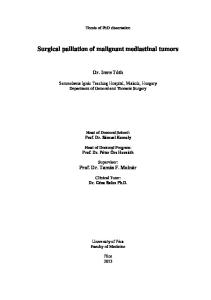
Polymorphous adenocarcinoma Evans and Batsakis88 described a malignant tumor arising in the intraoral minor salivary glands, very similar to lobular invasive carcinoma of the breast. Subsequent reports better defined the morphologic spectrum of this entity, leading to the term “polymorphous low grade adenocarcinoma (PLGA)” of salivary glands.89 Although polymorphous low grade adenocarcinoma mainly affects the intraoral salivary glands, occasional reports describe it in the parotid, lung, skin, vulva, and vagina. Recently, Asioli et al90 described 3 cases arising in the mammary gland. All 3 patients were adult females aged 37, 55, and 74 years, respectively. Tumors presented as firm nodules, ranging from 1.5 to 4 cm in maximum diameter. On histology, the tumors showed a central solid area surrounded by a peripheral rim of alveolar and cribriform structures as well as single neoplastic cells arranged in “Indian file” (Figure 8A and B). Neoplastic cells had round, central nuclei. Atypical mitotic figures were frequent. No necrosis was present. On immunohistochemistry neoplastic cells were strongly positive for BCL-2 (Figure 8C), whereas positivity for CK7 and E-cadherin was faint. Negative results were obtained with the following antibodies: EMA, CK14, CD117 (c-kit), HER-2, ER, and PR. Differential diagnosis includes invasive lobular carcinoma and AdCC. Invasive lobular carcinoma should be excluded because CK7 is weakly positive, E-cadherin, even though faint, is still maintained, whereas ER and PR receptors are negative in PLA. AdCC should be excluded because PLA lacks the classic biphasic components of epithelial and basal-myoepithelial cells; in addition, CD117 is consistently negative. Prognosis is difficult to establish as the number of the reported cases is very small. Case 1 of the original series died of widespread metastatic disease 3 years after diagnosis. Cases 2 and 3 were alive with no evidence of disease, but follow-up was limited to a few months. In view of the aggressive behavior demonstrated from case 1, at this stage, the term “low grade” should be avoided in cases occurring primarily in the breast. Further studies are necessary to determine the biology of PLA occurring in the breast.
Figure 8 Polymorphous adenocarcinoma. (A) The tumor is composed of a central solid area, surrounded by cells arranged in nests. (B) At higher power neoplastic cells are arranged in “indian file.” (C) Neoplastic cells are strongly positive for BCL-2.
88
Conclusions Salivary gland-type neoplasms of the breast are rare and include both benign and malignant tumors. In view of their rarity combined with broad morphologic spectrum, they often cause diagnostic dilemmas. Even though they are histologically analogous to the more common counterparts occurring in salivary glands, there are significant differences in their biological behavior. The correct diagnosis is important not only for prognostic purposes, but also for selecting the appropriate therapy. It is also important conceptually to try to place them in the relatively new, evolving “molecular classification” of breast tumors. During the last decade, molecular studies coupled with a more detailed immunohistochemical characterization of breast tumors have lead to a new classification having important implications for clinical management and therapeutic options. Six main types of breast carcinomas are emerging, each having a specific morphologic and immunohistochemical profile91 leading to specific therapeutic options. From the practical point of view, oncologists separate “luminal” and “basaloid” carcinomas. Basaloid carcinomas are characterized by a “triple negative” (negative for ER, PR, and HER-2) immunohistochemical profile. On H&E morphology, the “basaloid carcinomas” correspond more frequently to poorly differentiated, aggressive carcinomas. However, breast tumors analogous to those occurring more frequently in salivary glands usually do not express ER, PR, or HER-2 amplification and in contrast to other “triple negative” basal-like breast carcinomas, some of them behave in a low-grade manner. Therefore, “triple negativity” in breast carcinomas does not automatically signal a highly aggressive neoplasm because some of the salivary gland-type breast carcinomas like AdCC, majority of malignant adenomyoepitheliomas, and low-grade MEC should be regarded as low-grade neoplasms. In these cases, aggressive chemotherapeutic treatments are unnecessary. In summary, a careful and correct morphologic diagnosis of breast carcinomas of special types is important for correct management of the patients.
References 1. Azzopardi JG: Problems in breast pathology, in Azzopardi JG, Bennington JL (eds): Major Problems in Pathology. London, United Kingdom, WB Saunders, 1979 2. Foschini MP, Reis-Filho J, Eusebi V, et al: Salivary gland-like tumours of the breast: Surgical and molecular pathology. J Clin Pathol 56:497-506, 2003 3. Bennett AK, Mills SE, Wick MR: Salivary-type neoplasms of the breast and lung. Semin Diagn Pathol 20:279-304, 2003 4. Hamperl H: The myothelia (myoepithelial cells). Normal state; regressive changes; hyperplasia; tumors. Curr Top Pathol 53:161-220, 1970 5. Foschini MP, Eusebi V: Carcinomas of the breast showing myoepithelial cell differentiation. A review of the literature. Virchows Arch 432:303-310, 1998 6. Narita T, Matsuda K: Pleomorphic adenoma of the breast: Case report and review of the literature. Pathol Int 45:441-447, 1995
Seminars in Diagnostic Pathology, Vol 27, No 1, February 2010 7. Simha MR, Doctor VM, Udawadia TE. Mixed tumor of salivary gland type of the male breast. Indian J Cancer 28:14-17, 1992 8. Makek M, von Hochstetter AR: Pleomorphic adenoma of the human breast. J Surg Oncol 14:281-286, 1980 9. Agnantis NJ, Maounis N, Priovolou-Papaevangelou M, et al: Pleomorphic adenoma of the human female breast. Pathol Res Pract 188:235240, 1992 10. Mizukami Y, Takayama T, Takemura A: Pleomorphic adenoma of the breast. Radiat Med 26:442-445, 2008 11. Parham DM, Evans A: Pleomorphic adenoma of the breast, a potential for the misdiagnosis of malignancy on fine needle aspiration (FNA). Cytopathology 9:343-348, 1998 12. Iyengar P, Cody HS, Brogi E: Pleomorphic adenoma of the breast: Case report and review of the literature. Diagn Cytopathol 33:416-420, 2005 13. Lomax-Smith JD, Azzopardi JG: The hyaline cell: A distinctive feature of “mixed” salivary tumours. Histopathology 2:77-92, 1978 14. Geurts JM, Schoenmakers EF, Röijer E, et al: Identification of NFIB as recurrent translocation partner gene of HMGIC in pleomorphic adenomas. Oncogene 16:865-878, 1998 15. Åström AK, Voz AL, Kas K, et al: Conserved mechanism of PLAG1 activation in salivary gland tumors with and without chromosome 8q12 abnormalities: Identification of SII as a new fusion partner gene. Cancer Res 59:918-923, 1999 16. Voz ML, Agten NS, Van de Ven WJ, et al: PLAG1, the main translocation target in pleomorphic adenoma of the salivary glands, is a positive regulator of IGF-II. Cancer Res 60:106-113, 2000 17. Van Dyck F, Declercq J, Braem CV, et al: PLAG1, the prototype of the PLAG gene family: Versatility in tumour development [review]. Int J Oncol 30:765-774, 2007 18. Persson F, Andren Y, Winnes M, et al: High-resolution genomic profiling of adenomas and carcinomas in salivary glands reveals amplification, rearrangement, and fusion of HMGA2. Genes Chromosomes Cancer 48:69-82, 2009 19. Sato K, Ueda Y, Shimasaki M, et al: Pleomorphic adenoma (benign mixed tumor) of the breast: A case report and review of the literature. Pathol Res Pract 201:333-339, 2005 20. John BJ, Griffiths C, Ebbs SR: Pleomorphic adenoma of the breast should be excised with a cuff of normal tissue. Breast J 13:418-420, 2007 21. Hayes MM, Lesack D, Gilardet C, et al: Carcinoma ex-pleomorphic adenoma of the breast. Report of three cases suggesting a relationship to metaplastic carcinoma of matrix producing type. Virchows Arch 446:142-169, 2005 22. Elston CW, Ellis IO: Pathological prognostic factors in breast cancer. I. The value of histological grade in breast cancer: Experience from a large study with long-term follow-up. Histopathology 19:403-410, 1991 23. Di Palma S, Skalova A, Vanìèek T, et al: Non-invasive (intracapsular) carcinoma ex pleomorphic adenoma: Recognition of focal carcinoma by HER-2/neu and MIB1 immunohistochemistry. Histopathology 46: 144-152, 2005 24. Tavassoli FA, Eusebi V: Tumours of the Mammary Gland, Series 4. Washington, DC, Armed Forces Institute of Pathology, 2009 25. Lamovec J, Uz-Krazovec M, Zidar A, et al: Adenoid cystic carcinoma of the breast: A histologic, cytologic and immunohistochemical study. Semin Diagn Pathol 6:153-164, 1989 26. Bauer E, Sumpio E, Jennings TA, et al: Adenoid cystic carcinoma of the breast data from the Connecticut Tumour Registry and a review of the literature. Am J Surg 205:295-301, 1987 27. Arpino G, Clark GM, Mohsin S, et al: Adenoid cystic carcinoma of the breast. Molecular markers, treatment and clinical outcome. Cancer 94:2119-2127, 2002 28. Miliauskas JR, Leong AS: Adenoid cystic carcinoma in a juvenile male breast. Pathology 23:298-301, 1991 29. Okamoto Y, Sumiyama Y, Arima Y, et al: A case of adenoid cystic carcinoma (ACC) of the breast and review of the utility of preoperative imaging diagnose. Breast Cancer 8:84-89, 2001
Foschini and Krausz
Salivary Gland-Type Tumors of the Breast
30. Gupta RK, Green C, Naran S, et al: Cytology of adenoid cystic carcinoma of the breast. Diagn Cytopathol 20:82-84, 1999 31. Mastropasqua MG, Maiorano E, Pruneri G, et al: Immunoreactivity for c-kit and p63 as an adjunct in the diagnosis of adenoid cystic carcinoma of the breast. Mod Pathol 18:1277-1282, 2005 32. Azoulai A, Lae M, Freneaux P, et al: KIT is highly expressed in adenoid cystic carcinoma of the breast, a basal-like carcinoma associated with a favorable outcome. Mod Pathol 18:1623-1631, 2005 33. Shin SJ, Rosen PP: Solid variant of mammary adenoid cystic carcinoma with basaloid features: A study of nine cases. Am J Surg Pathol 26:413-420, 2002 34. Cheuk W, Chan JK, Ngan RK: Dedifferentiation in adenoid cystic carcinoma of salivary gland: An uncommon complication associated with an accelerated clinical course. Am J Surg Pathol 23:465-472, 1999 35. Chau Y, Hongyo T, Aozasa K, et al: Dedifferentiation of adenoid cystic carcinoma: Report of a case implicating p53 mutation. Hum Pathol 32:1403-1407, 2001 36. Nagao T, Gaffey TA, Serizawa H, et al: Dedifferentiated adenoid cystic carcinoma: A clinicopathologic study of 6 cases. Mod Pathol 16:1265-1272, 2003 37. Seethala RR, Hunt JL, Baloch ZW, et al: Adenoid cystic carcinoma with high-grade transformation: A report of 11 cases and a review of the literature. Am J Surg Pathol 31:1683-1694, 2007 38. Righi A, Morandi L, Lenzi M, et al: Adenoid cystic carcinoma of the breast associated with invasive ductal carcinoma: A case report. Int J Surg Pathol [Epub ahead of print] 39. Noske A, Schwabe M, Pahl S, et al: Report of a metaplastic carcinoma of the breast with multidirectional differentiation: An adenoid cystic carcinoma, a spindle cell carcinoma and melanoma. Virchows Arch 452:575-579, 2008 40. Ro JY, Silva EG, Gallager HS: Adenoid cystic carcinoma of the breast. Hum Pathol 18:1276-1281, 1987 41. Kleer CG, Oberman HA: Adenoid cystic carcinoma of the breast: Value of histologic grading and proliferative activity. Am J Surg Pathol 22:569-575, 1998 42. Kiaer H, Nielsen B, Paulsen S, et al: Adenomyoepithelial adenosis and low grade malignant adenomyoepithelioma of the breast. Virchows Arch A Pathol Anat Histol 405:55-67, 1984 43. Eusebi V, Casadei GP, Bussolati G, et al: Adenomyoepitheliom of the breast with a distinctive type of apocrine adenosis. Histopathology 11:305-315, 1987 44. Tamura G, Monma N, Suzuki Y, et al: Adenomyoepithelioma (myoepithelioma) of the breast in a male. Hum Pathol 24:678-681, 1993 45. Berna JD, Arcas I, Ballester A, et al: Adenomyoepithelioma of the breast in a male. Am J Roentegnol 169:917-918, 1997 46. Kurashina M: Fine needle aspiration cytology of benign and malignant adenomyoepithelioma of the breast. Report of two cases. Diagn Cytopathol 26:29-34, 2002 47. Yahara T, Yamaguchi R, Yokoyama G, et al: Adenomyoepithelioma of the breast diagnosed by a mammotome biopsy: Report of a case. Surg Today 38:144-146, 2008 48. McLaren BK, Smith J, Schuyler PA, et al: Adenomyoepithelioma clinical, histologic, and immunohistologic evaluation of a series of related lesions. Am J Surg Pathol 29:1294-1299, 2005 49. Ohi Y, Umekita Y, Rai Y, et al: Clear cell hidradenoma of the breast: A case report and review of the literature. Breast Cancer 14:307-311, 2007 50. Kazakov DV, Vanecek T, Mukensnabl P, et al: Skin-type hidradenoma of the breast parenchyma with t(11;19) translocation: Hidradenoma of the breast. Am J Dematopathol 29:457-461, 2007 51. Foschini MP, Pizzicannella G, Peterse JL, et al: Adenomyoepithelioma of the breast associated with low-grade adenosquamous and sarcomatoid carcinomas. Virchows Arch 427:243-250, 1995 52. Hikino H, Nagaoka S, Miura H, et al: Benign myoepithelioma of the breast: Origin and development. Pathol Int 59:422-426, 2009 53. Toth J: Benign human mammary myoepithelioma. Virchows Arch A Pathol Anat Histol 374:263-269, 1977
89 54. Enghardt MH, Hale JH: An epithelial spindle cell breast tumor of myoepithelial origin. An immunohistochemical and ultrastructural study. Virchows Arch A Pathol Anat Histol 416:177-184, 1989 55. Tavassoli FA: Myoepithelial lesions of the breast. Myoepitheliosis, adenomyoepithelioma and myoepithelial carcinoma. Am J Surg Pathol 15:554-568, 1991 56. Lakhani SR, O’Hare MJ, Managhan P, et al: Malignant myoepithelioma (myoepithelial carcinoma) of the breast: A detailed cytokeratin study. J Clin Pathol 48:164-167, 1995 57. Cartagena N Jr, Cabello-Inchausti B, Willis I, et al: Clear cell myoepithelial neoplasm of the breast. Hum Pathol 19:1239-1243, 1988 58. Damiani S, Dina RE, Eusebi V: Eosinophilic and granular cell tumors of the breast. Semin Diagn Pathol 16:117-125, 1997 59. Scarpellini F, Usellini L, Foschini MP: Malignant myoepithelioma associated with ductal carcinoma in situ and invasive. Description of a case and review of the literature. Pathologica 89:420-424, 1997 60. Jones C, Foschini MP, Chaggar R, et al: Comparative genomic hybridization analysis of myoepithelial carcinoma of the breast. Lab Invest 80:831-836, 2000 61. Jones C, Nonni AV, Fulford L, et al: CGH analysis of ductal carcinoma of the breast with basaloid/myoepithelial cell differentiation. Br J Cancer 85:422-427, 2001 62. Damiani S, Riccioni L, Pasquinelli G, et al: Poorly differentiated myoepithelial rich cell carcinoma of the breast. Histopathology 30: 542-548, 1997 63. Tsuda H, Tanabe T, Hasegowa T, et al: Myoepithelial differentiation in high grade invasive ductal carcinomas with large central acellular zones. Hum Pathol 30:1134-1139, 1999 64. Tsuda H, Tanabe T, Hasegowa T, et al: Large central acellular zones indicating myoepithelial tumor differentiation in high grade invasive ductal carcinomas as marker of predisposition to lung and brain metastases. Am J Surg Pathol 24:197-202, 2000 65. Stewart FW, Foote FW, Becker WF: Muco-epidermoid tumors of salivary glands. Ann Surg 122:820-844, 1945 66. Patchefsky AS, Frauenhoffer CM, Krall RA: Low-grade mucoepidermoid carcinoma of the breast. Arch Pathol Lab Med 103:196-198, 1979 67. Camelo-Piragua SI, Habib C, Kanumuri P, et al: Mucoepidermoid carcinoma of the breast share cytogenetic abnormality with mucoepidermoid carcinoma of the salivary gland: A case report with molecular analysis and review of the literature. Hum Pathol 40:1887-892, 2009 68. Horii R, Akiyama F, Ikenaga M, et al: Muco-epidermoid carcinoma of the breast. Pathol Int 56:549-553, 2006 69. Di Tommaso L, Foschini MP, Ragazzini T, et al: Mucoepidermoid carcinoma of the breast. Virchows Arch 444:13-19, 2004 70. Tavassoli FA, Devilee P (eds): World Health Organization Classification of Tumours: Tumours of the Breast and Female Genital Organs. Lyon, France, IARC Press, 2003 71. Rosen PP: Rosen’s Breast Pathology, (ed 3). Philadelphia, PA: Wolters Kluwer Lippincott Williams & Wilkins, 2009 72. Guo SP, Cheuk W, Chan JK: Pleomorphic adenoma with mucinous and squamous differentiation: A mimicker of mucoepidermoid carcinoma. Int J Surg Pathol 17:335-337, 2009 73. Foschini MP, Marucci G, Eusebi V: Low grade mucoepidermoid carcinoma of salivary glands: Characteristic immunohistochemical profile and evidence of striated duct differentiation. Virchows Arch 440:536-542, 2002 74. Luchtrach H, Moll R: Mucoepidermoid mammary carcinoma. Immunohistochemical and biochemical analyses of intermediate filaments. Virchows Arch 416:105-113, 1989 75. O’Neill ID: t(11;19) translocation and CRTC1-MAML2 fusion oncogene in mucoepidermoid carcinoma. Oral Oncol 45:2-9, 2009 76. Tjalma WA, Verslegers IO, De Loecker PA, et al: Low and high grade mucoepidermoid carcinomas of the breast. Eur J Gynaecol Oncol 23:423-425, 2002 77. Roncaroli F, Lamovec J, Zidar A, et al: Acinic cell-like carcinoma of the breast. Virchows Arch 429:69-74, 1996
90 78. Schmitt FC, Ribeiro CA, Alvarenga S, et al: Primary acinic-cell like carcinoma of the breast—A variant with good prognosis? Histopathology 36:286-289, 2000 79. Damiani S, Pasquinelli G, Lamovec J, et al: Acinic cell carcinoma of the breast: An immunohistochemical and ultrastructural study. Virchows Arch 437:74-81, 2000 80. Coyne JD, Dervon PA: Primary acinic carcinoma of the breast. J Clin Pathol 55:545-547, 2002 81. Foschini MP, Eusebi V: Rare (new) entities of the breast and medullary carcinoma. Pathology 41:48-56, 2009 82. Matoso A, Easley SE, Gnepp DR, et al: Salivary gland acinar-like differentiation of the breast. Histopathology 4:262-263, 2009 83. Lambros MB, Tan DS, Jones RL: Genomic profile of a secretory breast cancer with an ETV6-NTRK3 duplication. J Clin Pathol 67:604-612, 2009 84. Damiani S, Eusebi V, Losi L, et al: Oncocytic carcinoma (malignant oncocytoma) of the breast. Am J Surg Pathol 22:221-230, 1998 85. Tremblay G, Pearse AG: A cytochemical study of oxidative enzymes in the parathyroid oxyphil cell and their functional significance. Br J Exp Pathol 40:66-70, 1959
Seminars in Diagnostic Pathology, Vol 27, No 1, February 2010 86. Costa MJ, Silverberg SG: Oncocytic carcinoma of the male breast. Arch Pathol Lab Med 113:1396-1399, 1989 87. Ragazzi M, Reis-Filho JS, De Biase D, et al: Characterization of oncocytic breast carcinoma. Oral Presentation 22nd Congress European Congress of Pathology. Virchows Archives 455:S33, 2009 (suppl 1) 88. Evans HL, Batsakis JG: Polymorphous low-grade adenocarcinoma of minor salivary glands. A study of 14 cases of a distinctive neoplasm. Cancer 53:935-942, 1984 89. Evans HL, Luna MA: Polymorphous low-grade adenocarcinoma: A study of 40 cases with long-term follow up and an evaluation of the importance of papillary areas. Am J Surg Pathol 24:1319-1328, 2000 90. Asioli S, Marucci G, Ficarra G, et al: Polymorphous adenocarcinoma of the breast. Report of three cases. Virchows Arch 448:2934, 2006 91. Abd El-Rehim DM, Ball G, Pinder SE, et al: High-throughput protein expression analysis using tissue microarray technology of a large well-characterised series identifies biologically distinct classes of breast cancer confirming recent cDNA expression analyses. Int J Cancer 116:340-350, 2005