Human Reproduction vol.15 no.5 pp.1028–1036, 2000
Safety and efficacy of oestriol for symptoms of natural or surgically induced menopause
Kentaro Takahashi1, Masako Okada, Tomoya Ozaki, Hiroko Kurioka, Atsushi Manabe, Haruhiko Kanasaki and Kohji Miyazaki Department of Obstetrics and Gynecology, Shimane Medical University, 89-1, Enya-cho, Izumo 693-8501, Japan 1To
whom correspondence should be addressed
To assess the safety and efficacy of oestriol in relieving post-menopausal symptoms 53 post-menopausal Japanese women with climacteric symptoms, 27 with natural menopause (group I) and 26 with surgically induced menopause (group II), received oral oestriol, 2 mg daily for 12 months. Clinical parameters including Kupperman index (KI) and the degree of satisfaction with symptomatic relief; serum concentrations of oestradiol, FSH and LH; serum lipids; blood pressure; bone mineral density, serum calcium (Ca), alkaline phosphatase (ALP), and urinary Ca were compared between the two groups. Oestriol improved KI in groups I and II by 49 and 80% respectively. Satisfaction with treatment was 85% in group I and 93% in group II. For both parameters, values were significantly different between groups I and II (P < 0.05 for both). Serum concentrations of oestradiol, FSH and LH changed in group I versus group II 6 months after initiation. A significant decrease in serum ALP and Ca/Cr was observed in group I at 6 months. Except for serum triglycerides, oestriol had no significant effect on lipids. Systolic and diastolic blood pressures were significantly decreased in group I at 3 months versus baseline. Slight vaginal bleeding occurred in 14.3% of group I. Histological evaluation of the endometrium in all women of group I and ultrasound assessment of the breasts following 12 months of oestriol treatment found normal results in all women. Therefore, oestriol appeared to be safe and effective in relieving symptoms of menopausal women. The beneficial biochemical effects of oestriol were marked in the natural menopause. Overall, oestriol may serve as a good choice for hormone replacement therapy to protect against other climacteric symptoms in post-menopausal women who do not need medication for osteoporosis or coronary artery disease. Key words: bone metabolism/climacteric symptoms/hormone replacement therapy/lipid metabolism/oestriol
Introduction The benefits of hormone replacement therapy (HRT) are well established for the relief of climacteric symptoms in postmenopausal women as well as for the prevention of osteopor1028
osis and cardiovascular disease. In Japan, HRT has been used increasingly over the years for treating the climacteric. However, the prolonged exposure to unopposed oestrogens stimulates the growth of endometrium, thus increasing the risk of endometrial hyperplasia. An increasing number of women are receiving HRT with oestrogens plus added progestogens to prevent endometrial hyperplasia and neoplasia (Ettinger et al., 1998). Recently, it was reported (Hulley et al., 1998) that treatment with oral conjugated equine oestrogen plus medroxyprogesterone acetate did not reduce the overall rate of coronary heart disease events in post-menopausal women with established coronary disease. However, the debate on this regimen remains controversial regarding the development of breast cancer (Bergkvist et al., 1989; Colditz et al., 1995). The benefits of HRT must always be weighed against their risks. Oestriol, a weak oestrogen, has been successfully used in treating post-menopausal women in several countries for more than 30 years, and is considered to lack the potential risks of systemic oestrogen therapy (Esposito, 1991). Oestriol seems to present a logical alternative to standard oestradiol or other conjugated oestrogens used as HRT for women in the climacteric, and presents advantages to those at risk for endometrial cancer (Marslew et al., 1992). Oestriol succinate may have less thrombotic potential than synthetic oestrogens (Toy et al., 1978). Of the three human oestrogens, oestriol is the only one to lack mitogenic activity, while it protects the reproductive organs by means of its competition with endogenous oestrogens for the receptor (Lemon, 1975). However, it is still a subject of debate (Weiderpass et al., 1999). There are relatively few publications on the effects of oestriol on the symptoms of the climacteric (Tzingounis et al., 1978, 1980; Schneider, 1982; Kopera, 1983; Lauritzen, 1987; Bottiglione et al., 1995), especially studies lasting more than 12 months (Grasso et al., 1982; Yang et al., 1995; Melis et al., 1996). The present study was designed to assess and compare the efficacy and safety of oestriol for treating signs and symptoms related to the climacteric in women with natural menopause and surgically induced menopause, and to evaluate its effects on serum concentrations of oestradiol, gonadotrophins, lipids, as well as the biochemical markers of bone turnover, bone mineral density, and the blood pressure.
Materials and methods A total of 53 post-menopausal women (aged 40–62 years) with climacteric symptoms were included in this study. Each subject gave her written informed consent for participation. The study was approved by our hospital ethics committee. ‘Post-menopausal’ was defined as © European Society of Human Reproduction and Embryology
Oral oestriol for the climacteric women
amenorrhoea that had persisted for 艌 6 months. All patients had undergone either natural (n ⫽ 27) or surgically induced (n ⫽ 26) menopause for at least 6 months, and had not received HRT prior to study entry. They presented with such typical climacteric complaints as hot flushes, sweating, neurovegetative symptoms such as insomnia, depressive disturbance, and genito-urinary symptoms such as genital itching and dyspareunia. In all patients, no change or aggravation of climacteric complaints occurred, when 600 mg/day calcium L-aspartate (Aspara-Ca, Tanabe, Tokyo, Japan) as a placebo was orally administered for a few months prior to study entry. They were otherwise healthy according to results of clinical and laboratory evaluation. Excluded from study were women with thromboembolic disease, oestrogen-related neoplasms, breast disorder, urinary tract infection, uterine bleeding, abnormal uterine endometrial cytology, viral vaginal infection, and a body mass index (BMI) 艌 25. Patients received an oral dose of 2 mg oestriol (Estriel; Mochida, Tokyo, Japan) given daily for 12 months. The experimental design comprised two parallel groups, group I (n ⫽ 27) had undergone natural menopause, while group II (n ⫽ 26) had undergone surgically induced (hysterectomy with bilateral oophorectomy) menopause. The severity of the climacteric symptoms was evaluated according to the Kupperman index (KI) (Kupperman et al., 1959) each month of the study. Satisfaction with the hormone therapy was also evaluated at those times. Patients were asked to record the intensity of satisfaction on a 10 cm linear analogue scale marked from 0 to 100: 0 representing no change or aggravation of condition and 100 representing the perfect condition they had expected for this hormone therapy. Compliance with the regimen was checked by personal interviews and diary cards. Furthermore, serum concentrations of oestradiol, gonadotrophins, blood lipids, biochemical markers of bone turnover, bone mineral density, and blood pressure were measured. The presence of histological endometrial neoplasms was assessed using endometrial biopsy, and the presence of breast tumours by ultrasound before and after treatment. Endometrial biopsies were obtained with a curette without local anaesthesia. These specimens were fixed in neutral formalin, embedded in paraffin wax and sectioned at several levels. All biopsies were examined by a pathologist for signs of hormonal effects and atypia. Blood samples were collected from the subjects early in the morning after an overnight fast. Sera were stored at –30°C. To minimize variation, each subject’s sample was analysed at the same time. Pretreatment biometric data, reproductive history, endogenous sex hormones, estimates of calcium metabolism, bone mineral density, estimates of lipid metabolism, and the blood pressure at the outset of the study are shown in Table I. Except for age and time since occurrence of natural menopause or of hysterectomy with bilateral oophorectomy, the two groups showed no significant differences. Assay of serum concentrations of sex hormones Serum concentrations of oestradiol, FSH and LH were measured with commercially available kits before the initiation of the study and repeated every 6 months. Samples were obtained approximately 6 h after the intake of medication. Oestradiol concentrations were determined by radioimmunoassay using the Coat-A-Count oestradiol kit (Nippon DPC, Chiba, Japan), whose intra-assay and inter-assay coefficients of variation (CV) were ⬍7 and 6.2% respectively. The reference method has 10.0% cross-reactivity with oestrone and 0.32% with oestriol. FSH and LH were measured by radioimmunoassay using the SPAC-S FSH kit and SPAC-S LH kit (Daiichi Radioisotope Laboratory, Tokyo, Japan), whose intra-assay and inter-assay CV were 4.8 and 5.3%, and ⬍8 and 4% respectively.
Table I. Characteristics of women with natural menopause (group I) and women with surgically induced menopause (group II). Group I (n ⫽ 27) Age (years) Post-menopausal monthsa Weight (kg) Height (m) BMI (kg/m2) Kupperman index (points) Serum oestradiol Serum FSH (mIU/l) Serum LH (mIU/l) MIC GSmin sigma GS/D Serum ALP (IU/l) Serum Ca (mg/dl) Serum Pi (mg/dl) Urinary Ca/Cr Serum TC (mg/dl) Serum TG (mg/dl) Serum HDL-C (mg/dl) Serum LDL-C (mg/dl) Systolic blood pressure (mm Hg) Diastolic blood pressure (mm Hg)
53.6 68.4 52.1 152.0 22.5 18.4 13.9 94.8 34.9 0.53 0.24 2.31 124.4 9.5 3.5 0.25 205.3 105.6 60.6 120.9 129.9 79.4
⫾ ⫾ ⫾ ⫾ ⫾ ⫾ ⫾ ⫾ ⫾ ⫾ ⫾ ⫾ ⫾ ⫾ ⫾ ⫾ ⫾ ⫾ ⫾ ⫾ ⫾ ⫾
Group II (n ⫽ 26) 4.8 63.0 7.5 3.9 3.2 9.8 11.7 37.8 16.5 0.06 0.36 0.25 76.9 0.7 0.5 0.10 30.4 63.0 13.9 32.9 22.2 12.3
47.7 ⫾ 4.2* 19.7 ⫾ 16.0* 50.9 ⫾ 8.2 155.5 ⫾ 4.3 21.0 ⫾ 3.1 18.2 ⫾ 7.2 13.1 ⫾ 5.4 77.3 ⫾ 24.6 26.3 ⫾ 11.2 0.53 ⫾ 0.06 2.26 ⫾ 0.39 2.27 ⫾ 0.21 116.8 ⫾ 34.4 9.3 ⫾ 0.5 3.8 ⫾ 0.5 0.28 ⫾ 0.10 213.1 ⫾ 53.1 100.4 ⫾ 38.5 62.2 ⫾ 15.6 118.7 ⫾ 26.1 121.2 ⫾ 19.0 71.3 ⫾ 13.4
*P ⬍ 0.01. aPost-menopausal months in group I expressed as time since occurrence of natural menopause; those in group II expressed as time since occurrence of hysterectomy and bilateral oophorectomy. BMI, body mass index; MIC, metacarpal index; GSmin, index of densities of bone cortex and bone marrow; sigma GS/D, index of bone density per unit length; ALP, alkaline phosphatase; Ca, calcium; Pi, inorganic phosphate; Cr, creatinine; TC, total cholesterol; TG, triglycerides; HDL-C, high density lipoprotein cholesterol; LDL-C, low density lipoprotein cholesterol.
Determination of lipid profiles Total cholesterol (TC), triglycerides (TG) and high density lipoprotein cholesterol (HDL-C) were measured enzymatically with an automatic multi-purpose analyser (TBA 200 FR, Toshiba Medical Products, Tokyo, Japan). Total cholesterol was measured enzymatically using Determiner LTC-S (Kyowa Medex, Tokyo, Japan) (intra- and interassay CV, 0.96 and 0.94% respectively). Triglycerides were measured enzymatically using Autosera STGN (Daiichi Chemical, Tokyo Japan) (intra- and inter-assay CV of 0.66 and 1.02% respectively). The concentration of HDL-C was measured by a direct method (Nauck et al., 1996) with the use of Determiner L HDL-C (Kyowa Medex Tokyo, Japan) (intra- and inter-assay CV, 1.12 and 1.59% respectively). The concentration of low density lipoprotein cholesterol (LDL-C) was calculated from the formula TC – TG/5 – HDL-C (Friedewald et al., 1972). Bone mineral density and biochemical markers of bone turnover Bone mineral density (BMD) was assessed by use of the microdensitometry (MD) method (Inoue et al., 1983). The BMD of the second metacarpal bone of each subject was measured by X-ray using the MD method before and after treatment. An X-ray of the hand was also taken with a reference aluminium step-wedge. In the MD method, three indices, such as the metacarpal index (MCI), the index of the densities of bone cortex and bone marrow (GSmin) and the index of bone density per unit length (sigma GS/D), were calculated by computer analysis of the X-ray of each hand. In addition, serum concentrations of calcium (Ca), inorganic phosphate (Pi), alkaline phosphatase (ALP), and urinary Ca were measured to evaluate bone
1029
K.Takahashi et al.
metabolism. Serum concentrations of ALP and Pi were measured enzymatically with an automatic multi-purpose analyser (TBA200 FR, Toshiba Medical Products, Tokyo, Japan). Serum ALP was measured by the Quick Auto Neo ALP-JSII (Shino Test, Tokyo, Japan), intra- and inter-assay, 1.00 and 0.82% respectively. Serum Pi was measured with Determiner IP (Kyowa Medex, Tokyo, Japan), intra- and inter-assay CV, 1.02 and 4.53% respectively. Serum and urinary concentrations of Ca were measured by the orthocresolphthalein complexone (OCPC) colorimetric method using OCPC fluid (Eiken Chemical, Tokyo, Japan) (intra- and inter-assay, 0.71 and 1.37% respectively). The urinary excretion of Ca was corrected for urinary creatinine (Cr). Statistical analysis Data are expressed as the mean ⫾ SD. Data were analysed with the Statview software package (4.51.1). Differences between the groups were evaluated by either the Kolmogorov–Smirnov two-sample test, or Scheffe´ ’s multiple comparison test. One-way analysis of variance for repeated measures was used to evaluate the significance of any changes in parameters after the initiation of oestriol. A level of P ⬍ 0.05 was considered statistically significant.
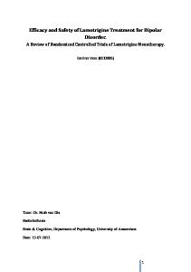
Results Three patients in group I reported slightly unexpected vaginal bleeding. The bleeding stopped of its own accord despite continuation of medication. Endometrial biopsy specimens taken during the bleeding incidences showed an atrophic endometrium without malignancy in one patient and weakly proliferative endometrium without malignancy in two patients. Furthermore, histological evaluation of the endometrium following 12 months of oestriol treatment found no atypical endometrium in all patients of group I including atrophic endometrium in 23 patients (85.2%) and weakly proliferative endometrium in four women (14.8%). Ultrasound assessment of the breasts following 12 months of treatment found no tumour in all women of both groups. Other adverse effects were observed in four patients: epigastralgia in three patients and mastodynia in one patient. However, no patient discontinued treatment due to side-effects. The menopausal index of Kupperman (KI) was employed to estimate subjective improvement. Both total KI scores in group I and group II prior to study entry (17.8 ⫾ 6.7 and 17.6 ⫾ 7.3 points respectively) were significantly increased at the start of this study. Only one patient in group I exhibited severe post-menopausal symptoms at the outset (KI ⬎35). The average scores and the percentage change in KI from baseline before and after treatment are shown for each group in Figure 1. At the end of the first month, the index was significantly reduced in both groups: 37.5% in group I and 28.8% in group II. Group I showed a gradual reduction in the KI over the 3 months after initiating oestriol, with a 49.8% reduction after 3 months of treatment. However, the continuation of treatment did not seem to induce any further substantial decreases in this index (8.6 ⫾ 7.8 points and 49.2 ⫾ 47.0% of mean KI and reduction rate respectively at the end of treatment). Group II showed a significant reduction in KI 3 months after initiating oestriol treatment, with a 56.8% reduction observed at 3 months. Significant substantial reductions in this index were also observed over the next 9 months (3.6 ⫾ 3.1 points and 1030
80.4 ⫾ 20.2% of mean KI and reduction rate respectively at the end of treatment; P ⬍ 0.01). These values differed significantly (P ⬍ 0.05) between the two groups after 12 months of treatment. Hot flushes, sweating and insomnia showed the most significant reduction in group I, reported by 2 months of treatment. Comparison of the feeling of satisfaction with this hormone therapy between the two groups is shown in Figure 2. At the end of the first month, the extent of satisfaction with the hormone therapy was ~75% in both groups. The continuation of treatment seemed to induce further increases in the extent of satisfaction: 85.2 ⫾ 8.4% in group I and 92.5 ⫾ 4.2% in group II at the end of treatment (P ⬍ 0.05). Serum concentrations of oestradiol, FSH and LH over the 12-month study are shown in Figure 3. No statistically significant changes in serum oestradiol were observed in either group during treatment with placebo prior to study entry (mean oestradiol values prior to study entry and at the start of this study: group I, 13.5 ⫾ 9.0 and 13.9 ⫾ 11.7 pg/ml respectively; group II, 12.0 ⫾ 2.9 and 13.1 ⫾ 5.4 pg/ml respectively). Statistical analysis of mean oestradiol values (21.8 ⫾ 12.2 pg/ml) at the 6 months of treatment showed a significant increase (P ⬍ 0.01) in group I as compared with mean pretreatment values. This effect continued until the end of the 12 month treatment period (17.1 ⫾ 7.4 pg/ml). No statistically significant changes in serum oestradiol were observed in group II during treatment. Mean serum concentrations of FSH (66.4 ⫾ 32.2 mIU/ml) and LH (23.7 ⫾ 13.0 mIU/ml) showed a significant decrease (P ⬍ 0.01) 6 months after starting treatment in group I as compared with pretreatment values. This decrease continued until the end of the 12 months of treatment (68.6 ⫾ 30.5 mIU/ml in FSH and 25.4 ⫾ 11.0 mIU/ml in LH). In group II, the mean FSH (59.6 ⫾ 15.7 mIU/ml) and mean LH (24.5 ⫾ 9.9 mIU/ml) at 12 months after the start of treatment showed a significant decrease as compared with pretreatment values (P ⬍ 0.01 and P ⬍ 0.05 respectively, while no statistically significant changes were observed in both serum FSH and LH concentrations 6 months after starting treatment. No statistically significant changes were seen in either group with regard to the markers of bone mineral density (MCI, GSmin and sigma GS/D). Concerning the biochemical markers of bone turnover, during the 12 months of treatment, no statistically significant changes were observed in any such markers in group II, and in the serum concentration of Ca in group I. In group I, the mean ALP (107.0 ⫾ 60.6 IU/l) and iP values (3.29 ⫾ 0.51 mg/dl) at the end of treatment showed a significant decrease (P ⬍ 0.05) as compared with pretreatment values respectively. Statistical analysis of mean urinary Ca/Cr values (0.21 ⫾ 0.10) at 6 months of therapy showed a significant decrease (P ⬍ 0.01) in group I as compared with pretreatment values. This decrease continued until the end of the 12 month study (0.19 ⫾ 0.10 pg/ml). This value after 12 months of treatment differed significantly from the value in group II (0.29 ⫾ 0.10) (Figure 4). No statistically significant changes were observed during treatment in serum concentrations of TG, HDL-C and LDL-C in either group. In group I, the mean TC (194.0 ⫾
Oral oestriol for the climacteric women
Figure 1. Mean scores (A) and percent change (B) in Kupperman index (KI). Mean scores before and after oestriol treatment in groups I (d—d) and II (s—s) are shown. Mean values are shown, SE is indicated by error bars. *P ⬍ 0.05; **P ⬍ 0.01 versus respective pretreatment values; †P ⬍ 0.05 versus group II group at that time.
Discussion
Figure 2. Satisfaction with oestriol therapy. Group I (d—d) and group II (s—s). Mean values are shown, SE is indicated by error bars. †P ⬍ 0.05 versus group I at that time.
37.1 mg/dl) at the end of treatment showed a significant (P ⬍ 0.05) decrease as compared with pretreatment values, whereas no statistically significant changes were observed in group II (Figure 5). Changes in blood pressure before and during treatment are shown in Figure 6. No statistically significant changes in blood pressure were observed in group II during the 12 months of treatment. However, in group I, the mean systolic pressure (114.2 ⫾ 15.2 mm Hg) and diastolic pressure (71.7 ⫾ 10.8 mm Hg) each showed a significant decrease (P ⬍ 0.05) at 3 months as compared with pretreatment values respectively.
Most studies on the treatment of complaints associated with the climacteric have utilized oestradiol or conjugated oestrogens as therapy, with relatively few reports on the effect of oestriol (Tzingounis et al., 1978, 1980; Schneider, 1982; Kopera, 1983; Lauritzen, 1987; Bottiglione et al., 1995). The use of oral oestriol as HRT remains controversial. For instance, a previous study reported that oestriol succinate administered at a dose of 2–4 mg/day orally for 1 year had little beneficial effect on climacteric symptoms, as shown by KI values (Grasso et al., 1982). Oral oestriol is rapidly inactivated by the liver to its glucuronide derivative, so that only 1–2% of the total dose administered enters the circulation in a bioactive form (Schiff et al., 1978; Heimer, 1987). The massive inactivation is influenced by the time of administration as well as by food intake, such that the circulating concentration of this hormone is difficult to maintain within the treatment range (Heimer, 1987). To solve this problem, high daily doses of oestriol given in divided amounts have been utilized. Heimer and Englund (Heimer and Englund, 1984) administered 6 and 12 mg of oestriol. A dose of 8 mg oestriol reportedly relieves hot flushes (Schneider, 1982). Nevertheless, such high doses of oestriol produce the same late responses, and the same endometrial and metabolic effects, as do the other oestrogens, because the nuclear permanence of oestriol receptors was double. Such high doses also induced side-effects such as nausea and mastalgia. The administration of oestriol by the vaginal route has been tried (Keller et al., 1981; Bottiglione et al., 1995; Melis et al., 1996). It was concluded that intra-vaginal oestriol is rapidly absorbed and is suitable for local and systemic oestrogen replacement therapy, and that it was more effective than the oral regimen 1031
K.Takahashi et al.
Figure 3. Changes in serum levels of oestradiol (A), FSH (B) and LH (C) during the 12 month period of oestriol treatment. Group I (d—d) and group II (s—s). Mean values are shown, SE is indicated by error bars. *P ⬍ 0.05; **P ⬍ 0.01 versus respective pretreatment values.
1032
Figure 4. Serum concentrations of alkaline phosphatase (A) and inorganic phosphate (B), and urinary calcium/creatinine (C) before and after oestriol treatment. Group I (d—d) and group II (s—s). Mean values are shown, SE is indicated by error bars. *P ⬍ 0.05; **P ⬍ 0.01 versus respective pretreatment values; †P ⬍ 0.05 versus group II at that time.
Oral oestriol for the climacteric women
(Keller et al., 1981). With 1 mg daily transvaginal oestriol the KI significantly decreased by ~50% after 2 weeks; KI scores were reduced by ~70% by the end of treatment (Bottiglione et al., 1995). However, long term therapy using the intravaginal route for oestriol administration has not received patient acceptance in Japan. The present study showed that oestriol at a dose of 2 mg/day significantly relieved climacteric symptoms in postmenstrual women. In those who had undergone natural menopause, the KI was significantly reduced by 37.5% at 1 month. The index then gradually declined over 3 months of treatment (49.8% reduction at 3 months). However, the continuation of treatment did not induce any further marked decrease in the KI, which is similar to earlier findings (Tzingounis et al., 1980). However, in women with surgically induced menopause, the KI showed a significant reduction at 3 months of treatment (56.8%
Figure 5. Changes in serum concentrations of total cholesterol (TC) during the 12 month treatment with oestriol. Group I (d—d) and group II (s—s). Mean values are shown, SE is indicated by error bars. *P ⬍ 0.05 versus pretreatment values.
reduction at 3 months). The continuation of treatment for 12 months was associated with a further significant decrease in the index (3.6 ⫾ 3.1 points and 80.4 ⫾ 20.2% of mean KI and reduction rate respectively). This value at 12 months was significantly lower than that reported in the women who had undergone natural menopause. In addition, the satisfaction with hormonal therapy was significantly higher in the women with surgically induced menopause than in those who had undergone natural menopause. The younger age and shorter time since menopause induced by hysterectomy and oophorectomy may perhaps explain the difference. Also, in women with early surgical menopause, treatment compliance may be easier. It is also possible that the same dose of oestriol has a stronger effect in women with surgically induced menopause. In the present study, a significant increase in serum oestradiol concentration was observed at 6 months of the oestriol treatment in women of the natural menopause group. This serum oestradiol elevation continued until the end of the 12 month period of treatment. However, this oestradiol increase was not seen in women who had oophorectomy. Although these elevated oestradiol concentrations might be accounted for by some residual ovarian activity, no statistically significant changes in serum oestradiol were observed in the natural menopause group during treatment by placebo prior to study entry. We also previously reported that the serum concentration of oestradiol increased transiently to be ⬎100 pg/ml in four women who were treated with oestriol within 5 years of the menopause (Okada et al., 1995). Therefore, it is considered that the administration of oestriol may have stimulated ovarian granulosa cells directly and/or indirectly to produce oestradiol, since oestriol itself cannot be converted to oestradiol. However, a detailed discussion of the oestradiol changes observed was not possible, since serum oestriol concentrations were not measured. Furthermore, the significant decreases in serum FSH and LH observed at the end of the 12 month study are more likely to be a physiological decline in direct pituitary production (the oestriol-induced inhibition of the hypophyseal release of FSH and LH) rather than a reduction due to oestradiol
Figure 6. Changes in systolic (A) and diastolic (B) blood pressure before and during oestriol treatment. Group I (d—d) and group II (s—s). Mean values are shown, SE is indicated by error bars. *P ⬍ 0.05 versus respective pretreatment values.
1033
K.Takahashi et al.
production by the ovary leading to negative feedback, as judged from the previous report of the lowered serum concentrations of FSH and LH in normal ovulatory women (Vahapassi and Adlercreutz, 1975), although it is well known that significant decreases in serum FSH and LH result from the negative feedback phenomenon produced by the elevation of oestradiol as a physiological phenomenon. Osteoporosis is common in post-menopausal women who have such risk factors as deficiencies of oestrogen and calcium. Oestrogen administration inhibits bone resorption, and can thus prevent further bone loss in such high-risk women (Lindsay and Cosman, 1990). Oestrogens suppress bone resorption at the time of accelerated bone turnover in the early stage after menopause and in elderly women, and suppress the reduction of bone mass, resulting in either the maintenance of, or an increase in, BMD (Lindsay and Tohme, 1990). However, oestriol therapy does not prevent post-menopausal osteoporosis, as revealed by evaluating the BMD of the lumbar spine using quantitative computed tomography (Yang et al., 1995). However, its effect on BMD has not been fully evaluated. The combined administration of intra-vaginal oestriol and of a nasal spray containing salmon calcitonin to postmenopausal women improved the neurovegetative symptoms and prevented a decrease in BMD (Melis et al., 1996). The rate of increase in the BMD of the lumbar vertebrae in the post-menopausal women administered orally 2 mg/day oestriol daily was recently evaluated. HRT with oestriol is effective in treating involutional osteoporosis, the bones of elderly women also maintain responsiveness to oestriol (Nishibe et al., 1998). One study (Minaguchi et al., 1996) has presented similar findings, that oestriol prevented post-menopausal bone loss; it concluded, based on the placebo results, that oestriol may be safe and efficacious. No significant change in BMD was observed in the present study. This differing result may be related to the methods of measurement. i.e. the BMD was measured at the middle of the metacarpal bone II with a densitometer on X-rays of the hands taken from the dorsal along an aluminium step-wedge. Although this method shows good reproducibility, the change in BMD of the metacarpal bone II as measured in this manner may be slower to show a change than when one evaluates the lumbar vertebrae (L2–L4) using dual-energy X-ray absorptiometry. However, it is reassuring that bone density did not decline over the 12 months. In the biochemical parameters of bone metabolism we examined, there were no significant decreases in all parameters in surgically induced menopausal women during treatment, while there were significant decreases in urinary Ca/Cr in natural menopausal women at 6 months and again at the end of 12 months administration. This decrease in markers of bone breakdown in the naturally menopausal women could be related to the higher oestradiol concentrations due to persistent ovarian activity and/or stimulating effect of the oestriol on ovary directly and/or indirectly. However, there was no significant change in serum Ca during treatment in the naturally menopausal women. This finding resembles that of a report in which no decrease in serum Ca was seen in post-menopausal women (Nishibe et al., 1998). However, it has also been 1034
reported that the serum Ca is significantly increased in postmenopausal women as compared with that in premenopausal women, and is reduced to the premenopausal value by HRT (Leino et al., 1994). Although oestriol might be clinically effective in maintaining normal bone metabolism in women with natural menopause from the current results, assessing biochemical markers of bone turnover (serum total and bonespecific ALP, serum C-terminal propeptide of type I collagen and serum osteocalcin as bone formation markers, and urinary cross-linked N-telopeptide of type I collagen as bone resorption index) must be more useful in the evaluation of oestriol treatment (Garnero et al., 1996). In general, oestrogen reduces TC and LDL-C, while increasing HDL-C (Knopp, 1988). However, no distinct effects of oestriol on lipids were observed in the menopausal women studied. Only serum TC showed a significant decrease in the naturally menopausal women. It is well known that several types of oestrogens are used clinically for HRT and their oestrogenic effects on the biological system may vary based on their chemical structures. Hence the outcome of the HRT may depend on the type of oestrogen used (The ESHRE Capri Workshop, 1998). In this regard, as compared to oestradiol, oestriol is a weak oestrogen that is known to have minimal uterotrophic effects (Anderson et al., 1975), and hence lacks the deleterious effects seen with oestradiol. On the other hand, oestriol may not be as effective in protecting post-menopausal women against cardiovascular disease as it has no antimitogenic effects on vascular smooth muscle cell growth, which plays a key role in vascular remodelling and neo-intima formation. Indeed, the same reason may be responsible for the lack of cardioprotective effect of conjugated equine oestrogens, as they contain less potent oestrogens such as oestrone and oestriol (ESHRE Capri Workshop, 1998). Similarly, the effects on bone density may vary. With regard to the potential mechanisms for the lack of effects on lipid, the binding affinity of oestriol to alpha or beta oestrogen receptor (ER), its antioxidant potential as well as antagonistic effects on oestradiol mediated effects may be considered. The effects of oestrogen and oestrogen agonists can be mediated by ER. Recent discovery of a new ER (Kuiper et al., 1996) has led to further questions regarding separate roles for these two receptors, termed ERα and ERβ. In the rat the highest expressions of ERα occur in the testes, pituitary gland, uterus, kidneys and adrenal glands; highest expressions of ERβ occur in the ovary and the prostate (Kuiper et al., 1997). Some data suggest that oestradiol activates transcription with ERα and inhibits at ERβ, whereas tamoxifen, raloxifene, and ICI 164 384 activate transcription with ERβ (Paech et al., 1997). Further work in the field of molecular and genetic endocrinology has shed more light on the complex subject of anti-oestrogenic agonist and antagonist modes of action. It has been proposed (Petersen et al., 1998) that ERβ2 should be considered in addition to ERβ1 and ERα when describing the effects of oestrogen, oestrogen agonists/antagonists, or environmental oestrogens. ERβ2 bound oestradiol with a lower affinity than either ERα or ERβ1. The binding of ERβ2 was selective in that cortisol, testosterone, aldosterone, and progesterone among other agents did not compete for oestradiol
Oral oestriol for the climacteric women
binding. However, a variety of known oestrogenic agents, including physiological oestrogens (oestrone and oestriol), plant and environmental oestrogens (genistein, coumestrol, bisphenol A, methoxychlor), and pharmocological agents (tamoxifen, α-hydroxytamoxifen) effectively competed for oestradiol binding to both ERβ1 and ERβ2. Thus, although it has been demonstrated that ERβ2 possesses altered binding of oestradiol, and in addition, some ligands exhibit potent agonism of ERβ2 while antagonizing ERδ and ERβ1 (Petersen et al., 1998), it is not yet known how ERβ1 or ERβ2 respond to oestriol. Further investigation may increase the understanding of the mechanisms of action of medically important oestrogenic compounds. Post-menopausal women have an increased incidence of cardiovascular disease; it is well known that HRT with oestradiol or conjugated oestrogens reduces this risk (Dubey et al., 1998; Modena et al., 1998). Both the systolic and diastolic blood pressure are higher in post-menopausal women than in men of same age or in premenopausal women, suggesting that oestrogen deficiency may influence the age-related increase in blood pressure (Bairey et al., 1998; Mercuro et al., 1998). Although it has been reported that the administration of oestradiol for relatively short periods (4–12 months) has no effect on blood pressure (Serup et al., 1981; Christiansen et al., 1982; Blum et al., 1986), in recent studies, the administration of oestradiol significantly reduced the 24-h systolic and diastolic blood pressures (Danser et al., 1998). Concentrations of prorenin and renin in women who received oestrogen were significantly lower than those in women without oestrogen administration (Danser et al., 1998). HRT using oestradiol and progesterone may protect post-menopausal women against cardiovascular disease by inhibiting the growth of cardiac fibroblasts and of cardiac remodelling (Dubey et al., 1998). Nevertheless, the effect of oestriol on cardiac risk factors is somewhat equivocal. Oestriol does not seem to affect hypertension (Erkkola et al., 1978; Head, 1998). However, in the present study, both the systolic and diastolic blood pressures in the naturally menopausal women showed a significant decrease 3 months after starting treatment as compared with mean pretreatment values. The reason for these results is not known, and further investigation in a larger number of patients is indicated. The administration of oestriol may stimulate the ovarian granulosa cells directly and/or indirectly, stimulating oestradiol production, causing oestradiol elevation and a decrease in serum ALP, urinary Ca/Cr, and serum TC, as observed in the naturally menopausal women in the present study, although it is possibly due to persistent ovarian activity. It is well known that there is a marked placebo response in terms of improvement of hot flushes and of KI (Saletu et al., 1995). Furthermore, the reduction of climacteric symptoms could still be an effect of time since the menopause. However, in this study the Ca tablets as placebo were given at the start of the preliminary study when the symptoms would not have been expected to decline. A placebo effect had therefore been evaluated prior to this study as no changes of clinical symptoms occurred after the administration of Ca tablets for 2–3 months. Therefore, any changes in clinical symptoms of both groups were considered to be attributed to the administration of
oestriol. The present study suggests that oestriol may be a safe and effective alternative for the relief of climacteric symptoms in post-menopausal women who reject, or who have contraindications, to conventional HRT with oestradiol or conjugated oestrogens. Few side-effects were observed, and endometrial examination revealed no abnormalities. All patients completed the 12-month study. About 90% were satisfied with the results at the end of hormonal treatment. The beneficial biochemical effects of oestriol were marked in the natural menopausal women. It is possible that these effects are related to the stimulation of oestradiol production from ovaries which may retain some partial function activity. However, a recent paper (van Haaften et al., 1997) showed that similar signs of oestrogen stimulation of the endometrium were seen following oestradiol and oestriol medication in the histological studies. Furthermore, another study (Weiderpass et al., 1999) warned that close surveillance of patients is needed and addition of a progestagen should be considered, since oral use of oestriol 1–2 mg daily increased the relative risk of endometrial cancer and endometrial hyperplasia: the odds ratios for at least five years of use compared with never use were 3.0 and 8.3 respectively. Therefore, the final results must be regarded with caution until confirmed on the basis of a double-blind, placebo-controlled trial in a large number of patients. In conclusion, the efficacy and safety of oestriol in relieving climacteric symptoms was investigated in Japanese women with natural menopause and with surgically induced menopause. Although no distinct effects of oestriol on lipids and bone were observed, the effects of oestriol on the parameters studied in the two groups of post-menopausal women were not identical. It was demonstrated that the hormonal effects of oestriol were stronger in women with natural menopause, while the relieving effects for climacteric symptoms were stronger in women with surgically induced menopause. However, the overall effects in both groups appear to be safe and effective in relieving menopausal symptoms. In postmenopausal women who do not need medication for osteoporosis or coronary artery disease, oestriol may serve as a good choice for HRT to protect against other climacteric symptoms. References Anderson, J.N., Peck, E.J. Jr and Clark, J.H. (1975) Estrogen-induced uterine responses and growth: relationship to receptor estrogen binding by uterine nuclei. Endocrinology, 96, 160–167. Bairey, M.C.N., Kop, W., Krantz, D.S. et al. (1998) Cardiovascular stress response and coronary artery disease: evidence of an adverse postmenopausal effect in women. Am. Heart J., 135, 881–887. Bergkvist, L., Adami, H-O., Persson, I. et al. (1989) The risk of breast cancer after estrogen and estrogen–progestin replacement. N. Engl. J. Med., 321, 293–297. Blum, M., Assa, S., Bacalu, B. et al. (1986) The influence of short-term estrogen replacement therapy (ERT) on the blood pressure and daily urinary catecholamine excretion in a small group of post-menopausal women. Eur. J. Obstet. Gynecol. Reprod. Biol., 23, 195–199. Bottiglione, F., Volpe, A., Esposito, G. et al. (1995) Transvaginal estriol administration in postmenopausal women: a double blind comparative study of two different doses. Maturitas, 22, 227–232. Christiansen, M.S., Hagen, C., Christiansen, C. et al. (1982) Dose-response evaluation of cyclic estrogen/gestagen in postmenopausal women: placebocontrolled trial of its gynecologic and metabolic actions. Am. J. Obstet. Gynecol., 144, 873–879.
1035
K.Takahashi et al. Colditz, G.A., Hankinson, S.E., Hunter, D.J. et al. (1995) The use of oestrogens and progestins and the risk of breast cancer in postmenopausal women. N. Engl. J. Med., 332, 1589–1593. Danser, A.H., Derkx, F.H., Schlekamp, M.A. et al. (1998) Determinants of interindividual variation of renin and prorenin concentrations: evidence for a sexual dimorphism of (pro)renin levels in humans. J. Hypertens., 16, 853–862. Dubey, R.K., Gillespie, D.G., Jackson, E.K. et al. (1998) 17β-Estradiol, its metabolites, and progesterone inhibit cardiac fibroblast growth. Hypertension, 31, 522—528. Erkkola, R., Lammintausta, R., Punnonen, R. et al. (1978) The effect of estriol succinate therapy on plasma renin activity and urinary aldosterone in postmenopausal women. Maturitas, 1, 9–14. Esposito, G. (1991) Estriol: a weak estrogen or a different hormone? Gynecol. Endocrinol., 5, 131–53. Ettinger, B., Li, D.-K. and Klein, R. (1998) Unexpected vaginal bleeding and associated gynecologic care in postmenopausal women using hormone replacement therapy: comparison of cyclic versus continuous combined schedules. Fertil. Steril., 69, 865–869. Friedewald, W.T., Levy, R.I. and Fredrickson, D.S. (1972) Estimation of lowdensity lipoprotein cholesterol in plasma without use of the preparative ultracentrifuge. Clin. Chem., 18, 499–450. Garnero, P., Sornay-Rendu, E., Chapuy, M.C. et al. (1996) Increased bone turnover in late postmenopausal women is a major determinant of osteoporosis. J. Bone Miner. Res., 11, 337–349. Grasso, A., Baraghini, F., Barbieri, C. et al. (1982) Endocrinological features and endometrial morphology in climacteric women receiving hormone replacement therapy. Maturitas, 4, 19–26. Head, K.A. (1998) Estriol: safety and efficacy. Alternative Medicine Review, 3, 101–113. Heimer, G.M. (1987) Estriol in the postmenopause. Acta Obstet. Gynecol. Scand. Suppl., 139, 5–23. Heimer, G.M. and Englund, D.E. (1984) Enterohepatic recirculation of oestriol studied in cholecystectomized and non-cholecystectomized menopausal women. Upsala J. Med. Sci., 89, 107–115. Hulley, S., Grady, D., Bush, T. et al. (1998) Randomized trial of estrogen plus, progestin for secondary prevention of coronary heart disease in postmenopausal women. JAMA, 280, 605–613. Inoue, T., Kusida, K., Miyamoto, S. et al. (1983) Quantitative assessment of bone density on X-ray picture. J. Jpn Orthop. Assoc., 57, 1923–1936. Keller, P.J., Riedmann, R., Fischer, M. et al. (1981) Oestrogens, gonadotropins and prolactin after intra-vaginal administration of oestriol in postmenopausal women. Maturitas, 3, 47–53. Knopp, R.H. (1988) The effects of postmenopausal estrogen therapy on the incidence of arteriosclerotic vascular disease. Obstet. Gynecol., 72, 23S–30S. Kopera, H. (1983) Actions and potencies of estriol in the human. In Jasonni, V.M., Nenci, J. and Flamigni, C. (eds), Steroids and Endometrial Cancer. Raven Press, New York, pp. 175–183. Kuiper, G., Enmark, E., Pelto-Hhuikko, M. et al. (1996) Cloning of a novel estrogen receptor expressed in rat prostate and ovary. Proc. Natl Acad. USA, 93, 5925–5930. Kuiper, G., Carlsson, B. and Grandien, K. (1997) Comparison of the ligand binding specificity and transcript tissue distribution of estrogen receptors alpha and beta. Endocrinology, 138, 863–870. Kupperman, H.S., Wetchler, B.B. and Blatt, M.H.G. (1959) Contemporary therapy of the menopausal syndrome. JAMA, 171, 1627–1637. Lauritzen, C. (1987) Results of a 5-year prospective study of estriol succinate treatment in patients with climacteric complaints. Horm. Metab. Res., 19, 579–584. Leino, A., Jarvisalo, J., Impivaara, O. et al. (1994) Ovarian hormone status, life-style factors, and markers of bone metabolism in women aged 50 years. Calcif. Tissue Int., 54, 262–267. Lemon, H.M. (1975) Estriol prevention of mammary carcinoma induced by 7,2,dimethyl-benzanthracene and procarbazine. Cancer Res., 35, 1341–1353. Lindsay, R. and Cosman, F. (1990) Estrogen in prevention and treatment of osteoporosis. Ann. N. Y. Acad. Sci., 592, 326–33. Lindsay, R. and Tohme, J.F. (1990) Estrogen treatment of patients with established postmenopausal osteoporosis. Obstet. Gynecol., 76, 290–295. Marslew, U., Overgaard, K., Riis, B.J. et al. (1992) The new combinations of estrogen and progestogen for prevention of postmenopausal bone loss: longterm effect on bone, calcium and lipid metabolism, climacteric symptoms, and bleeding. Obstet. Gynecol., 79, 202–210.
1036
Melis, G.B., Cagnacci, A., Bruni, V. et al. (1996) Salmon calcitonin plus intravaginal estriol: effective treatment for the menopause. Maturitas, 24, 83–90. Mercuro, G., Zoncu, S., Piano, D. et al. (1998) Estradiol-17β reduces blood and restores the normal amplitude of the circadian blood pressure rhythm in postmenopausal hypertension. Am. J. Hypertens., 11, 909—913. Minaguchi, H., Uemura, T., Shirasu, K. et al. (1996) Effect of estriol on bone loss in postmenopausal Japanese women: a multicenter prospective open study. J. Obstet. Gynaecol. Res., 22, 259–265. Modena, M.G., Rossi, R., Muia, N. Jr et al. (1998) Short-term results of transdermal estrogen replacement therapy in cardiovascular disease-free postmenopausal females with and without hypertension. Giornale Italiano di Cardiologia, 28, 636–644. Nauck, M., Marz, W., Haas, B. et al. (1996) Homogeneous assay for direct determination of high-density lipoprotein cholesterol evaluated. Clin. Chem., 42, 424–429. Nishibe, A., Morimoto, S., Hirota, K. et al. (1998) Comparison of effects of estriol on bone mineral density of vertebrae between elderly and postmenopausal women. J. Bone Miner. Metab., 16, 21–26. Okada, M., Takahashi, K., Kurioka, H. et al. (1995) Atypical uterine bleeding and serum estradiol level. J. Jpn. Menopause Soc., 3, 211–215. Paech, K., Webb, P. and Kuiper, G. (1997) Differential ligand activation of estrogen receptors ER alpha and ER beta at AP1 sites. Science, 277, 1508–1510. Petersen, D.N., Tkalcevic, G.T., Koza-Taylor, P.H. et al. (1998) Identification of estrogen receptor β2, a functional variant of estrogen receptor β expressed in normal rat tissues. Endocrinology, 139, 1082—1092. Saletu, B., Brandstatter, N., Metka, M. et al. (1995) Double-blind, placebocontrolled, hormonal, syndromal and EEG mapping studies with transdermal oestradiol therapy in menopausal depression. Psychopharmacology, 122, 321–329. Schiff, I., Wentworth, B., Koos, B. et al. (1978) Effect of estriol administration on the hypogonadal women. Fertil. Steril., 30, 278–282 Schneider, H.P.G. (1982) Oestriol and the menopause: clinical results from a prospective study. In Fioretti, P., Martini, L., Melis, G.B. and Yen, S.S.C. (eds), Serono Symposium, Vol. 39, The Menopause: Clinical, Endocrinological and Pathophysiological Aspects. Academic Press, London, pp. 523–533. Serup, J., Bostofte, E., Larsen, S. et al. (1981) Effectivity and acceptability of oral contraceptives containing natural and artificial oestrogens in combination with a gestagen. A controlled double-blind investigation. Acta Obstet. Gynaecol. Scand., 60, 203–206. The ESHRE Capri Workshop (1998) Hormones and cardiovascular disease: oral contraceptives and hormonal replacement therapy: differential effects on coronary heart disease, deep venous thrombosis and stroke. Hum. Reprod., 13, 2325–2333. Toy, J.L., Davies, J.A., McNicol, G.P. et al. (1978) The effects of longterm therapy with oestriol succinate on the haemostatic mechanism in postmenopausal women. Br. J. Obstet. Gynaecol., 85, 363–366. Tzingounis, V.A., Aksu, F. and Greenblatt, R.B. (1978) Estriol in the management of the menopause. JAMA, 239, 1638–1641. Tzingounis, V.A., Aksu, F. and Greenblatt, R.B. (1980) The significance of oestriol in the management of the post-menopause. Acta Endocrinol., 233, 45–50. Vahapassi, J. and Adlercreutz, H. (1975) Effect of continuous oral administration of estriol on corpus luteum function. Contraception, 11, 427–439. van Haaften, M., Donker, G.H., Gie-Go, D.M. et al. (1997) Biochemical and histological effects of vaginal estriol and estradiol applications on the endometrium, and vagina of postmenopausal women. Gynecol. Endocrinol., 11, 175–185. Weiderpass, E., Baron, J.A., Adami, H-O. et al. (1999) Low-potency oestrogen and risk of endometrial cancer: a case-control study. Lancet, 353, 1824–1828. Yang, T.S., Tsan, S.H., Chang, S.P. et al. (1995) Efficacy and safety of estriol replacement therapy for climacteric women. Chin. Med. J. (Taipei), 55, 386–391. Received on November 1, 1999; accepted on February 10, 2000