Reduced Eccentric Loading of the Knee with the Pose Running Method REGAN E. ARENDSE1, TIMOTHY D. NOAKES1, LIANE B. AZEVEDO1, NICHOLAS ROMANOV1, MARTIN P. SCHWELLNUS1, and GRAHAM FLETCHER2 1
MRC/UCT Exercise Science and Sports Medicine Research Unit, Department of Human Biology, Faculty of Health Sciences, University of Cape Town, SOUTH AFRICA; and 2University College of the Fraser Valley, British Columbia, CANADA
ABSTRACT ARENDSE, R. E., T. D. NOAKES, L. B. AZEVEDO, N. ROMANOV, M. P. SCHWELLNUS, and G. FLETCHER. Reduced Eccentric Loading of the Knee with the Pose Running Method. Med. Sci. Sports Exerc., Vol. 36, No. 2, pp. 272–277, 2004. Purpose: The aim of this study was to compare the biomechanical changes during natural heel-toe running with learned midfoot and Pose running. Methods: Twenty heel-toe runners were instructed in midfoot running and a novel running style in which the acromium, greater trochanter, and lateral malleolus are aligned in stance (Pose running). Clinical gait analysis was performed for each running style and the biomechanical variables compared. Results: In comparison with midfoot and heel-toe running Pose running was characterized by shorter stride lengths and smaller vertical oscillations of the sacrum and left heel marker. Compared with midfoot and Pose running heel-toe running was characterized by greater magnitudes and loading rates of the vertical impact force. In preparation for initial contact, the knee flexed more in Pose than in heel-toe and midfoot running. The ankle at initial contact was neutral in Pose compared with a dorsiflexed and plantarflexed position in heel-toe and midfoot running, respectively. The knee power absorption and eccentric work were significant lower (P ⬍ 0.05) in Pose than in either heel-toe or midfoot running. In contrast, there was a higher power absorption and eccentric work at the ankle in Pose compared with heel-toe and midfoot running. Conclusions: Pose running was associated with shorter stride lengths, smaller vertical oscillations of the sacrum and left heel markers, a neutral ankle joint at initial contact, and lower eccentric work and power absorption at the knee than occurred in either midfoot or heel-toe running. The possibility that such gait differences could be associated with different types and frequencies of running injuries should be evaluated in controlled clinical trails. Key Words: KINETICS, KINEMATICS, MIDFOOT, RUNNING STYLE MODIFICATION
R
with forward running, the knee is more flexed in terminal swing, initial contact, and stance (7). One of the consequences of this running style is that the peak ground reaction force is only 25–33% of that measured during forward running, suggesting that the calf musculature absorbs more of the impact forces during backward running (15). The peak patellofemoral compressive force is also reduced with backward running (3.0 ⫾ 0.6 body weight (BW) compared with 5.6 ⫾ 1.3 BW for forward running (8)). These biomechanical characteristics of backward running may be more beneficial in the treatment of running injuries, as suggested by anecdotal reports (15). But backward running is an impractical method for the treatment or prevention of running injuries. Running in the forward direction with similar flexed knee geometry and midfoot contact to that of backward running may be hypothesized to offer equivalent treatment benefits. Midfoot running is, however, not associated with a lower risk of injury (3). Although the stance phase knee geometry of midfoot running is not described, it appears that foot contact is unlikely to be the exclusive determinant of the risk of injury. A novel running style with a midfoot strike pattern and a flexed knee in stance has been developed and is called Pose running. The Pose running lower-limb geometry instance is achieved by forward lean of the trunk and vertical alignment of the ipsilateral shoulder, hip, and heel of the supporting
unning style is described as a “learned response to a given set of anthropometric and physiological constraints” (4) so that the movement of the body components minimizes the amount of mechanical work performed (1). Running style may be described by the overall action, body angle, arm swing, foot placement, rear leg lift, and length of stride (19). The biomechanical variables associated with specific running styles change with running speed (16), inclination of the running surface (2,20), the use of running shoes (6), and the use of treadmills (24). Biomechanical variables also differ between different running styles for example between forward and backward running (7). Backward running is characterized by initial contact with the midfoot, with the ankle plantarflexed (7). Compared Address for correspondence: Dr. Regan E. Arendse, MB.ChB., M.Sc., MRC/UCT Research Unit for Exercise Science and Sports Medicine, Department of Human Biology, University of Cape Town, Sports Science Institute of South Africa, P. O. Box 115 Newlands 7725, South Africa; E-mail:
[email protected]. Submitted for publication May 2003. Accepted for publication October 2003. 0195-9131/04/3602-0272 MEDICINE & SCIENCE IN SPORTS & EXERCISE® Copyright © 2004 by the American College of Sports Medicine DOI: 10.1249/01.MSS.0000113684.61351.B0
272
limb. Pose running therefore appears to have a similar lower-limb geometry to backward running. It is intuitive that Pose running may have a role in the treatment of running injuries equivalent to backward running. Accordingly, the aims of this study were to determine whether clinical gait analysis can distinguish between midfoot and Pose running in natural heel-toe recreational runners and whether the Pose method produces biomechanical changes that might be of value in the treatment or prevention of running injuries.
METHODS Subjects. Twenty (20) male and female natural heel-toe recreational runners (height: 1.62 ⫾ 0.29 m, mass: 75.9 ⫾ 16.6 kg, age: 33.2 ⫾ 12.7 yr), free of physical deformity or neurological abnormality, were recruited from running clubs in the Cape Town, South Africa, region. The mean ⫾ SD 10-km time for the group was 54.3 ⫾ 22.5 min (11.1 km·h⫺1). Written informed consent was obtained from the runners before their participation in the study. Ethical approval for the study was obtained from the Research and Ethics Committee of the Faculty of Health Sciences, University of Cape Town. Test procedure. A repeated measures experimental design (within-subjects) was employed. The runners were tested in each of three running styles. Initial biomechanical data were collected for the natural heel-toe running style. The runners were then instructed in midfoot and Pose running. The data collection procedure was repeated once the runners were able to run confidently with the two novel running styles. The running trials were conducted barefoot to aid reliability of marker placement on successive test sessions and reduce the effect differences in footwear between runners. Running style. All runners naturally employed a heeltoe running style in which initial contact with the supporting surface was made with the heel followed by the mid and anterior portions of the foot (3). The runners were instructed in midfoot and Pose running (Fig. 1). Midfoot running instruction consisted of practical demonstration and verbal instruction to the runners to alter the
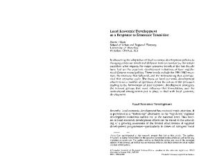
FIGURE 1—Representative heel-toe, midfoot and pose running styles at initial contact. RUNNING STYLE AND KNEE ECCENTRIC LOAD
point of first contact of the foot with the supporting surface from the heel to the midfoot area. The runners were instructed to avoid contact with the heel of the foot with the supporting surface in stance. The runners were allowed to practice midfoot running until they volunteered that they were confident with the running style and the investigators were satisfied that there was no contact of the heel with the supporting surface in stance. None of the runners required more than 15 min to become confident in the midfoot running style. In contrast, learning the Pose method required a total instruction of 7.5 h and comprised 1.5-h sessions daily on five consecutive days. The runners were encouraged to achieve the following postural changes and actions: 1. Align the acromium, the greater trochanter, and lateral malleolus in stance. 2. Lean forward in the above posture and allow the body to fall forward and thereby initiate movement. 3. At initiation of movement, lift the supporting foot by flexing the knee and avoid pushing away from the supporting surface. 4. At successive stance phases, contact with the midfoot (ball of foot, not the toes) and avoid contact of the heel with the supporting surface. 5. Maintain a flexed knee throughout the gait cycle. Running speed. The trials in each running style were undertaken at self-selected speeds. The runners were encouraged to maintain a constant speed for all three styles. A self-selected running speed was described as a running speed that he or she could select independently, that felt comfortable, and that would be representative of the running speed they each followed in an easy training run. The running speed for each trial was determined from the displacement of the sacrum marker in the x-axis of the laboratory co-ordinate system during the processing of the biomechanical data for each trial. Data collection. Biomechanical data were collected with a strain gauge force plate (1000 Hz) (AMTI Inc., Newton, MA) that was mounted flush with the surface of the running track and a six-camera Vicon-370 Motion Analysis system (120 Hz) (Oxford Metrics, Oxford, UK). A modified Helen Hayes marker set (22) was used to collect kinematic data. Biomechanical data were collected from 10 barefoot running trials in each style. The 10 trials were scrutinized for errors, and five complete trials of each runner in each running style were processed. The runners were unaware of the position of the force plate. A trial was considered successful when the runner made contact exclusively with the force plate with the right foot, all retro-reflective markers were tracked for the duration of the right-foot stance phase on the force plate, and there was no alteration in running style as observed from the runner with the naked eye and on review of the animation from the collected gait analysis data. All biomechanical variables were collected and processed to C3D files with the Workstation® program by Oxford Metric (Oxford, UK). The C3D files were further converted Medicine & Science in Sports & Exercise姞
273
to DST files using the Rdata2® program by Motion Lab Systems (Baton Rouge, LA). Processing of the raw data files was accomplished with the Bodybuilder® program by Oxford Metrics and the GaitLab® program by Kiboho Publishers (Cape Town, South Africa) to calculate the temporaldistance variables, ground reaction forces, knee and ankle joint angles, and power-time curves. All data were exported as text files for analysis in Excel® (Microsoft Corporation, Redmond, WA). Selection of biomechanical variables. The biomechanical variables selected for this study were based on selected previous studies (6,14). Temporal-distance parameters such as stride and step length and vertical displacement of the sacrum and left heel marker during a gait cycle were determined. The average horizontal (x-axis) and vertical (z-axis) displacements of the left heel marker over five running trials were used to determine the average stride length (m) and vertical foot displacement (m), respectively. The average vertical displacement of the sacral marker in the z-axis of the global reference system over five running trials, determined the average vertical body displacement (m). The ground reaction forces of the five trials per runner per running style were averaged and specific variables selected (5,6,12). These included: the loading rate and magnitude of the vertical impact force peak, the vertical propulsive force peak, and the horizontal braking and propulsive force peaks. Due to the inconsistent presence of the vertical impact force peak in the midfoot style previously reported (3) the loading rate and magnitude of the vertical impact force at 25 ms of stance were determined. All ground reaction forces were expressed in multiples of BW. The knee and ankle joint angles were limited to those about the y-axis through the knee and ankle joint centers averaged for five running trials per runner per style and analyzed (22). Kinematic data included the peak knee flexion angle in swing, the peak knee extension angle, and the accompanying ankle angle immediately before and at initial contact, and the peak knee flexion and ankle dorsiflexion angles in stance phase (°). The knee and ankle power (W·kg⫺1) curves during stance phase were determined with an inverse dynamics method and a three degrees of freedom joint model in GaitLab® (Kiboho Publishers, Cape Town, South Africa). The peak negative and positive knee and ankle power values were determined from the data in an Excel® (Microsoft Corporation) spreadsheet. Negative (eccentric) and positive (concentric) work (J·kg⫺1) values were calculated from the trapezoidal integration of the area below the negative and positive portions of the power-time curve, respectively. Statistical analysis. Statistical tests were performed with a commercial statistical program (StatSoft, Inc. (2000); STATISTICA for Windows [Computer program manual]; Tulsa, OK). Detailed descriptive statistical analysis was undertaken of the demographic and training variables. The means of the running speed and biomechanical variables in each test condition were compared with the repeated mea274
Official Journal of the American College of Sports Medicine
TABLE 1. A comparison of the temporal-distance variables between heel-toe, midfoot, and Pose running (N ⫽ 20). VSDsp VHDsp StrideLn StepLn
Heel-Toe
Midfoot
Pose
F-Ratio
P
0.09 ⫾ 0.03 0.38 ⫾ 0.11 2.20 ⫾ 0.56 1.09 ⫾ 0.33
0.08 ⫾ 0.03 0.36 ⫾ 0.14 2.17 ⫾ 0.71 1.09 ⫾ 0.36
0.05 ⫾ 0.04* 0.28 ⫾ 0.21* 1.48 ⫾ 1.04* 0.83 ⫾ 0.60*
22.421 6.491 11.478 4.417
0.001 0.003 0.001 0.017
* Significantly different from heel-toe and midfoot running. Vertical sacrum marker displacement (m) [VSDsp], vertical left heel marker displacement (m) [VHDsp], stride length (m) [StrideLn], step length (m) [StepLn].
sures ANOVA, the level of significance posted at P ⬍ 0.05. Significant relationships between the means as indicated by significant F-ratios were explored with the Scheffe´ post hoc analysis test.
RESULTS Running speed. The running speed of the heel-toe running style (2.98 ⫾ 0.42 m·s⫺1) was similar to that have midfoot (P ⫽ 0.066) and Pose running (P ⫽ 0.089). The running speed for midfoot running (3.06 ⫾ 0.42 m·s⫺1) was greater than for Pose running (2.90 ⫾ 0.37 m·s⫺1) (P ⫽ 0.001). Temporal-distance biomechanical variables. Pose running was characterized by shorter mean stride and step lengths and smaller vertical oscillations of the sacrum and left heel marker, compared with both heel-toe and midfoot running (Table 1). Ground reaction forces. Heel-toe running caused greater magnitudes and loading rates of the vertical impact force at 25 ms of stance and at peak magnitude compared with midfoot and Pose running (Table 2). The vertical propulsive force was similar between the heel-toe, midfoot, and Pose running. The horizontal braking and propulsive forces were less in Pose than heel-toe and midfoot running. Knee and ankle joint angles. The peak knee flexion during the swing phase was the same in all three running styles (Table 3). The knee flexed more in preparation for initial foot contact in Pose compared with heel-toe and midfoot running. The ankle in terminal swing was neutral in Pose compared with a dorsiflexed and plantarflexed position in heel-toe and midfoot running, respectively. The knee and ankle geometries in terminal swing of the respective running styles were maintained at initial foot contact. The peak knee TABLE 2. A comparison of the ground reaction force variables between heel-toe, midfoot, and Pose running (N ⫽ 20). HBF HPF VIF25 VLR25 VIF VLR VPF
Heel-toe
Midfoot
Pose
F-Ratio
P
0.22 ⫾ 0.05 0.18 ⫾ 0.04 1.20 ⫾ 0.24‡ 49.39 ⫾ 8.98‡ 1.21 ⫾ 0.24‡ 49.89 ⫾ 9.18‡ 2.22 ⫾ 0.17
0.22 ⫾ 0.07 0.18 ⫾ 0.05 0.87 ⫾ 0.26 37.09 ⫾ 7.68 0.87 ⫾ 0.25 37.27 ⫾ 7.35 2.25 ⫾ 0.51
0.15 ⫾ 0.07* 0.15 ⫾ 0.07* 0.78 ⫾ 0.39 36.89 ⫾ 9.94 0.78 ⫾ 0.39 36.88 ⫾ 9.94 1.97 ⫾ 0.78
18.045 3.588 28.191 35.131 28.907 34.193 2.426
0.001 0.035 0.001 0.001 0.001 0.001 0.099
* Significantly different from heel-to and midfoot running. ‡ Significantly different from midfoot and Pose running. Horizontal braking force (BW) [HBF], horizontal propulsive force (BW) [HPF], vertical impact force at 25 ms (BW) [VIF25], vertical impact force at 25-ms loading rate (BW䡠s)⫺1 [VR25], vertical impact force peak (BW) [VIF], vertical impact force peak loading rate (BW䡠s)⫺1 [VLR], vertical propulsive force peak (BW) [VPF].
http://www.acsm-msse.org
TABLE 3. A comparison of the knee and ankle joint angles between heel-toe, midfoot, and Pose running (N ⫽ 20). KswF KswE Kic Kst Asw Aic Ast
Heel-Toe
Midfoot
Pose
F-Ratio
P
85.6 ⫾ 2.4 23.3 ⫾ 6.5 27.3 ⫾ 5.3 49.4 ⫾ 3.8 ⫺18.3 ⫾ 9.8‡ ⫺13.2 ⫾ 4.5‡ ⫺26.1 ⫾ 2.0‡
85.0 ⫾ 5.6 23.6 ⫾ 6.1 27.2 ⫾ 6.4 47.6 ⫾ 6.9 11.00 ⫾ 5.9 11.5 ⫾ 5.8 ⫺19.7 ⫾ 3.2
83.1 ⫾ 6.2 29.5 ⫾ 7.8* 31.5 ⫾ 8.1* 43.9 ⫾ 6.5* ⫺0.4 ⫾ 4.9* ⫺0.2 ⫾ 6.4* ⫺20.6 ⫾ 3.1
1.274 10.460 4.304 5.819 29.899 138.548 32.594
0.291 0.001 0.020 0.006 0.001 0.001 0.001
* Significantly different from heel-toe and midfoot running. ‡ Significantly different from midfoot and Pose running. Knee swing phase peak flexion angle (°) (KswF), knee angle in terminal swing phase (°) (KswE), knee angle at initial supporting surface contact (°) (Kic), knee angle in midstance (°) (Kst), ankle angle in terminal swing phase (°) (Asw), ankle angle at initial supporting surface contact (°) (Aic), ankle angle in midstance (°) (Ast).
flexion in stance was greater in the heel-toe and midfoot compared with Pose running. The peak ankle dorsiflexion in stance was greater in the heel-toe than midfoot and Pose running. Knee and ankle work and power. The knee power absorption (Table 4) and eccentric work (Fig. 2) were less in Pose running compared with heel-toe and midfoot running. The ankle power absorption (Table 4) and eccentric work (Fig. 3) were greater in Pose compared with heel-toe and midfoot running. The knee power generation and concentric work were less in Pose compared with either heel-toe or midfoot running (Table 4). There were no differences in ankle power generation and concentric work between the running styles.
DISCUSSION The first relevant finding of this study was that clinical gait analysis was able to identify the biomechanical differences in midfoot and Pose running in natural heel-toe runners. Thus, it was found that Pose running is characterized by shorter stride lengths, lower magnitudes of the vertical impact forces, greater knee flexion in preparation for and at initial contact, and less eccentric work at the knee and more eccentric work at the ankle compared with midfoot and heel-toe running. Previous attempts to modify running style have measured the efforts of changing stride length (26), vertical displacement of the body (18), trunk inclination, arm swing, and leg mechanics (13). Although each of these studies has demonstrated changes in individual kinematic variables, these changes have been inconsistent, showing the resistance of
FIGURE 2—A comparison of the knee eccentric work between heeltoe, midfoot and Pose running (N ⴝ 20). *Significantly different from heel-toe and midfoot running.
the naturally chosen running style to change. In contrast, marked changes in running biomechanics have been measured with changes in running speed (16), gradient of the supporting surface (20), and the use of running shoes (6). We now show that Pose running can produce marked changes in biomechanical variables. Pose running produced significantly smaller vertical displacement of the body; the feet are kept close to the supporting surface and the stride and step lengths are shorter, compared with heel-toe and midfoot running (Table 1). Like midfoot running, but in contrast to heel-toe running, Pose running reduced the magnitudes and loading rates of the vertical impact force at 25 ms of stance, as well as the peak value (Table 2). However, Pose and midfoot running cause different lower-limb geometry during the running stride (Table 3). The horizontal braking and propulsive forces appeared to be the only components of the ground reaction force that may distinguish between Pose and midfoot running. This suggests that the ground reaction forces may not be the most valuable measurements for evaluating
TABLE 4. A comparison of the knee and ankle dynamic variables between heel-toe, midfoot and Pose running (N ⫽ 20). KPA APA KPG APG CKW CAW
Heel-toe
Midfoot
Pose
F-Ratio
P
7.01 ⫾ 1.75 5.85 ⫾ 1.39 6.27 ⫾ 1.44 8.49 ⫾ 1.54 0.48 ⫾ 0.13 0.53 ⫾ 0.11
7.40 ⫾ 1.93 5.87 ⫾ 1.40 6.34 ⫾ 1.55 8.57 ⫾ 1.56 0.49 ⫾ 0.13 0.54 ⫾ 0.11
3.70 ⫾ 1.40* 6.91 ⫾ 1.93* 5.01 ⫾ 2.49* 8.29 ⫾ 1.94 0.30 ⫾ 0.16* 0.50 ⫾ 0.13
64.320 4.629 7.089 0.405 25.198 1.711
0.000 0.016 0.003 0.669 0.000 0.195
* Significantly different from heel-toe and midfoot running. Knee power absorption (W䡠kg⫺1) (KPA), Ankle power absorption (W䡠kg⫺1) (APA), knee power generation (W䡠kg⫺1) (KPG), ankle power generation (W䡠kg⫺1) (APG), concentric knee work (J䡠kg⫺1) (CKW), concentric ankle work (J䡠kg⫺1) (CAW).
RUNNING STYLE AND KNEE ECCENTRIC LOAD
FIGURE 3—A comparison of the ankle eccentric work between heeltoe, midfoot and Pose running (N ⴝ 20). *Significantly different from heel-toe and midfoot running. Medicine & Science in Sports & Exercise姞
275
modification of running style but that the other biomechanical variables need also to be considered. A change in knee and ankle geometry with the transition from barefoot to shod running (6), and between running at different speeds on treadmills (10,11) and overground (16), are well described. The results of the present study indicate the knee is more flexed in preparation for and at initial contact in Pose compared with heel-toe and midfoot running (Table 3). This may be a function of the short stride length characteristic of Pose running (Table 1). Ankle position also distinguished between the three running styles in terminal swing and at initial contact, with the ankle dorsiflexed in heel-toe, plantarflexed in midfoot, and neutral in Pose running. Ankle dorsiflexion in heel-toe running appears to be associated with the greater magnitude of the vertical impact force compared with midfoot and Pose running. The difference in ankle angles between Pose and midfoot running does not appear to have any association with any other variable measured. Downhill running has been shown to increase knee eccentric work (2). This study shows that knee power absorption and eccentric work were significantly reduced with Pose compared with heel-toe and midfoot running (Fig. 1). In contrast, ankle power absorption and eccentric work were increased with Pose compared with heel-toe and midfoot running (Fig. 2). This suggests that there is redistribution of the work activity between the knee and ankle joints with different running styles. However, the effects of a different running style on the hip dynamics were not studied. The hip is the more important source of power for forward propulsion during running than during walking (16,17). In contrast, the contribution of the knee and ankle power is minimized as running speed increases. The reduction in knee power generation and concentric work with Pose running was not accompanied by a complementary increase in the ankle power generation and concentric work. Presumably that this was provided by changes in the hip as a source of power and work for forward propulsion, which was likely increased in Pose running. This would explain the constant ankle dynamics in propulsion in all three running styles. To clarify this relationship, the hip biomechanics should be evaluated in future studies of running style modification. The magnitude of the moment arm at the joint of interest is an important determinant of the magnitude of the mechanical joint work calculated with the inverse dynamics approach (21). The low work values at the knee and high work values at the ankle in Pose running suggest that the magnitude of the moment arm at the knee is least but is greatest at the ankle. This observation may be due to a combination of the lower-limb geometry and the position of the torso in stance. The similarity of knee and ankle work and power between the heel-toe and midfoot running suggests that the different ankle joint positions do not change the moment arms. Positioning of the torso over the supporting limb may influence the direction of the ground reaction force in relation to the lower limb joint centers. The posture of the whole body throughout the gait cycle should be 276
Official Journal of the American College of Sports Medicine
included in future studies to fully understand the reasons for the changes in knee and ankle joint power and work. In this study, the runners were evaluated in the barefoot condition, to which they were not habituated. Although standard to all three test conditions, it is possible that the barefoot condition may prevent the adoption a conventional heel-toe running style. Runners protect the heel during barefoot running and reduce distortion of the heel fat pad (6). Subsequently, initial contact occurs predominantly with the anterior portion of the foot. This may explain the similar biomechanical variables between heel-toe and midfoot running in the barefoot condition. The runners in this study naturally employed a heel-toe running style. The duration of instruction in midfoot running was no greater than 15 min. In contrast, Pose running was instructed over a total of 7.5 h. The prolonged time to teach Pose running was devoted mainly to achieving the desired alignment of the trunk and supporting limb in stance. It is unlikely that a longer duration of instruction in midfoot running may have elicited differences in the biomechanical variables between midfoot and heel-toe running. This opinion is based on the results of a study in which six male and three female heel-toe runners readily changed to midfoot contact with verbal instruction by the investigators with similar instruction time to the present study (25). More importantly, the biomechanical variables of the natural heeltoe runners with the changed landing technique were similar to that of natural midfoot runners (25). Accordingly, because the kinetics and kinematics of Pose running were different from those of midfoot running, we conclude that the biomechanical characteristics of midfoot running were faithfully reproduced in this study and were not an artifact of too-short familiarization period. An increase in walking and running speed is known to significantly increase the magnitude of the ground reaction forces (6,23). Although the running speed in midfoot running was significantly greater than that of Pose, the ground reaction forces were similar. Kinematic adaptations may produce similar ground reaction forces with different running conditions (9). In this regard, the differences in ankle kinematics between midfoot and Pose running may have minimized any differences in the ground reaction forces between the two, despite a difference in running speed. Our results suggest that runners can be taught to run with the novel running style of Pose running which has biomechanical characteristics different to that of midfoot and heel-toe running. Pose running is characterized by a shorter stride length, lower vertical impact forces, a greater knee flexion in preparation for and at initial contact, less eccentric work at the knee, and more eccentric work at the ankle compared with midfoot and heel-toe running. The position of the torso during the gait cycle may explain the reduced knee eccentric work and increased ankle eccentric work in Pose running compared with the heel-toe and midfoot running styles. This suggests that the position of the torso and the center of mass should be included in future studies of running style modification. http://www.acsm-msse.org
We did not evaluate the clinical or performance consequences of the biomechanical differences between heel-toe, midfoot, and Pose running. Nevertheless, the possibility that the reduced knee eccentric work and increased ankle eccen-
tric work in Pose running may be associated with or have a role in the treatment and rehabilitation of different types and frequencies of running injuries should be evaluated in controlled trials.
REFERENCES 1. BECKETT, R., and K. CHANG. An evaluation of the kinematics of gait by minimum energy. J. Biomech. 1:147–149, 1968. 2. BUCZEK, F. L., and P. R. CAVANAGH. Stance phase knee and ankle kinematics and kinetics during level and downhill running. Med. Sci. Sports Exerc. 22:669 – 677, 1990. 3. CAVANAGH, P. R., and M. A. LAFORTUNE. Ground reaction forces in distance running. J. Biomech. 13:397– 406, 1980. 4. CAVANAGH, P. R., M. L. POLLOCK, and J. LANDA. A biomechanical comparison of elite and good distance runners. In: The Marathon: Physiological, Medical, Epidemiological and Psychological Studies, P. Milvy (Ed.). New York: Annals of the New York Academy of Sciences, 1977, pp. 328 –345. 5. CLARKE, T. E., E. C. FREDERICK, and L. B. COOPER. Effects of shoe cushioning upon ground reaction forces in running. Int. J. Sports Med. 4:247–251, 1983. 6. DE WIT, B., D. DE CLERCQ, and P. AERTS. Biomechanical analysis of the stance phase during barefoot and shod running. J. Biomech. 33:269 –278, 2000. 7. DEVITA, P., and J. STRIBLING. Lower extremity joint kinematics and energetics during backward running. Med. Sci. Sports Exerc. 23: 602– 610, 1991. 8. FLYNN, TW, and R. W. SOUTAS-LITTLE. Patello-femoral joint compressive forces in forward and backward running. J. Orthop. Sports Phys. Ther. 21:277–282, 1995. 9. FREDERICK, E. C. Kinematically mediated effects of sports shoe design: a review. J. Sports Sci. 4:169 –184, 1986. 10. KIVI, D. M. R., B. K. V. MARAJ, and P. GERVAIS. A kinematic analysis of high-speed treadmill sprinting over a range of velocities. Med. Sci. Sports Exerc. 34:662– 666, 2001. 11. MANN, R. A., and J. HAGY. Biomechanics of walking, running, and sprinting. Am. J. Sports Med. 8:345–350, 1980. 12. MCCAW, S. T., M. E. HEIL, and J. HAMILL. The effect of comments about shoe construction on impact forces during walking. Med. Sci. Sports Exerc. 32:1258 –1264, 2000. 13. MESSIER, S. P., and K. J. CIRILLO. Effects of verbal and visual feedback system on running technique, perceived exertion and running economy in novice female runners. J. Sports Sci. 7:113–126, 1989.
RUNNING STYLE AND KNEE ECCENTRIC LOAD
14. MESSIER, S. P., S. E. DAVIS, W. W. CURL, R. B. LOWERY, and R. J. PACK. Etiologic factors associated with patellofemoral pain in runners. Med. Sci. Sports Exerc. 23:1008 –1015, 1991. 15. MORTON, C. Running backward may help athletes move forward. Phys. Sportsmed. 14:149 –152, 1986. 16. NOVACHECK, T. F. Walking, running and sprinting: a three-dimensional analysis of kinematics and kinetics. Instr. Course Lect. 44:497–506, 1995. 17. OUNPUU, S. The biomechanics of running: a kinematic and kinetic analysis. Instr. Course Lect. 39:305–318, 1990. 18. PETRAY, C. K., and G. S. KRAHENBUHL. Running training, instruction on running technique, and running economy in 10-year old males. Res. Q. Exerc. Sport 56:251–255, 1985. 19. SUBOTNICK, S. I. The biomechanics of running: implications for the prevention of foot injuries. Sports Med. 2:144 –153, 1985. 20. SWANSON, S. C., and G. E. CALDWELL. An integrated biomechanical analysis of high speed incline and level treadmill running. Med. Sci. Sports Exerc. 32:1146 –1155, 2000. 21. VAUGHAN, C. L. Are joint torques the Holy Grail of human gait analysis? Hum. Mov. Sci. 15:423– 443, 1996. 22. VAUGHAN, C. L., B. L. DAVIS, and J. O’CONNOR. Dynamics of Human Gait, 2nd Ed. Cape Town, South Africa: Kiboho Publishers, 1999, pp. 29 –33. 23. VAUGHAN, C. L., L. L. DU TOIT, and M. ROFFEY. Speed of walking and forces acting on the feet. In: International Series on Biomechanics X-A, B. Jonsson (Ed.). Champaign, IL: Human Kinetics, 1987, pp. 349 –354. 24. WANK, V., U. FRICK, and D. SCHMIDTBLEICHER. Kinematics and electromyography of lower limb muscles in overground and treadmill running. Int. J. Sports Med. 19:455– 461, 1998. 25. WILLIAMS, D., I. MCCLAY, and K. MANAL. Lower extremity mechanics in runners with a converted forefoot strike pattern. J. Appl. Biomech. 16:210 –218, 2000. 26. WILLIAMS, K. R., J. E. JONES, and R. E. SNOW. Mechanical and physiological adaptations to alterations in running stride length. Med. Sci. Sports Exerc. 23:S6, 1991.
Medicine & Science in Sports & Exercise姞
277