Eur. J. Biochem. 110, 599-604 (1980)
0by FEBS 1980
Role of 16-S RNA in Ribosome Messenger Recognition Claude BACKENDORF, Gerrit P. OVERBEEK, Jacques H. VAN BOOM, Gijs VAN DER MAREL, Gerrit VEENEMAN, and Jan VAN D U I N Department of Biochcrnistry and of Organic Chemistry, University of Leiden (Received March 5 /June 6, 1980)
The deoxyoctanucleotide (5’-3’)d(A-A-G-G-A-G-G-T), which is complementary to the 3‘ end of 16-S RNA, inhibits the formation of the complex between the 30-S subunit and MS2 RNA described in the preceding paper. If the complex is preformed, the octanucleotide cannot prevent entry of the complex into the ribosome cycle upon supplementation with the components for protein synthesis. The subunit. MS2-RNA complex is unable to bind the octanucleotide. It is concluded that in the subunit . phage-RNA initiation precursor the 1 6 3 terminus is base-paired with a complementary MS2 RNA sequence. Edeine, aurintricarboxylic acid and antibodies against ribosomal protein S1 prevent the association of phage RNA with 30-S subunits. These compounds to 30-S subunits. It is concluded do not, however, inhibit the binding of (5‘-3’)d(A-A-G-G-A-G-G-T) that the formation of the complex between MS2 RNA and 30-S subunits does not depend solely on the Shine and Dalgarno base-pairing reaction. In the preceding paper we have shown the existence of an initiation intermediate consisting of the small ribosomal subunit of Escfzeuic/ziu coli bound to MS2 or QP RN A [I]. In this study we will analyze some of the molecular interactions that hold the two structures together. In particular it is of interest to examine whether the ‘Shine and Dalgarno interaction’ [2], whose importance for the formation of 3 0 3 or 7 0 3 initiation complexes was recently shown [3 - 61, exists already at the level of the precursor. If so, we expect that oligonucleotides complementary to the 16-S RNA 3‘ terminus will inhibit the formation of the precursor. Accordingly we have synthesized such an oligonucleotide and its sequence and base-pairing scheme with 16-S RNA are presented in Fig.l. The t , of the hybrid structure with the colicin fragment was determined as 61 “C under our experimental conditions (van Charldorp and van Knippenberg, personal communication). ( 5 ’ ) d ( A-A-G-G-A-G-G-T ) . . . . . . . . ( 3’ )oHA- U - U -C-C-UC - C-A - C-U-A- - --16 - S RNA Fig. 1. UuscyxMng scheme hetween the 3‘ lerminus of 16-S K N A and octuc~[~oxynuc.lt.oliJe
We will show that 3 0 4 subunits, to which l.he octanucleotide is bound, are unable to form the precursor. Ahhreviution. Poly(X), poly(xan1hydilic acid). Enzymes. (IUB Recommendations, 1978) Polynucleotide kinase (EC 2.7.1.78); RNA ligase (EC 6.5.1.3); RNase H (EC 3.1.26.4).
On the other hand, once the precursor is formed the octanucleotide cannot prevent its entry into the ribosome cycle. These data constitute additional evidence for the precursorship of subunit . phage-RNA complexes as described also in the preceding paper. It is concluded that an interaction between the 16-S RNA 3’ terminus and a complementary sequence in the phage RNA is formed during the initial ribosome messenger contact. The relation between these data and the structural analysis of Taniguchi and Weissmann [7] of a similar complex is discussed. We have also asked whether the initial ribosome messenger interaction is restricted to RNA . RNA base-pairing. This appears not to be so. Conditions that inhibit ribosome messenger association do not necessarily affect binding to the octanucleotide to 30-S subunits. Several observations implicate the existence of protein RNA interactions in the complex between MS2 RNA and the 30-S subunit. MATERTALS AND METHODS Synthesis oj0ligonuclrotide.v The deoxyoligonucleotide (5’-3‘)d(A-A-G-G-A-GG-T)oll and the ribooligonucleotide (5’-3’)A-C-C-UC-C-U-U-AoH were prepared according to previously published methods [8 - lo]. The molar absorbance of the octanucleotide as based on the published values for the mononucleotides [ I l l was &260 = 102200 M-’ cm-’. Hypochromicity was taken to be 20%. Thus
600
Role of the 16-S RNA in Phage RNA Recognition
-
Sedlmentation
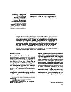
Fig. 2. Sucrose gradient analysis of the binding of oligonucleotides to ribosomes. (A) 50 pmol 30-S subunits, 30 pmol [32P]octanucleotide. (B) 50 pmol 30-S subunits, 30 pmol [32P]nonanucleotide.(C) 45 pmol 50-S subunits, 30 pmol [32P]octanucleotide.(D) 50 pmol 3 0 3 subunits and 45 pmol 50-S subunlts preincubated at 37°C for 10 min, then 30 pmol of [32P]octanucleotide were added. All samples were In lop1 standard buffer and incubation with the oligonucleotides was at 0 "C. Sedimentation was for 90 min at 40000 rev./min in the SW 56 rotor in 5 - 20 % sucrose
1 ,4260 unit corresponds to 11 nmol octanucleotide. Similarly the nonanucleotide had a calculated &260 = 89500 M - l cm-'. Corrected for 10% hypochromicity this corresponds to 12 nmol in 1 A260 unit.
-cE .
15
Y)
.u c 3
-e
10
0
0 c 0
Luheling of Oligonucleotides
(5'-3')d(A-A-G-G-A-G-G-T) and (5'-3')A-C-C-UC-C-U-U-A were labeled at their 5' ends with 32P as described by Donis-Keller et al. [12]. Following kination the fragments were reisolated by polyacrylamide electrophoresis and finally supplemented with excess non-radioactive fragment of known concentration. Preparation of Components Used for Translation of M S 2 R N A Ribosomes, initiation factors and other translation factors used were prepared as described in [I]. The assay for the functional activity of the complex between 30-S subunits and MS2 RNA is also described in [l]. Standard buffer is 10 mM Tris-HCI, 10 mM magnesium acetate, 60 mM NH4Cl and 6 mM 2mercaptoethanol, pH 7.4. RESULTS Binding of ( 5 '-3')d(A-A-G-G-A-G-G-T) to Ribosomes The binding of 32P-labeledoctanucleotide to ribosomes is easily detected either on sucrose gradients or on nitrocellulose filters. Binding to 30-S subunits on sucrose gradients is shown in Fig. 2A. The binding does not require incubation at 37 "C and is sequence-
-
"3
L m
-
-5a 5
N
5 0
'0 Sedimentation
Fig. 3. Exchange of' free and ribosome-hound octunucleotidr. (a) 50 pmol 30-S subunits and 30 pmol 32P-labeled octanucleotide incubated in 15 ~1 standard buffer for 10 min at 37 "C. (b) As (a) but 300 pmol unlabeled octanucleotide included. (c) as (a) but a second incubation at 0 "C is carried out after addition of 300 pmol unlabeled octanucleotide. The sucrose gradient analysis was performed as in Fig. 2, but centrifugation was for 2 h. Arrows indicate the position of the 30-S subunits
specific, as the oligonucleotide (5'-3')A-C-C-U-C-CU-U-A does not bind (Fig.2B). In addition (5'-3')d(A-A-G-G-A-G-G-T) will not associate with 50-S subunits (Fig. 2 C). Binding to 70-S ribosomes, on the other hand, is about the same as to 30-S particles (Fig. 2D). In general about 60 % of the 30-S particles are active in binding the octanucleotide. Octanucleotide bound to 30-S subunits is completely exchangeable with free octamer at 37 'C and at 0 "C. This is shown in Fig. 3 by sucrose gradient analysis. Similarly, octanucleotide bound to the colicin fragment of 16-S RNA is also fully exchangeable with its free counterpart (not shown). For this reason hybridisation in situ of the octaplet to the 3' terminus
C. Backendorf, G. P. Overbeek, J. H. van Boom, G. van der Marel, G. Veeneman, and J. van Duin
60 1
of 16-S RNA, as outlined in Fig. 1, cannot be proven by showing its association with the colicin fragment upon diassembly of colicin-cleaved ribosomes in sodium dodecylsulphate [5,7]. We have noticed that such association can occur after ribosome disassembly. In a forthcoming paper we will show that hybridisation of the octanucleotide to the 16-S RNA 3' terminus in situ does indeed take place. It is possible to1 cut off this terminus of 16-S RNA upon incubation with the octamer and 'RNase H' (hybrid RNase). This enzyme cuts RNA when hybridized to DNA [13- 171. (5'-3)d(A-A-G-G-A-G-G-T)and MS2 RNA Compete f o r the Same Ribosomal Site To determine whether the 3' terminus of 16-S RNA contributes to the formation of the initiation intermediate discussed in the preceding paper, 30-S subunits were preincubated without octanucleotide or with a fivefold and a tenfold excess of octanucleotide. A second incubation at 37 "C with MS2 RNA followed and the subunit . MS2 RNA precursor forma1,ion was assayed as before [ l ] by measuring coat protein synthesis upon completion with missing translational components. Edeine was included to permit one ribosomal cycle (compare preceeding paper for details). The result is shown in Fig.4 (slots 2, 3, and 4). It is clear that the octanucleotide interferes with coat protein production and thus with precursor formation. This indicates that the 3' end of 16-S RNA is directly involved in messenger binding at the level of precursor formation. To verify this point further, the order of additions was reversed; 30-S subunits were preincubated with MS2 RNA at 37 "C followed by a second incubation at 37°C with a fivefold and tenfold excess of octanucleotide. In this case the octanucleotide is no longer inhibitory (slots 5 and 6). Once the messenger is bound, the 30-S subunits are protected against inactivation by the DNA fragment. Apparently the messenger is bound to the 16-S RNA 3' terminus and this end is now no longer available for binding the octanucleotide. Binding of the messenger to 30-S ribosomes is such that it is hardly displaced by a tenfold excess of the DNA fragment over a 5-min period at 37°C. In slots 7 and 8 30-S ribosomes were preincubated with a mixture of MS2 RNA and octanucleotide to allow competition between the two components for the ribosome. We observe a strongly reduced coat protein synthesis, comparable to that in slots 3 and 4 where the octanucleotide was preincubated with the ribosomes. In view of the complete exchangealbility of bound and free octanucleotide (Fig. 3) the results of slots 3, 4, 7, and 8 indicate that the association rate of 30-S ribosomes with the octanucleotide is much greater than with MS2 RNA. It may be noted that our finding that 30-S subunits can be rescued from inactivation by (5'-3')d(A-A-G-G-
Fig. 4. Effect of (5'-3')d(A-A-G-G-A-G-G-T)on the formation of the subunit MS2-RNA initiation intermediate. 30-S subunits, MS2 RNA and the octanucleotide were mixed and preincubated in different orders. The formation of the subunit. MS2-RNA complex was then assayed by its ability to synthesize MS2 coat protein in the presence of inhibitors of initiation, i.e. edeine. The coat protein produced is visualized by autofluorography. See [l] for details. Slot 1 : MS2 coat protein marker. Slot 2: 30-S subunits preincubated with MS2 RNA for 10 min at 37 "C. Slot 3: 3 0 4 subunits preincubated for 10 rnin at 0°C with 400 pmol octanucleotide, then MS2 RNA was added and incubation continued (5 rnin at 37 "C). Slot 4: as slot 3 but 800 pmol octanucleotide were added. Slot 5 : 30-S subunits preincubated with MS2 RNA (5 min at 37 "C) then incubated with 400 pmol octanucleotide (5 min at 37 "C). Slot 6: as slot 5 but 800 pmol octanucleotide added. Slot 7: 30-S preincubated with a mixture of MS2 RNA and 400 pmol octanucleotide (10 min at 37°C). Slot 8 : as slot 7 but 800 pmol octanucleotide added. Slot 9 : no octanucleotide and no MS2 RNA present. Each slot contained 80 pmol 30-S subunits and, except slot 9, 16 pmol MS2 RNA
A-G-G-T) via a preincubation with phage RNA represents additional evidence for the precursor status of the subunit . MS2-RNA complex. Our interpretation of the results leads to the prediction that the complex between the 30-S subunit and MS2 RNA can no longer bind the octanucleotide. This was tested directly by sucrose gradient analysis of a preformed complex to which 32P-labeled octanucleotide was added at 37 "C. As shown in Fig. 5 A the subunit . MS2-RNA complex sediments slightly ahead of the 30-S subunit peak. When labeled octamer is added to a preformed subunit . MS2-RNA complex no 32P label is associated with it (Fig.5C). Similarly preincubation of the 30-S subunits with the DNA fragment prevents formation of the precursor, as evident from the non-appearance of the leading peak (Fig. 5 B). The inhibitory effect of the octanucleotide on ribosome messenger interaction is sequence-specific. The
602
Role of the 16-5 RNA in Phage RNA Recognition
-Sedimentation
Fig. 5 . Cornpetition between MS2 R N A und j S 8 - 3 ' J d ( A - A - G - G A G-G-T) for 30-S suhunit binding. (A) 50 pmol 3 0 3 subunits incubated with 50 pmol MS2 RNA for 15 min at 37 "C in standard buffer. (B) As (A) but 30-S subunits were preincubated with a 70-1i~1~1 excess of octanucleotide. (C) As (A) but 100 pmol "Plahclcd octanucleotide added during the last 5 min of the incubation. Further details of the sucrose gradient analysis are as in the legend to Fig.3. Arrows indicate from right to left the positions of MS2 RNA, 30-S subunits and the subunit - MS2-RNA complex
decadeoxynucleotide dAlo has no effect when assayed (5'-3')A-C-C-U-C-C-U-U-A, representing the last nine bases of 16-S RNA, had a slight inhibitory effect, probably by competing with 30-S subunits for complcmentary sequences in the MS2 RNA. As we attribute the inhibitory action of (5'-3')d(A-A-G-G-A-G-G-T) on ribosome messenger recognition to base-pairing with the 16-S RNA, we expect to reverse its effect by the complementary fragment (5'-3')A-C-C-U-C-C-U-U-A. In Fig. 6 we show this is indeed so. The design of the experiment is identical to that of Fig. 4. No oligonucleotide is added in slot 1. In slot 2 30-S ribosomes and a 15-fold excess octanucleotide were preincubated. Slot 3 is identical to slot 2 except that preincubation includes also a 15-fold excess of (5'-3')A-C-C-U-C-C-U-U-A over ribosomes. The reappearance of coat protein in slot 3 as compared to slot 2 demonstrates that the nonanucleotide competes successfully with the 30-S subunits for binding the DNA fragment. This competition can also be shown by direct physical measurement on sucrose gradients. The analysis is presented in Fig. 7 and illustrates that in the presence of nonanucleotide less octanucleotide is bound to 30-S subunits than in its absence. This result is independent of the order of mixing the three components, again indicating the rapid exchange between bound and free octanucleotide. Its final distribution over the two substrates suggests that its affinity for 30-S ribosomes and the nonanucleotide is not very different . Ribosome . Messenger Interaction is not Identical to Ribosome . Octunucleotide Interaction A relevant question is whether the interaction between the 30-S subunit and MS2 RNA in the pre-
Fig.6. Inhibition of suhunit . MS2-RNA complex formution by (5'-3')d(A-A-G-G-A-G-G-T) and reversul of the inhibition by ( 5 '-3'IA-C-C-U-C-C-U-U-.4.Precursor formation between 3 0 4 subunits and MS2 RNA was assayed by its ability to synthesize MS2 coat protein in the prcsence of edeine, as described before [2]. Slot 1 : 30 pmol 30-S subunits preincubated at 37 "C with 16 pmol MS2 RNA. Slot 2: as slot 1 except that 30-S subunits were reacted with 500 pmol octanucleotide before MS2 RNA was added. Slot 3 : 30 prnol 30-5 subunits were incubated with a mixture of 450 pmol octanucleotide and 450 pmol nonanucleotide and then further incubated with MS2 RNA (10 min at 37°C). All three mixtures were processed for protein synthesis and the products analyzed by polyacrylamide gel electrophoresis. The arrow indicates the position of the MS2 coat protein. The origin of the protein band above the coat protein in slot 1 is unknown
0
-I 0
W N
P
"
-Sedimentatton
Fig. 7. Competition between 30-S subunits and nonunucleotide f o r (5 '-3')d(A-A-G-G-A-G-G-T), (A) 50 pmol 30-S subunits and 30 pmol "P-labeled octanucleotide. (B) 50 pmol 3 0 4 subunits, 30 pmol 3ZP-labeledoctanucleotide and 40 pmol unlabeled nonanucleotide. All incubations were in 10 pl standard buffer at 0 'C. The sucrose gradient analysis was performed as in Fig. 3
C. Backendorf, G. P. Overbeek, J. H. van Boom, G. van der Marel, G. Veeneman, and J. van Duin A 1
0
603
DISCUSSION
In the present study we have analyzed the initiation intermediate between 30-S subunits and MS2 RNA in greater detail. In particular we have asked if in this complex the 16-S RNA 3' end is base-paired to an MS2 RNA sequence. Our results strongly suggest this to be true. If the octanucleotide (5'-3')d(A-A-G-G-A-G-G-T) is hybridized to the 16-S RNA terminus, the 30-S subunits fail to form the precursor. m Moreover, once the precursor is preformed, the octa'0 nucleotide can no longer prevent entry of the complex 0 Sedimentation into the ribosome cycle. In addition, we have shown Fig. 8. Binding of octanuclrolidr to 30-S subunits in the presence by sucrose gradient analysis that the octanucleotide of edeine. (A) 50pniol 3 0 3 subunits incubated with 3Opinol of does not bind to 30-S subunits that are already com[32P]octanuckotide for 10 inin at 37 'Tin 10 pl standard buffer. plexed with MS2 RNA. We conclude that in the sub(B) As (A) but 3 0 3 subunits preincubated in 2 pM edeine. The sucrose gradient analysis was performed as in Fig.3. When 30-S u n it. MS2-RNA precursor the 3' end of 16-S RNA subunits were preincubated with 0.3 mM aurintricarboxylic acid interacts directly with the message and this interaction the binding of lhe octanucleotide was the same as in (A) or (B) is indispensable for the formation and maintenance of the complex. For our understanding of the translation of RNA initiation complex is confined to two complementary phages it is important to known which MS2 RNA RNA sequences. Additional contacts are conceivable. sequence is actually base-paired to 16-S RNA in the To examine this point we have asked if conditions that inhibit ribosome . messenger interaction also initiation precursor. It is generally assumed that this prevent binding of (5'-3')d(A-A-G-G-A-G-G-T) to is the so-called 'Shine and Dalgarno sequence' located the 30-S particles. This appears not to be so. For some ten nucleotides before the first AUG codon to be translated. However, the recent analysis of instance edeine, aurintricarboxylic acid as well as Taniguchi and Weissmann [7] shows that 30-S sublow temperatures (0 "C) prevent formation of the initiation precursor [l]. However, none of these condiunits are bound to so-called 'non-initiator regions' in QP RNA that show good complementarity to the tions interferes with the binding of the fragment to ribosomes (Fig. 2 and 8). Simikarly antibodies against 16-S RNA terminus, but are not located at the start ribosomal protein S1 do no inhibit binding of the of the coat cistron [7]. If the complexes analyzed by octamer to 30-S ribosomes (to be published), although these authors are identical to our subunit. phage-RNA 30-S messenger interaction is abolished by anti-S1 precursor complexes, this suggests that such 'non[1]. We infer that the complementary RNA . RNA initiator regions' are transiently used in initiation of interaction is just part of the forces that couple the protein synthesis. Although there is no obvious reason messenger to the small subunit in their first encounter. why Taniguchi and Weissmann's complexes should differ from our precursor, caution should be taken An additional argument stressing a difference between since in both studies the fraction of ribosomes exaribosome . messenger and ribosome . octanucleotide mined is well below 50%. binding can be derived from Fig.4 (slots 5 and 6). These experiments show that ribosome-bound MS2 Finally we have examined whether the initial RNA is not exchanged with free octanucleotide. On ribosome messenger interaction is nothing but an RNA . RNA double-helix formation. All our results the other hand, ribosome-bound otanucleoi.ide exchanges rapidly and completely with the free fragment show this to be very unlikely. Conditions that exclude as shown in Fig. 3. the formation of a functional complex between 30-S At this moment we cannot be certain what causes subunits and MS2 RNA, e.g. the presence of anti-S1, the greater stability of the subunit. MS2-RNA as edeine and aurintricarboxylic acid, have no measurable compared to the subunit . octaplet complex. Aurineffect on the binding of the octanucleotide to the 30-S tricarboxylic acid has been shown to prevent intersubunit. Another argument is that the initiation preactions between protein and nucleic acid [18]. As we cursor cannot form at 0 "C whereas the octanucleotide have shown that aurintricarboxylic acid interferes binds equally well to 30-S particles at 0 "C or at 37 "C. with formation of the first but not with formation of Furthermore we have shown that exchange between the last complex, we suggest that interaction(s) beribosome-bound and free octanucleotide is a fast tween ribosomal protein(s) and MS2 RNA provide process even at 0°C. In contrast MS2 RNA bound additional contact points. This suggestion is consistent to 30-S subunits is not rapidly exchanged for free with earlier findings that ribosomal protein S1 plays octanucleotide. These data show clearly that addia decisive role in the initial recognition event 119-211. tional forces stabilize the complex. The inhibitory
-
604
C. Backendorf et al.: Role of the 16-S RNA in Phage RNA Recognition
effect of aurintricarboxylic acid on interactions between 30-S subunits and MS2 RNA hints directly at the involvement of protein in the reaction. The requirement for ribosomal protein S1 is well established [19-211. The possible contribution of several other proteins in binding QP RNA to 30-S subunits has recently been reported [22]. At present it seems unlikely that S1 would contribute to the ribosome . messenger interaction by unfolding the 16-S RNA 3’ terminus as suggested by Dahlberg and Dahlberg [23]. Binding of d(A-A-G-G-A-G-G-T) to 30-S subunits occurs irrespective of the presence of S1 (to be published). C. Backendorf is grateful to EMBO (European Molecular Biology Organisation) for a long-term fellowship. We thank Drs P. H. van Knippenberg and L. Bosch for critical reading of the manuscript. Special appreciation is expressed to Dr G. van Dieijen for many valuable discussions. Mrs J. Talens is acknowledged for preparing MS2 phage, and Dr T. Blumenthal for valuable comments.
REFERENCES van Duin, J., Overbeek, G. P. & Backendorf, C. (1980) Eur. J . Biochem. 110,593-597. Shine, J . & Dalgarno, L. (1975) Nature (Lond.) 254, 34-38. Taniguchi, T. & Weissmann, C. (1978) Nature (Lond.) 275, 770-772. Eckhardt, H. & Luhrmann, R. (1979) J . Biol. Chem. 254, 11185-11188. Steitz, J. A. & Jakes, K . (1975) Proc. Nut1 Acad. Sci. USA, 72, 4734 - 4738.
6. Dunn, J. J., Burash-Pollert, E. & Studier, F. W. (1978) Proc. Natl Acad. Sci. USA, 75, 2741 - 2745. 7. Taniguchi, T. & Weissmann, C. (1979) J . Mol. B i d . 128, 481 500. 8. Arentzen, R., van Boeckel, C. A. A,, van der Marel, G. & van Boom, J. H. (1979) Synthesis, 137-138. 9. de Rooij, J. F. M., Wille-Hazeleger, G., van Deursen, P. H., Serdijn, J. & van Boom, J. H. (1979) Recl. Trav. Cliim. Paysbas, 98,537 - 548. 10. van Boom, J . H., Burgers, P. M. J., Verdegaal, C. H. M. & Wille, G. (1978) Tetrahedron, 34, 1999-2007. 11. Handbook of Biochemistry (Sober, H. A,, ed.) Chemical Rubber Co., Ohio. 12. Donis-Keller, H., Maxam, A. M. & Gilbert, W. (1977) Nucleic Acids Res. 4, 2527-2538. 13. Berkower, I., Leis, J. & Huruitz, J. (1973) J. Biol. Chem. 248, 5914-5921. 14. Dorlix, J. L. (1975) Eur. J . Biochem. 51, 369-376. 15. Stavrianopoulos, J. G. & Chargaff, E. (1973) Proc. Nut1 Acad. Sci. USA, 70,1959-1963. 16. Haberkern, R. C. & Cantoni, G. L. (1973) Biochemistry, 12, 2389 - 2395. 17. Henry, C. M., Ferdinand, F. J. & Knippers, R. (1973) Biochem. Biopliys. Res. Commun. 50, 603 - 61 1. 18. Grollman, A. P. & Huang, M. T. (1976) in Protein Synthesis (McConkey, E. H., ed.) vol. 11,pp. 125 - 134, Marcel Dekker, New York. 19. van Dieijen, G., Zipori, P., van Prooijen, W. & van Duin, J. (1978) Eur. J . Biochem. Y2, 235-241. 20. Szer, W. & Leffler, S. (1974) Proc. Natl Acad. Sci. USA, 71, 361 1 - 3615. 21. Isono, S. & Isono, K. (1975) Eur. J. Biochern. 56, 15-22. 22. Chang, C. & Craven, C. R. (1977) J . Mol. Biol. 117, 401 -418. 23. Dahlberg, A. E. & Dahlberg, J. E. (1975) Proc. Nut1 Acad. Sci. USA, 72,2940 - 2944.
C. Backendorf, G. P. Overbeek, and J. van Duin*, Biochemisch Laboratorium, Rijksuniversiteit te Leiden, Postbus 9505, NL-2300-RA Leiden, The Netherlands J. van Boom, G. van der Marel, and G. Veeneman, Organisch-Chemisch Laboratorium, Rijksuniversiteit de Leiden, Wassenaarseweg 76, NL-2333-AL Leiden, The Netherlands
-__ * To whom correspondence should be addressed