REVIEW ARTICLE
Acta Orthop. Belg., 2010, 76, 289-297
Rheumatoid forefoot reconstruction Amit AMIN, Nicholas CULLEN, Dishan SINGH
From the Royal National Orthopaedic Hospital, Stanmore, U.K.
Management of the patient with rheumatoid forefoot deformity requires a multidisciplinary integrated approach for a successful outcome. Despite recent advances in the pharmacological management of rheumatoid arthritis and its impact upon disease progression, forefoot deformity and pain remain common manifestations requiring input from orthopaedic surgeons. The typical deformities encountered include hallux valgus, with subluxation or frank dislocation at the lesser metatarsophalangeal (MTP) joints. Surgical intervention is directed at correcting and decompressing these deformities, with the ultimate goal of a stable, painless, functional plantigrade foot. Although a variety of surgical options exist, fusion of the 1st MTP joint with lesser MTP joint excision arthroplasty remains the gold standard, upon which newer procedures should be judged. This article reviews the pathophysiology of forefoot deformity in rheumatoid arthritis with special emphasis on recent advances in surgical management.
most frequently, particularly earlier in the course of the disease process (27). The commonly encountered deformities in the forefoot include hallux valgus and dorsal subluxation or dislocation of the lesser metatarsophalangeal (MTP) joints, with or without fixed claw toe deformity (fig 1). Pathophysiology of MTP joint deformity RA is characterised by chronic synovial inflammation, which results in capsular distension, joint destabilisation (due to attenuation of collateral ligaments and plantar plate), articular cartilage destruction and erosive bone loss. The relatively small 1st MTP joint contact area and shallow articular profile of the proximal phalanx provide little inherent stability, most of which is provided by the capsuloligamentous and tendinous structures which surround and traverse
Keywords : rheumatoid arthritis ; forefoot ; deformity ; reconstruction. ■ Amit Amin, FRCS (Tr and Orth), SpR Trauma and
Orthopaedics (NE Thames, Percivall Pott Rotation). ■ Nicholas Cullen, FRCS (Tr and Orth), Consultant
INTRODUCTION Rheumatoid arthritis (RA) is a chronic, systemic, inflammatory disorder characterised by symmetric polyarthritis. Approximately 20% of patients initially present with foot and ankle symptoms (54). The overall prevalence of symptoms in the foot and ankle approaches 90%, with the forefoot affected No benefits or funds were received in support of this study
Orthopaedic Surgeon. ■ Dishan Singh, FRCS (Tr and Orth), Consultant Orthopaedic
Surgeon. Foot and Ankle Unit, Royal National Orthopaedic Hospital, Stanmore, U.K. Correspondence : Amit Amin, 39 Church Drive, North Harrow, Middx HA2 7NR, U.K. E-mail :
[email protected] © 2010, Acta Orthopædica Belgica.
Acta Orthopædica Belgica, Vol. 76 - 3 - 2010
290
A. AMIN, N. CULLEN, D. SINGH
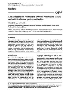
Fig. 1. — Typical forefoot deformity associated with RA : hallux valgus with lesser hammer toe deformities. Note plantar callosities as a result of MTP joint dislocation and plantar displacement of metatarsal heads.
over the dorsal proximal interphalangeal joint and underneath the now prominent metatarsal heads. Finally, the plantar plate is displaced distally, with failure often occurring at its weaker proximal attachment (38). The thicker cartilaginous attachment to the proximal phalanx subluxes dorsally, along with the plantar fat pad, effectively uncovering the metatarsal head. Plantar callosities occur underneath the metatarsal heads as a result of increased pressure during gait. NON-SURGICAL MANAGEMENT
the joint. Disruption of these soft tissue structures results in progressive joint instability and usually valgus drift of the great toe, or very rarely, a dorsal or varus deformity. As a result, the proportion of forces transferred through the hallux are reduced, leading to greater load through the lesser rays. The combination of capsular and ligamentous attenuation and increased loading of the lesser rays results in dorsal subluxation of the proximal phalanges, distal displacement of the plantar fat pad and a resultant plantar displacement of the metatarsal heads. Progressive dorsiflexion at the lesser MTP joints during gait perpetuates the deformity. The primary plantar flexors of the MTP joints, the interossei and the lumbricals, act through their insertion onto the proximal phalanx, plantar to the axis of rotation. With dorsal subluxation of the proximal phalanx, the axis moves dorsally, relegating the interossei to weak, effectively functionless extensors (38,49). The lumbricals by virtue of their course beneath the deep transverse metatarsal ligament do not subluxate, but lose their mechanical advantage and are essentially defunctioned due to the change in the direction of action that occurs with dorsal subluxation of the proximal phalanx (38). Flexor-extensor tendon imbalance worsens the deformity. The extensor digitorum brevis and longus function best with the toes in a neutral or slightly flexed position, however with chronic dorsiflexion at the MTP joints, they become inefficient in relation to the flexors (49). Without treatment the initial flexible claw toe deformity quickly becomes fixed, with patients developing corns and calluses Acta Orthopædica Belgica, Vol. 76 - 3 - 2010
Conservative measures and medical therapy play an important part in the management of the rheumatoid forefoot. Administration of non-steroidal antiinflammatory drugs (NSAIDs), corticosteroids, disease modifying anti-rheumatic drugs (DMARDs) and newer biological agents under the guidance of a rheumatologist represents the standard of care. NSAIDs do not prevent disease progression, in contrast to DMARDs which should be instituted at the earliest opportunity. Typical DMARDs include methotrexate, sulfasalazine, cyclosporine, penicillamine, leflunomide and azathioprine. If employed, corticosteroid use should be discontinued when DMARDs have gained control of the disease. Increasing evidence supports the use of cytokine modulators targeting tumour necrosis factor alpha (42). Orthotic management includes shoe wear modification, with wide and/or high toe boxes, accommodative insoles and rocker soles. They form the mainstay of treatment for the elderly infirm patient, those with significant co-morbidities or those who are poor surgical candidates (56). In the ankle and hindfoot, fluoroscopic guided injections have been shown to be superior to ‘blind’ injections for diagnostic purposes and the temporary symptomatic relief of synovitis or arthritis (7). Pre and post gadolinium enhanced MRI scans performed following corticosteroid injection in patients with juvenile chronic arthritis confirm long-lasting suppression of inflammation and negligible toxic effects on host tissues (45). The role of such injections in the forefoot remains less well defined.
RHEUMATOID FOREFOOT RECONSTRUCTION
SURGICAL MANAGEMENT Perioperative management The systemic involvement, characteristic of RA, should alert the clinician to potential pre-operative problems. A thorough examination of the entire limb is necessary to rule out proximal deformity, which generally should be corrected first. This allows one to accurately correct true forefoot deformity with reference to a normally aligned ankle, hindfoot and lower limb, reducing the possibility of recurrence or the uncovering of forefoot malalignment if the proximal limb is corrected at a later date. Instability of the atlanto-axial junction maybe assessed with radiographs and with flexion-extension views. Rheumatoid vasculitis can predispose to accelerated atherosclerosis and an overall increased cardiovascular risk during the peri-operative period. Patients often have thin, friable skin, which demands careful soft-tissue handling to prevent infection and wound-healing complications. Temporary discontinuation of drug therapy remains a controversial topic and is the focus of ongoing trials. Methotrexate has been shown to be safe, and one large study even demonstrated a lower infection rate in those continuing therapy, compared to those who never received it and those who temporarily stopped therapy two weeks before and after surgery (21). Little has been published to guide our management of the other common DMARDs. Divided opinions exist regarding the use of antitumour necrosis factor alpha inhibitors, with some studies demonstrating safety, whilst others reported an increased infection risk (8,16,46). Inherent difficulties comparing these studies, based on differing populations and varied surgical intervention means that no clear conclusion can be drawn as to whether these drugs should be discontinued or not. The role of synovectomy Little attention has been directed to the role of synovectomy in the management of early disease. Often, patients present to an orthopaedic surgeon towards the end of the inflammatory cascade, with-
291
out synovitis but with significant deformity. Aho reported significant pain relief in a small series of patients treated with early synovectomy of the lesser MTP joints, although the long-term radiographic results were not disclosed (1). Whilst it has been suggested that the natural history of the condition may be altered, a true joint preserving effect is yet to be clearly demonstrated (2,11,40). Early advances in the management of forefoot deformity Table I summarises the important early contributions to our understanding and management of the rheumatoid forefoot (12,18,25,28,29,30). Dwyer first noticed that simple pan-metatarsal head excision, whilst initially relieving the patient of their symptoms, appeared only to provide a short lived effect and recurrence was common. He advocated arthrodesis of the 1st MTP joint and of the lesser toe proximal interphalangeal joints, together with metatarsal head excisions (17). Watson observed, in a long-term follow-up of 46 patients undergoing forefoot arthroplasty, poor results associated with resection arthroplasty of the 1st MTP joint, even when stabilised with a Kirshner wire. Conversely all patients, who either presented with a 1st MTP joint arthrodesis, had it performed as part of the forefoot arthroplasty, or developed a spontaneous ankylosis after surgery, had good results (56). Modern Concepts A great deal of debate continues regarding the optimal treatment of rheumatoid forefoot deformity. Many regard arthrodesis as the benchmark for the treatment of 1st MTP joint arthritis (13,32,33). However, there are proponents of hallux 1st MTP joint correction (3) and replacement (15,23,47) within the literature. Recent attention has been directed to preservation of the lesser metatarsal heads with procedures such as Weil’s osteotomy (4) or the Stainsby procedure (10). A variety of options exist for correction of lesser toe deformity, including closed osteoclasis, arthroplasty and fusion. It has been suggested that the Acta Orthopædica Belgica, Vol. 76 - 3 - 2010
292
A. AMIN, N. CULLEN, D. SINGH
Table I. — Historical overview of approaches to forefoot reconstruction AUTHOR
YEAR
CONTRIBUTION
Hoffman (25)
1912
Fowler (18)
1959
Clayton (12)
1960
Kates et al. (28)
1967
Lipscomb (29)
1968
Described pan-metatarsal head excision through a single plantar apex-distal incision just proximal to the interdigital web spaces, without extensor tenotomies. Argued that relaxation of the soft tissues by generous metatarsal resection obviated the need for soft tissue correction. The disadvantage of the distal plantar approach was injury to the neurovascular bundles. Described a combined dorsal and plantar approach. The dorsal incision included short longitudinal extensions along the 1st and 5th metatarsals. He advocated excision of the proximal halves of the phalanges, together with metatarsal head excisions. The plantar incision was used to excise the callus and perform a dermoplasty to relocate the fat pad beneath the metatarsal remnants. Suggested using a single straighter dorsal incision, with bony resection as described by Fowler, but with extensor tendon transections if required. Promoted a single curved plantar incision, excising an ellipse for further deformity correction. A pan-metatarsal head excision was performed, with the 1st MTP joint stabilised with a Kirshner wire and a plaster boot applied with the lesser toes plantar flexed. Popularised three dorsal longitudinal incisions, with metatarsal head excisions. Originally suggested by Larmon (22) in 1951.
variety of procedures commonly used reflects the population being treated, whilst others consider it a reflection of each individual surgeon’s experience. Comparison of the available literature is difficult in view of the paucity of prospective randomised studies, small patient numbers and variability in follow-up and reported outcome measures. Management of the 1ST MTP joint : Arthrodesis It is generally well accepted that few indications exist for resection of the 1st MTP joint as part of forefoot arthroplasty. Poor long-term results are attributed to defunctioning of the first ray, with recurrent hallux valgus and transfer metatarsalgia, plantar keratoses, lack of plantar flexion, and weakened push-off (19,34,36,41,52,53). Henry and Waugh demonstrated this dysfunction using Harris-mat studies (24). They evaluated pre and post-operative weight-bearing, in two groups of patients treated for hallux valgus with either excision arthroplasty or arthrodesis. Increased 1st MTP joint weight-bearing was reported in 80% of the arthrodesis group compared to 40% of the excision group. More recent pedobarographic studies by Coughlin (13), Beauchamp et al (6), Mann and Thompson (32) and Mulcahy et al (37), support these early findings. Arthrodesis is the most widely used procedure in the treatment of 1st MTP joint in rheumatoid arthriActa Orthopædica Belgica, Vol. 76 - 3 - 2010
tis. Coughlin reported his long-term follow-up of forefoot arthroplasty in 58 feet, using 1st MTP joint arthrodesis and lesser metatarsal head resection. According to the AOFAS scoring system, an excellent or good result was obtained in 90% of patients. He recommended stable realignment of the first ray, to allow permanent correction of the hallux deformity. This improved the weight-bearing capacity of the medial column protecting the lateral rays from recurrent deformity by reducing lesser metatarsal stress and maintaining correct alignment of the plantar fat pad (13). Fixation Numerous biomechanical studies have been performed to elucidate the optimal fixation method to secure a successful arthrodesis. Reported techniques include intramedullary Steinman pins, crossed Kirshner wires, staples, crossed or parallel screws, dorsal locked or standard plating, with or without supplementary lag screw fixation (50). Patients with RA typically exhibit poor bone quality and in the very frail, elderly patient, who presents for surgery, arthrodesis may not be technically feasible. Hughes et al suggested that failure to achieve union of the 1st MTP joint produced greater dissatisfaction than reported with resection arthroplasty (26). However, they disclosed a 33% failure
RHEUMATOID FOREFOOT RECONSTRUCTION
rate of fusion in their study, a nonunion rate that is far higher than reported elsewhere in the literature, where fusion rates of greater than 90% are consistently documented (14,50), with 100% success reported by Coughlin in his landmark study (13). He attributed this to using a dorsal plate and crosscompression screw, which has been shown to be the most stable construct (43). He also used specially designed ball and socket reamers, to promote congruent, domed fusion surfaces. The position of arthrodesis remains critical to a successful outcome, and should not be underestimated. The recommended ideal position of arthrodesis is 10 to 15° valgus, 20° to 30° of dorsiflexion relative to metatarsal shaft, and neutral rotation. Technically, ball and socket joint preparation allows accurate joint positioning and flexibility with adjustment, as opposed to planar resection using a saw blade (13). Excessive dorsiflexion can impair the ability of the toe to engage the ground resulting in transfer metatarsalgia and leads to the dorsum of the toe rubbing on footwear. Excessive plantar flexion predisposes to accelerated degenerative change at the interphalangeal joint, which may necessitate subsequent fusion. Arthritis of the interphalangeal joint is common following a well aligned 1st MTP joint arthrodesis, and has been reported to occur in up to 90% of cases in long-term studies, although most patients remain asymptomatic (13,33). The 1st MTP joint : Alternative techniques There are limited numbers of studies reporting various alternative surgical techniques used in the management of 1st ray involvement, including metatarsal osteotomy (3), Heuter-Mayo resection arthroplasty (22) and joint-replacement procedures (15,23,47). Grondal et al have published the only prospective randomised study comparing differing reconstructive modalities. The Heuter-Mayo resection arthroplasty (resection of the metatarsal head of the 1st MTP joint) was compared with hallux arthrodesis in 33 rheumatoid forefeet. Twentynine patients were followed up after a mean of 72 months (57-80 months). There was no statistically significant difference in deformity recurrence, patient satisfaction, shoe wear or parameters of gait
293
analysis. No difference was found with respect to lateral forefoot pressure measurements. The authors contended that the Heuter-Mayo resection avoids the shortcomings of the Keller procedure, as the proximal phalanx and plantar plate are preserved, providing inherent stability to the joint and maintaining flexion ability (22). Whilst considered Level 1 evidence, small patient numbers and limited power suggest that the conclusions should be interpreted with caution. These results have not been reproduced elsewhere in the literature. Fuhrmann and Anders reported a deterioration of results in the long term associated with the HeuterMayo resection arthroplasty (19) in keeping with the poor results generally associated with resection arthroplasties of the 1st MTP joint (36,41,52,53). Replacement arthroplasty of the 1st MTP joint using silicone implants was developed in the late sixties, as a treatment for hallux rigidus and hallux valgus. In theory, this form of arthroplasty maintains flexibility as compared to arthrodesis, and provides more stability than resection. Despite improvements in material properties over the years, including the addition of titanium grommets to add strength, reported complication rates were relatively high (20,44), notably implant failure (as a result of wear and repeated abrasion with microparticle release initiating synovitis and bony cystic osteolysis), cock-up deformity, stiffness and transfer metatarsalgia. Implant fracture with associated cystic osteolysis makes revision surgery challenging, with most cases requiring interpositional bone block arthrodesis. There are isolated reports of good results, despite the preponderance of poor results in the literature and these merit discussion. Cracchiolo et al reported the results of 86 silicone double stemmed implants followed for an average 6.8 years, with over half in patients with RA. Lesser metatarsal head excision was performed in conjunction in the RA group. Overall, 84% of patients were completely satisfied with treatment. Only two implant failures were noted out of 49 procedures in the rheumatoid group (15). Hanyu et al reported the results of silicone hinge interpositional arthroplasty with concomitant shortening osteotomies of the lesser metatarsals. Of Acta Orthopædica Belgica, Vol. 76 - 3 - 2010
294
A. AMIN, N. CULLEN, D. SINGH
60 feet followed for an average of 12 years, 74% of patients were satisfied with treatment. Using revision as an end-point, the implant survival rate was 93%, and with radiologically proven fracture as an end-point, the survival rate was 87% (23). Moeckle et al reported on 67 silicone doublestemmed implants followed for an average of 6 years. Eighty-seven percent of patients had excellent or good results. Only 4% of patients experienced complications related to the implant, which required revision surgery. Ten percent of patients had silent radiographic fractures, or fragmentation of the implant (35). This study and that by Hanyu et al (23), highlight an interesting conundrum. They have shown that fracture or fragmentation per se does not necessarily dictate a poor result or necessitate revision surgery. It has been suggested that the implant functions as a spacer in this situation and a fibrous reaction occurs which stabilises the joint, imparting pain relief (48). Rahman and Fagg however have reported synovitis occurring in up to 72% of cases in their series and on the basis of their findings suggested that the procedure should be abandoned (44). Consistent results using replacement arthroplasty of the 1st MTP joint have not been documented in the literature, and whilst a successful outcome may be more likely in the older rheumatoid patient, the complications reported and the complexity of salvage surgery, suggest caution should be exercised when using this treatment modality to correct rheumatoid 1st MTP joint deformity. Lesser MTP joint excision or preservation Resection of the lesser MTP joints has been considered the standard of care in rheumatoid forefoot reconstruction, largely based upon Coughlin’s longterm follow-up study (13). The amount of resection should be tailored to the individual patient, although a thorough decompression should be performed to allow wound closure and relocation of the plantar fat pad. Fowler first recognised that patients in whom the metatarsal heads were irregularly trimmed often returned with pain (18). Reconstitution of the metatarsal parabola into a smooth arc assumes Acta Orthopædica Belgica, Vol. 76 - 3 - 2010
equal importance, as it provides an even distribution of weight-bearing forefoot stress, and prevents recurrence of pain and deformity. Stainsby took this concept one step further by suggesting preservation of the metatarsal head with joint arthroplasty (excision of proximal half of proximal phalanx and interpositional tenodesis of the toe extensor to the flexor tendon) (10). His technique, whilst not strictly considered to be joint-preserving, allows relocation of the plantar plate and fat pad beneath the corresponding metatarsal head and maintains the normal weight-bearing parabola. Whilst commonly performed by many surgeons in the UK, few long-term outcome studies have been published. Stainsby himself reported an 88% excellent or good result, in a study of 20 feet followed for an average of 20 months (10). Dorsal, plantar or combined approaches to the lesser MTP joints using either transverse or longitudinal incisions have been described. The combined approach originally described by Fowler (18) is no longer deemed necessary. The Kates, Kessel and Kay complete plantar approach allows both excellent direct visualisation of the metatarsal heads, and the ability to perform a dermoplasty to relocate the plantar fat pad (28). Disadvantages include an increased risk of keratotic scar formation and wound-healing complications, given the location and orientation of the scar. Transverse incisions demand an adequate decompression to allow tension-free wound closure. Barton reported a 46% rate of wound healing complications with the plantar wound when performed as part of Fowler’s original reconstruction (5). The ability to off-load the forefoot, and allow early weight-bearing using heelwedge shoes, has reduced the incidence of complications, although a lower rate of wound problems has been reported with longitudinal dorsal incisions (36). Some argue that exposure with this technique can occasionally be inadequate, although exposure can be improved by providing generous incisions to avoid over-retraction of the soft tissues. True joint-preserving surgery requires an intact metatarsal head and can be achieved with shortening osteotomies as described by Weil (4). Bolland et al described their results of 1st MTP joint arthrodesis and multiple lesser metatarsal Weil’s
RHEUMATOID FOREFOOT RECONSTRUCTION
osteotomies, in 26 feet followed for an average of 26.2 months. Subjectively, 23 feet were reported as having an excellent or good result. A 12% rate of recurrent metatarsalgia was noted, requiring revision shortening or resection, with symptomatic resolution in all cases (9). The option of preserving the lesser MTP joint is clearly appealing, both in terms of maintaining function and widening the revision options, when faced with recurrent deformity. Lesser toe deformity Following correction of the lesser MTP joint deformity, lesser toe deformity can be addressed. Flexible deformities, can be corrected with soft-tissue rebalancing (extensor tendon lengthening and/or plantar plate release) with or without closed osteoclasis. A Girdlestone flexor to extensor transfer can be used. Rigid deformities require joint arthroplasty or arthrodesis. Myerson suggested reserving arthrodesis for revision surgery and recommended performing joint resection with interpositional extensor to flexor tenodesis (36). Regardless of the chosen technique Kirshner wire stabilisation should be employed for 3-6 weeks, driving the wires into the metatarsal shafts, when the metatarsal heads have been resected. Pan MTP joint preserving surgery Taking the concept of joint preserving surgery further, Barouk and Barouk (3) recently reported their preliminary mid-term results of pan MTP joint preserving forefoot surgery in a continuous series of 60 feet. Fifty five feet (92%) underwent a shortening and corrective scarf osteotomy, whereas only 5 patients (8%) had an arthrodesis. Ninety five percent of hallux valgus corrections were maintained, with only one failure requiring arthrodesis. The lesser metatarsals were treated similarly with joint preserving surgery when the quality of metatarsal head allowed. The severity of dislocation did not influence the ability to preserve the metatarsal head. Eighty-six percent of the lateral rays were treated with Weil’s double cut osteotomies, whereas only 14% underwent resection. A more detailed analysis of these patients with validated outcome measures
295
is awaited. Important features of successful surgery described by Barouk and Barouk include shortening of all metatarsals respecting the MS (metatarsal shortening) point as described by Maestro et al (31). This represents the most proximal aspect of the proximal phalanx of the most deformed ray. The 1st ray is corrected and shortened to the MS point and the metatarsal parabola follows, with the second metatarsal shortened to the same length, followed by 3 mm, 6 mm, and 12 mm recessions of the 3rd, 4th and 5th rays respectively. Initial poor results in Barouk’s study were noted with over aggressive translation of the scarf osteotomy (resulting in hallux varus), and when metatarsal shortening was not aggressive enough. As a result they report an average of 11 mm and 13 mm shortening of the 1st and lateral rays respectively. Although the early results appear promising, few others have tried to replicate their technique and thus little support exists in the literature and more work is required. Nagashima et al reported on the modified Hohman osteotomy for hallux valgus correction and lesser MTP joint shortening osteotomies in RA patients, with 78% achieving complete pain resolution (39). Conversely, Thordarson et al noted poor results albeit in a small study which included patients undergoing 1st metatarsal osteotomy or no osteotomy and lesser MTP joint resection (51). AUTHORS’ PREFERRED METHOD We perform a 1st MTP joint fusion for all patients with rheumatoid forefoot disease. The procedure reliably and permanently eliminates forefoot deformity, and is well supported in the literature as the gold standard of treatment. We typically use a dorsal plate and plantar transarticular screw for fixation, given the often poor bone quality. For the lesser MTP joints we individualise treatment based on the severity of the disease. Our philosophy is to try and preserve the metatarsal head if possible, and therefore we perform Weil’s osteotomies for patients with early disease. As the disease progresses, we opt to perform the Stainsby procedure, on the premise that preservation of the metatarsal head may improve function. Additionally, an excisional Acta Orthopædica Belgica, Vol. 76 - 3 - 2010
296
A. AMIN, N. CULLEN, D. SINGH
arthroplasty, can be performed should the index procedure fail to relieve forefoot metatarsalgia. REFERENCES 1. Aho H. Synovectomy of MTP joints in rheumatoid arthritis. J Rheumatol 1987 ; 11 : 126-130. 2. Akagi S, Sugano H, Ogawa R. The long-term results of ankle joint synovectomy for rheumatoid arthritis. Clin Rheumatol 1997 ; 16 : 284-290. 3. Barouk LS, Barouk P. Joint-preserving surgery in rheumatoid forefoot : preliminary study with more than two year follow-up. Foot Ankle Clin North Am 2007 ; 12 : 435-454. 4. Barouk LS. [Weil’s metatarsal osteotomy in the treatment of metatarsalgia] (in German) Orthopäde 1996 ; 25 : 338343. 5. Barton NJ. Arthroplasty of the forefoot in rheumatoid arthritis. J Bone Joint Surg 1973 ; 55-B : 126-133. 6. Beauchamp CG. Kirby T, Rudge SR, Worthington BS, Nelson J. Fusion of the first metatarsophalangeal joint in forefoot arthroplasty. Clin Orthop Relat Res 1984 ; 190 : 249-253. 7. Beukelman T, Arabshahi B, Cahill A, Kaye R, Cron R. Benefit of intraarticular corticosteroid injection under fluoroscopic guidance for subtalar arthritis in juvenile idiopathic arthritis. J Rheumatol 2006 ; 33 : 2330-2336. 8. Bibbo C, Goldberg JW. Infectious and healing complications after elective orthopaedic foot and ankle surgery during tumor necrosis factor-alpha inhibition therapy. Foot Ankle Int 2004 ; 25 : 331-335. 9. Bolland BJRF, Sauve PS, Taylor GR. Rheumatoid forefoot reconstruction : First metatarsophalangeal joint fusion combined with Weil’s metatarsal osteotomies of the lesser rays. J Foot Ankle Surg 2008 ; 47 : 80-88. 10. Briggs PJ, Stainsby GD. Metatarsal head preservation in forefoot arthroplasty and the correction of severe claw toe deformity. Foot Ankle 2001 ; 7 : 93-101. 11. Carl HD, Swoboda B. [Effectiveness of arthroscopic synovectomy in rheumatoid arthritis.] (in German). Z Rheumatol 2008 ; 67 : 485-490. 12. Clayton ML. Surgery for the forefoot in rheumatoid arthritis. Clin Orthop Relat Res 1960 ; 16 : 136-140. 13. Coughlin MJ. Rheumatoid forefoot reconstruction. A long-term follow-up study. J Bone Joint Surg 2000 ; 82-A : 322-341. 14. Coughlin MJ. Arthrodesis of the first metatarsophalangeal joint with mini-fragment plate fixation. Orthopedics 1990 ; 13 : 1037-1044. 15. Cracchiolo A, Weltmer J, Lian G. Arthroplasty of the first metatarsophalangeal joint with double-tem silicone implant. J Bone Joint Surg 1992 ; 74-A : 552-556. 16. den Broeder AA, Creemers MC, Fransen J et al. Risk factors for surgical site infections and other complications in elective surgery in patients with rheumatoid arthritis
Acta Orthopædica Belgica, Vol. 76 - 3 - 2010
17. 18. 19.
20.
21.
22.
23.
24.
25. 26.
27. 28. 29.
30.
31.
32.
33.
with special attention for anti-tumor necrosis factor : a large retrospective study. J Rheumatol 2007 ; 34 : 689-695. Dwyer AF. Correction of severe toe deformities. J Bone Joint Surg 1970 ; 52-B : 192. Fowler AW. A method of forefoot reconstruction. J Bone Joint Surg 1959 ; 41-B : 507-513. Fuhrmann RA, Anders JO. The long-term results of resection arthroplasties of the first metatarsophalangeal joint in rheumatoid arthritis. Int Orthop 2001 ; 25 : 312316. Granberry WM, Noble PC, Bishop JO, Tullos HS. Use of a hinged silicone prosthesis for replacement of the first metatarsophalangeal joint. J Bone Joint Surg 1991 ; 73-A : 1453-1459. Grennan D, Gray J, Loudon J, Fear S. Methotrexate and early post-operative complications in patients with rheumatoid arthritis undergoing elective orthopaedic surgery. Ann Rheum Dis 2001 ; 60 : 214-217. Grondal L, Brostrom E, Wretenberg P, Stark A. Arthrodesis versus Mayo resection : the management of the first metatarsophalangeal joint in reconstruction of the rheumatoid forefoot. J Bone Joint Surg 2006 ; 88-B : 914919. Hanyu T, Yamazaki H, Ishikawa H et al. Flexible hinge toe implant arthroplasty for rheumatoid arthritis of the first metatarsophalangeal joint : long-term results. J Orthop Sci 2001 ; 6 : 141-147. Henry APJ, Waugh W. The use of footprints in assessing the results of operations for hallux valgus. A comparison of Keller’s operation and arthrodesis. J Bone Joint Surg 1975 ; 57-B : 478-481. Hoffman MD. An operation for severe grades of contracted or clawed toes. Am J Orthop Surg 1912 ; 9 : 441-449. Hughes J, Grace D, Clark P, Klenerman L. Metatarsal head excision for rheumatoid arthritis. 4 year follow-up of 68 feet with and without hallux fusion. Acta Orthop Scand 1991 ; 62 : 63-66. Jaakola JI, Mann RA. A review of rheumatoid arthritis in the foot and ankle. Foot Ankle Int 2004 ; 25 : 866-874. Kates A, Kessel L, Kay A. Arthroplasty of the forefoot. J Bone Joint Surg 1967 ; 49-B : 552-557. Lipscomb PR. Surgery for rheumatoid arthritis-timing and techniques : summary. J Bone Joint Surg 1968 ; 50-A : 614-617. Larmon WA. Surgical treatment of deformities of rheumatoid arthritis of the forefoot and toes. Q Bull Northwest Univ Med Sch 1951 ; 25 : 39. Maestro M, Besse JL, Ragusa M, Berthonnaud E. Forefoot morphotype study and planning method for forefoot osteotomy. Foot Ankle Clin 2003 ; 8 : 695-710. Mann RA, Thompson FM. Arthrodesis of the first metatarsophalangeal joint for hallux valgus in rheumatoid arthritis. J Bone Joint Surg 1984 ; 66-A : 687-692. Mann RA, Schakel ME II. Surgical correction of rheumatoid forefoot deformities. Foot Ankle Int 1995 ; 16 : 1-6.
RHEUMATOID FOREFOOT RECONSTRUCTION
34. McGarvey SR, Johnson KA. Keller arthroplasty in combination with resection arthroplasty of the lesser metatarsophalangeal joints in rheumatoid arthritis. Foot Ankle 1988 ; 9 : 75-80. 35. Moeckel BH, Sculco TP, Alexiades MM et al. The double stemmed silicone rubber implant for rheumatoid arthritis of the first metatarsophalangeal joint. Long-term results. J Bone Joint Surg 1992 ; 74-A : 564-570. 36. Molloy AP, Myerson MS. Surgery for the lesser toes in rheumatoid arthritis : Metatarsal head resection. Foot Ankle Clin North Am 2007 ; 12 : 417-433. 37. Mulcahy D, Daniels TR, Lau JT, Boyle E, Bogoch E. Rheumatoid forefoot deformity : a comparison of 2 functional methods of reconstruction. J Rheumatol 2003 ; 30 : 1440-1450. 38. Myerson MS. Arthroplasty of the second toe. Semin Arthroplasty 1992 ; 3 : 31-38. 39. Nagashima M, Kato K, Miyamoto Y, Takenouchi K. A modified Hohman method for hallux valgus and telescoping osteotomy for lesser toe deformities in patients with rheumatoid arthritis. Clin Rheumatol 2007 ; 26 : 39-43. 40. Nakamura H, Tanaka H, Yoshino S. Long-term results of multiple synovectomy for patients with refractory rheumatoid arthritis : effects on disease activity and radiologic progression. Clin Exp Rheumatol 2004 ; 22 : 151-157. 41. Patsalis T, Georgousis H, Gopfert S. Long-term results of forefoot arthroplasty in patients with rheumatoid arthritis. Orthopedics 1996 ; 19 : 439-447. 42. Pieringer H, Stuby U, Biesenbach G. Patients with rheumatoid arthritis undergoing surgery : how should we deal with antirheumatic treatment ? Semin Arthritis Rheum 2007 ; 36 : 278-286. 43. Politi J, Hayes J, Njus G, Bennett GL, Kay DB. First metatarsophalangeal joint arthrodesis : a biomechanical assessment of stability. Foot Ankle Int 2003 ; 24 : 332-337. 44. Rahmann H, Fagg PS. Silicone granulomatous reactions after first metatarsophalangeal joint hemiarthroplasty. J Bone Joint Surg 1993 ; 75-B : 637-639.
297
45. Remedios D, Martin K, Kaplan G et al. Juvenile chronic arthritis : diagnosis and management of tibiotalar and subtalar disease. Br J Rheumatol 1997 ; 36 : 1214-1217. 46. Ruyssen-Witrand A, Gossec L, Salliot C et al. Complication rates of 127 surgical procedures performed in rheumatic patients receiving tumor necrosis factor alpha blockers. Clin Exp Rheumatol 2007 ; 25 : 430-436. 47. Sebold EJ, Cracchiolo A 3rd. Use of titanium grommets in silicone implant arthroplasty of the hallux metatarsophalangeal joint. Foot Ankle Int 1996 ; 7 : 145-151. 48. Senthil Kumar C, Holt G. Hallux metatarsophalangeal arthroplasty in the rheumatoid forefoot. Foot Ankle Clin N Am 2007 ; 12 : 405-416. 49. Stainsby GD. Pathological anatomy and dynamic effect of the displaced plantar plate and the importance of the plantar plate-deep transverse metatarsal ligament tie-bar. Ann R Coll Surg Engl 1997 ; 79 : 58-68. 50. Stevens BW, Anderson JG, Bohay DR. Hallux metatarsophalangeal joint fusion for the rheumatoid forefoot. Foot Ankle Clin North Am 2007 ; 12 : 395-404. 51. Thordarson D, Aval S, Krieger L. Failure of hallux MTP preservation surgery for rheumatoid arthritis. Foot Ankle Int 2002 ; 23 : 486-490. 52. Trieb K. Management of the foot in rheumatoid arthritis. J Bone Joint Surg 2005 ; 87-B : 1171-1177. 53. van der Heijden KW, Rasker JJ, Jacobs JW, Dey K. Kates forefoot arthroplasty in rheumatoid arthritis. A 5-year follow-up study. J Rheumatol 1992 ; 19 : 15451550. 54. Vainio K. Rheumatoid foot. Clinical study with pathological and roentgenological comments. Ann Chir Gynaecol Fenn Suppl 1956 ; 45 (Suppl) : 1-107. 55. Woodburn J, Barker S, Helliwell P. A randomized controlled trial of foot orthoses in rheumatoid arthritis. J Rheumatol 2002 ; 29 : 1377-1383. 56. Watson MS. Long-term follow-up of forefoot arthroplasty. J Bone Joint Surg 1974 ; 56-B : 527-533.
Acta Orthopædica Belgica, Vol. 76 - 3 - 2010