UvA-DARE (Digital Academic Repository)
Rheumatoid arthritis : of mice and men : towards the development of innovative therapies for rheumatoid arthritis Gerlag, D.M.
Link to publication
Citation for published version (APA): Gerlag, D. M. (2008). Rheumatoid arthritis : of mice and men : towards the development of innovative therapies for rheumatoid arthritis
General rights It is not permitted to download or to forward/distribute the text or part of it without the consent of the author(s) and/or copyright holder(s), other than for strictly personal, individual use, unless the work is under an open content license (like Creative Commons).
Disclaimer/Complaints regulations If you believe that digital publication of certain material infringes any of your rights or (privacy) interests, please let the Library know, stating your reasons. In case of a legitimate complaint, the Library will make the material inaccessible and/or remove it from the website. Please Ask the Library: http://uba.uva.nl/en/contact, or a letter to: Library of the University of Amsterdam, Secretariat, Singel 425, 1012 WP Amsterdam, The Netherlands. You will be contacted as soon as possible.
UvA-DARE is a service provided by the library of the University of Amsterdam (http://dare.uva.nl)
Download date: 29 Jan 2017
Chapter 6 Synovial biopsy
Danielle Gerlag, M.D., and Paul P. Tak, M.D., PhD. Div. of Clinical Immunology and Rheumatology, Academic Medial Center, University of Amsterdam, Amsterdam, The Netherlands. Best Pract Res Clin Rheumatol 2005 Jun;19(3):387-400.
Abstract
Chapter 6
In patients with arthritis, synovial tissue is easily accessible for analysis. Blind needle biopsy is a simple and safe procedure. Arthroscopic biopsy is also safe, it allows access to most sites in the joint and it can provide adequate tissue for extensive laboratory investigations, both before and after successful therapy. Synovial tissue analysis has been successfully applied to the study of disease mechanisms and response to treatment. In addition, there may be an indication for diagnostic synovial biopsy in selected cases.
Synovial biopsy
94
Introduction
Blind needle biopsy Early studies have used blind needle biopsy techniques with, for instance, the ParkerPearson needle, which is a simplified 14-gauge biopsy needle that does not require a skin incision (12). Using standard aseptic techniques, the skin and subcutaneous tissue over the biopsy area, usually the lateral aspect of the knee joint, is infiltrated to the capsule with 1% local anaesthetic after aspirating the synovial fluid. The trochar is inserted into the joint through the anaesthetized skin and the biopsy needle is introduced through
95 Synovial biopsy
Biopsy techniques
Chapter 6
The synovium lines the non-cartilaginous surfaces of the synovial joints and provides nutrients to avascular structures such as cartilage. Synovial tissue is also found in tendon sheaths and bursae. Since the synovium is the key target of a variety of arthritides, it is to be expected that examination of synovial tissue may provide insight into the pathogenesis of the disease. Thus, descriptive studies of rheumatoid synovium contribute to an understanding of the events that take place in vivo and complement experimental animal studies and in vitro studies (1). Many of the older studies have examined synovial tissue obtained at surgery. In these patients inflammation is not necessarily a prominent feature. Moreover, patients requiring joint surgery obviously represent a highly selected group in whom specific pathogenetic mechanisms might be operative that are associated with the process of destruction. Recent work has shown that the features of surgical specimens may indeed differ from synovial biopsies obtained during the active stage of the disease (2). In recent years, examination of serial arthroscopic synovial biopsy samples has also been very useful in understanding the mechanism of action of targeted therapies (3). In the clinic, synovial tissue analysis may assist in the diagnostic work-up when synovial fluid cannot be aspirated, or in cases of suspicion of neoplastic or granulomatous disease, deposition disease, or infection in spite of negative synovial fluid culture (4-6). Synovial biopsy is a safe and generally well-tolerated procedure (7-9). Synovial tissue is most easily acquired from the inflamed knee joint by needle biopsy under local anaesthesia. The development of needle arthroscopy has considerably enhanced the opportunities for selecting biopsy samples from other joints, such as the ankle, wrist and metacarpophalangeal joints (10;11). Skill in synovial biopsy techniques is a valuable research resource, which can be exploited when studying disease mechanisms, the effects of therapeutic intervention or when attempting to identify prognostic indicators of the clinical course of the disease.
Chapter 6 Synovial biopsy
96
the trochar. Multiple tissue samples may be obtained by altering the angle of the biopsy needle. Suction can be applied through the needle using a 20 ml syringe in order to acquire larger tissue samples. Biopsy of knee joint synovium demonstrating minimal or no clinical evidence of inflammation or effusion may be facilitated by introducing 10-20 ml isotonic saline to expand the joint prior to introducing the trochar (13;14). Closed needle biopsy has, in some cases, also been performed on other joints including the shoulder, elbow, wrist and ankle. A modified short needle may be used to obtain synovial tissue from the small joints of the hands (15). Many investigators have used blind needle biopsy to obtain synovial tissue samples, mostly for diagnostic reasons. It is a well-tolerated, safe, inexpensive and technically easy method that yields adequate tissue samples in most of the cases. It is important to note that measures of inflammation in tissue samples taken from clinically inflamed joints using the blind needle technique are generally similar to samples obtained by arthroscopy under vision (14). In our experience with more than 800 Parker-Pearson biopsy procedures, we have obtained sufficient synovial tissue for histological examination in about 85% of the patients with various forms of arthritis. The procedure failed especially in joints which were not swollen. In this series, the procedure was never complicated by haemarthros or infection. However, there are also limitations and disadvantages of the use of blind needle biopsy. It is usually restricted to larger joints, such as the knee joint, the operator is not able to visually select the tissue, causing potential sampling error, and as described above, it is not always possible to obtain adequate tissue samples. This is especially true when clinically quiescent joints are investigated, for example after successful therapy.
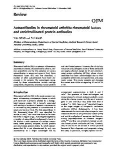
Arthroscopic biopsy During the last 30 years arthroscopy made its entrance into rheumatology. Technology has considerably improved over time and the use of local and regional anaesthetics have made this a safe, office-based procedure. Small-bore arthroscopes may be employed under local anaesthesia (8;11;16). After introducing the arthroscope, a second portal is required for the introduction of the grasping forceps. Knee arthroscopy can be performed using a small-bore, 2.7-mm arthroscope and utilising an infrapatellar skin portal for macroscopic examination of the synovium and a second suprapatellar portal for the biopsy procedure (Figure 1). This makes it possible to obtain synovial biopsy samples under direct vision from the suprapatellar pouch, the cartilage-pannus junction, the patellar gutters, and the tibiofemoral junction. Similarly, arthroscopy of the wrist joint can be performed using a small-bore, 1.9-mm arthroscope through a radial and an ulnar skin portal at the dorsum of the wrist at the proximal and/or distal carpal row.
Chapter 6
Figure 1. Knee arthroscopy using a small-bore arthroscope and a second suprapatellar portal for synovial biopsy under visual inspection.
Synovial biopsy
A recent survey showed a rapid increase in the number of arthroscopies performed by rheumatologists in an out-patient setting (17). The arthroscopic procedure is well tolerated and has a low complication rate, with 35-36% of patients reporting minimal pain or discomfort during the procedure (9). Minor complications, such as vasovagal reactions and temporary swelling of the joint, were mentioned in 5-10% of the cases. In a recent survey, in which information of 15,682 arthroscopies performed by rheumatologists was collected, the complication rate of haemarthros was 0.9%, deep vein thrombosis 0.2% and wound and joint infection 0.1%. The total complication rate reported was 15.1/1000 arthroscopies, which is comparable to the figures reported in the orthopaedic literature (17). The irrigation volume of the knee joint was positively correlated with the rate of wound infection and the total complication rate. A possible explanation for this is the extended length of the procedure. In our own series of more than 2,000 arthroscopic synovial biopsy procedures over the last 5 years, the complication rate was < 0.3% (haemarthros, portal infection and septic arthritis). The procedure was generally welltolerated. Deep vein thrombosis did not occur. There was no permanent damage or loss of function after infection in any of the patients as a result of aggressive joint drainage and prompt antibiotic therapy. Obviously, strict instructions to the patients after the arthroscopy procedure are essential to prevent any delay in case of complications.
97
Chapter 6 Synovial biopsy
98
Although the technique is more complicated and more expensive than the blind needle biopsy, there are several advantages. First, it allows macroscopic evaluation of the synovium. A positive correlation between the macroscopic signs of inflammation at the biopsy site and the immunohistological features of inflammatory activity in the corresponding tissue sample has been observed (18). It is not possible, however, to predict in a reliable way the microscopic features of the inflamed synovium on the basis of the macroscopic appearance at arthroscopy (19). Interestingly, previous reports have suggested that macroscopic scoring systems could be used to distinguish different patient groups (20;21). The visual evaluation needs more extensive validation, however, before it can be used in the diagnostic work-up. Using narrow-diameter arthroscopes, small joints, such as ankles, wrists and even metacarpophalangeal and proximal interphalangeal joints, can be evaluated. Another advantage of arthroscopy over blind needle biopsy is the fact that evaluation of the articular cartilage may be used for the evaluation of the effects of treatment (22). Moreover, it is basically always possible to obtain tissue in adequate amounts, even when the synovial tissue volume has decreased as a result of effective treatment.
Synovial tissue samples: how many and from which patients? Several studies have suggested a certain degree of morphological heterogeneity in synovial samples taken from one joint (18). However, it is possible to quantify several markers of inflammation in a reliable way examining a limited area of tissue (23-25). For T cell infiltration and expression of activation antigens in rheumatoid arthritis (RA) synovium, a variance of less than 10% can be reached when at least six biopsy specimens are examined (26), suggesting that representative data can be obtained when a limited number of biopsy samples from different areas within one joint are investigated. Consistent with these data, it has been demonstrated that using about six tissue samples allows for the detection of twofold differences in gene expression by quantitative polymerase chain reaction (PCR) (27). Therefore, we recommend obtaining at least six biopsies for each technique used in research. When the biopsies are taken from an actively inflamed joint there is, on average, no clear cut difference in the features of synovial inflammation or the expression of mediators of inflammation and destruction at the pannus-cartilage junction compared with other regions away from the pannus-cartilage junction (28-30). In addition, the signs of inflammation in tissue samples taken from inflamed small joints compared to larger joints, such as the knee, are generally comparable (11), indicating that the inflammatory process in one inflamed joint is representative of that in other inflamed joints.
In contrast, there is large variability of synovial inflammation between different patients in all phases of the disease (31-34). Therefore, it is essential that the number of patients is sufficient when different arthritides are compared. In addition, patient cohorts should be stratified for disease activity and use of medication to allow meaningful conclusions, since both variables influence the features of the synovium (31;35).
Processing of synovial tissue
Differential diagnosis In rheumatology it is usually possible to make a diagnosis on the basis of the history, clinical examination, routine laboratory tests, radiographical examination, and synovial fluid analysis. When synovial fluid cannot be aspirated, or in cases of suspicion of infection in spite of negative synovial fluid culture, examination of synovial biopsies may
99 Synovial biopsy
Indications of synovial tissue analysis
Chapter 6
For routine pathological examination by light microscopy, the tissue samples are fixed in 4% formalin and embedded in paraffin. This can be used for haematoxylin and eosin staining and certain immunohistochemical stainings. When gout is suspected, the biopsy samples should be conserved in absolute alcohol because the monosodium urate crystals will dissolve in most other fixatives. Unstained sections can be examined with the polarization microscope. It is also possible to use the DeGolanthal stain to detect urate crystals. In the case of a suspected infection, the tissue should be kept in suitable culture media. When tissue samples are to be examined for the presence of bacterial DNA by PCR, contaminating DNA needs be avoided and the samples should be snap frozen in liquid nitrogen. For research purposes, synovial tissue can be processed for histology, immunohistochemistry, immunofluorescense, in situ hybridization, PCR, micro-array, tissue enzyme-linked immunoassay (ELISA), proteomic profiling and cell or tissue culture. Immunohistochemistry can be performed on formalin fixed, paraffin-embedded material or on samples that were snap-frozen in optimum cutting temperature (OCT) compoundembedding medium and stored in liquid nitrogen. mRNA can be extracted directly from fresh biopsy samples, but detection of mRNA is also possible in samples that were snap frozen immediately after the tissue was obtained. Tissue samples can be used to culture synovial cell populations (36) and whole biopsy samples (37) when appropriate tissue culture media are used.
Chapter 6 Synovial biopsy
100
be of additional value (38-40). The mainstay of the diagnosis is a positive culture, but this may take days or even weeks for slow-growing pathogens. Cultures can also be negative when the pathogen is difficult to culture or due to prior exposure to antibiotics. Recently, the use of PCR has been evaluated in detecting bacterial DNA in the synovium for differential diagnosis as well as monitoring therapy of infectious arthritis. By applying broad-range bacterial primers to the analysis and amplification of genes coding for ribosomal RNA, most bacterial species can be detected. This method has been used successfully, even when culture failed (41;42). PCR might be especially helpful when antibiotic treatment has been initiated before synovial fluid was obtained for culture (41), in case of slow-growing micro-organisms such as mycobacteria (38), or when Borrelia, Chlamydia or Neisseria species are suspected (39;43;44). Very stringent conditions are required because of the risk of false positive results caused by contamination (4;45). It is important to note that a positive result does not necessarily proof the presence of a viable or proliferating organism. Examination of synovial biopsies may also help to make a diagnosis in some relatively rare infiltrative and deposition diseases of the joints. In gout and pseudogout, tophuslike deposits can be found in the synovial membrane and cartilage (46). This may reveal the diagnosis in occasional cases when synovial fluid analysis is negative. Other relatively rare diagnoses that can be made on the basis of synovial tissue analysis include amyloidosis, ochronosis, haemochromatosis, proliferative disorders such as pigmented villonodular synovitis (PVNS), synovial chondromatosis, synovial chondrosarcomas, synovial haemangiomas, lipoma aborescens, intracapsular chondromas, synovial metastases from distant neoplasms and synovial involvement of lymphoreticular malignancy (47). Synovial tissue analysis might also assist in the diagnostic process in early phases of immune-mediated forms of arthritis before the patients fulfil classifying diagnostic criteria. It would be particularly useful to predict which patients will have a persistent and destructive disease, such as RA. However, many of the pathological changes in the rheumatoid synovium, including vascular congestion, intimal lining layer hyperplasia, mononuclear cell infiltration and fibrin depositions commonly occur in disorders other than RA. Still, synovial tissue analysis might have diagnostic potential in distinguishing RA from other forms of arthritis. Multivariate models can predict a diagnosis of RA solely on the basis of examination of synovial biopsy specimens with an accuracy of 85% when massive infiltration by plasma cells and macrophages in the synovial sublining is present. A diagnosis other than RA can be predicted in > 95% of the cases when there is only minimal infiltration by these cells (5). Previous work has suggested that the presence of citrullinated proteins could be specific for RA (48), but this could not be confirmed in subsequent studies (49;50); citrullination of synovial antigens may occur in any form of joint inflammation (51). The induction of autoantibodies directed to these proteins,
however, appears to be specific for RA (51). Taken together, examination of synovial tissue may be useful for diagnosing infectious, infiltrative and deposition diseases of the joints, but the role in the differential diagnosis of immune-mediated disease is limited.
Pathogenetic studies
Chapter 6
101 Synovial biopsy
The synovium is a primary site of inflammation and a major effector organ in a variety of joint diseases, including RA. As a result, there has been increased interest in studies of the pathological changes of the synovium (52). Rheumatoid synovial tissue is characterised by marked intimal hyperplasia and by accumulation of T cells, plasma cells, macrophages, B cells, mast cells, natural killer cells and dendritic cells in the synovial sublining layer, but only few neutrophils are found (1). It is generally believed that large numbers of neutrophils diffuse through the intimal lining layer into the synovial fluid, where they form the predominant cell population (53). Many pro-inflammatory mediators and tissue degrading products such as reactive oxygen species and reactive nitrogen species, prostaglandins, cytokines, auto-antibodies and proteases are secreted into the synovial compartment by the infiltrating cell populations. Of interest, the synovial cell infiltrate, as well as the expression of adhesion molecules, cytokines, chemokines and matrix metalloproteinases, already exhibit all features of chronic synovial inflammation very early after the clinical onset of the disease (21;31;54-58). This is consistent with the notion that the development of signs and symptoms is preceded by a phase characterized by immunological abnormalities and asymptomatic synovitis (54;59-61). As indicated above, it is essential to stratify patient cohorts for disease activity and use of medication when comparing the tissue characteristics from early RA patients to those with longstanding disease, since these variables represent an important source of bias. Synovial tissue analysis has also shown that there is marked variation between different individuals in all phases of RA (31;32;62). It has been suggested that RA patients display reproducible patterns in the organization and activity of synovial infiltrates, which are associated with the level of cytokine expression in the tissue (33). Recent work using complementary DNA microarray analysis to profile gene expression in rheumatoid synovial tissue also showed considerable variability, resulting in the identification of molecularly distinct subsets of RA tissues (34). Thus, using different methodologies, these studies have consistently indicated that RA may comprise different pathogenetic mechanisms leading to a common clinical syndrome. Conceivably, more insight into these distinct subsets may help to define homogeneous groups for clinical studies and the evaluation of targeted therapies. Together, studies on rheumatoid synovial tissue have provided important insight into the key factors involved in the pathogenesis of the disease and they have revealed the heterogeneity of RA as well as the chronicity of the inflammatory process in what we in the clinic define as ‘early RA’.
Although the data are even more limited, similar studies are being undertaken in other arthritides, such as psoriatic arthritis (63-65), reactive arthritis (66;67) and ankylosing spondylitis (21;68).
Chapter 6
Synovial tissue response to treatment
Synovial biopsy
102
Analysis of serial synovial tissue samples is increasingly used in the evaluation of targeted therapies in an early stage of drug development (3). This approach may help to increase our understanding of the mechanism of action of the therapeutic intervention. Furthermore, examination of the effects at the site of inflammation in phase I/II studies may be used for target validation and to demonstrate proof of principle. In addition to studying the specific effects of the intervention, sensitive biomarkers associated with clinical efficacy may be used for selection purposes during the development process (35;69;70). It has been suggested that our usual thinking about the use of biomarkers in clinical trials is about to change dramatically (71). Clinical investigations will increasingly consist of small trials with a high density of data. The importance of including the analysis of synovial tissue samples has been underscored by the observation that the activity of clinical arthritis is accompanied by histological signs of synovitis after treatment of RA patients with the monoclonal antibody Campath-1H, despite profound depletion of circulating lymphocytes (72). A recent study was designed to identify the optimal synovial biomarker associated with clinical efficacy following a short treatment duration (69). This study demonstrated the status of sublining macrophages as an optimal biomarker associated with clinical response. Subsequently, the merit of using this biomarker was tested across a range of discrete interventions and kinetics (26). Eighty-eight patients who participated in various randomised clinical trials were evaluated in the same centre, using standardised techniques. The treatments evaluated included methotrexate, leflunomide, prednisolone, infliximab, a specific chemokine receptor-1 (CCR-1) inhibitor and placebo. All patients had baseline and follow-up biopsies and disease activity scores (DAS) obtained. There was a significant correlation between the change in the number of macrophages and the change in DAS28. The change in sublining macrophages could explain 76% of the variation in the change in DAS28. Interestingly, this close correlation was independent of the primary mode of action of the individual therapy, indicating that the effect of treatment is associated with an ultimate effect on common final pathways. The sensitivity to change of the biomarker was high in actively treated patients, while no significant changes were detected in placebo-treated patients. These data are consistent with other studies showing that serial biopsy samples from RA patients who received either placebo or unsuccessful treatment did not reveal synovial changes in the relevant biomarkers (35;73-77). Thus, changes in serial biopsy samples cannot
The synovial membrane is a major target tissue in RA and other arthropathies. Recent technical developments have allowed synovial biopsy samples to be obtained in a safe and generally well-tolerated way. Synovial tissue analysis can be used for diagnostic purposes in selected cases. In addition, the evaluation of inflamed synovial tissue plays an increasingly important role in pathogenetic studies as well as in the identification of novel therapeutic targets. Experimental studies evaluating the effects of targeted therapeutic interventions at the site of inflammation may further increase our insight into the pathogenesis of a variety of arthritides. This approach can also be used for proof of principle studies and to screen for potential efficacy of new compounds. Recent work has identified synovial biomarkers that are associated with clinical efficacy independent
103 Synovial biopsy
Concluding remarks
Chapter 6
be explained by placebo effects, regression to the mean, expectation bias, or by the arthroscopy procedure itself. Rather, they reflect biological effects of the treatment. Therefore, this approach can be used as a screening method to test new drug candidates requiring relatively small numbers of subjects. The absence of changes after treatment would suggest that the therapy is probably not effective. However, the demonstration of biological changes at the site of inflammation may provide the rationale for larger, placebo-controlled trials. Using this approach, successful treatment with disease-modifying anti-rheumatic drugs, such as gold (78), methotrexate (79-81) and leflunomide (81), was shown to be associated with decreased mononuclear cell infiltration. Similarly, successful treatment of RA patients with infliximab (82-85) and anakinra (74) resulted in reduced synovial inflammation. Of interest, the number of macrophages in the synovium was already significantly decreased 48 hours after initiation of infliximab treatment (85). After 1 month, the most pronounced reduction of macrophage numbers was found in the patients who fulfilled criteria for clinical improvement at 12 weeks, suggesting that this approach might be used to predict the clinical response. Most of the biopsy studies have been performed in RA patients, but recent work has shown that the same approach can be used for the evaluation of novel therapies in patients with other rheumatological disease, such as spondylarthropathies (86-89). Serial synovial biopsy is now widely used by a limited number of centres with standardised methodology. The advances in the evaluation of the synovial tissue response to targeted therapy will challenge academic rheumatology to optimise the clinical resources and expertise in both arthroscopy and sophisticated tissue analysis. Moreover, there will be interesting opportunities for collaboration with pharmaceutical industries in early phases of drug development.
Chapter 6
of the primary mechanism of action of treatment. It is likely that more sophisticated biomarkers, including gene expression profiles, will be developed and validated in the near future. It can be anticipated that future work will also include the identification of predictive indices of outcome in patients with arthritis. Since studies of synovial tissue samples will increasingly be used in multi-centre studies, further standardisation and validation of the methodology will be essential.
Synovial biopsy
104
Reference List 1.
2.
3. 4. 5.
6.
8. 9.
12. 13.
14.
15. 16. 17. 18. 19.
105 Synovial biopsy
10. 11.
Chapter 6
7.
Tak PP. Examination of the synovium and synovial fluid. In: Firestein GS, Panayi GS, Wollheim FA, editors. Rheumatoid arthritis. Frontiers in pathogenesis and treatment. New York: Oxford University Press, Inc., 2000: 55-68. Smeets TJ, Barg EC, Kraan MC, Smith MD, Breedveld FC, Tak PP. Analysis of the cell infiltrate and expression of proinflammatory cytokines and matrix metalloproteinases in arthroscopic synovial biopsies: comparison with synovial samples from patients with end stage, destructive rheumatoid arthritis. Ann Rheum Dis 2003; 62(7):635-638. Tak PP. Lessons learnt from the synovial tissue response to antirheumatic treatment. Rheumatology (Oxford) 2000; 39:817-820. Johnson JS, Freemont AJ. A 10 year retrospective comparison of the diagnostic usefulness of synovial fluid and synovial biopsy examination. J Clin Pathol 2001; 54(8):605-607. Kraan MC, Haringman JJ, Post WJ, Versendaal J, Breedveld FC, Tak PP. Immunohistological analysis of synovial tissue for differential diagnosis in early arthritis. RHEUMATOLOGY 1999; 38:10741080. Baeten D, Kruithof E, De Rycke L, Vandooren B, Wyns B, Boullart L et al. Diagnostic classification of spondylarthropathy and rheumatoid arthritis by synovial histopathology: a prospective study in 154 consecutive patients. Arthritis Rheum 2004; 50(9):2931-2941. Szachnowski P, Wei N, Arnold WJ, Cohen LM. Complications of office based arthroscopy of the knee. J Rheumatol 1995; 22(9):1722-1725. Reece R, Emery P. Needle arthroscopy. Br J Rheumatol 1995; 34:1102-1104. Baeten D, Van den Bosch F, Elewaut D, Stuer A, Veys EM, De KF. Needle arthroscopy of the knee with synovial biopsy sampling: technical experience in 150 patients. Clin Rheumatol 1999; 18(6):434-441. Rozmaryn LM, Wei N. Metacarpophalangeal arthroscopy. Arthroscopy 1999; 15(3):333-337. Kraan MC, Reece RJ, Smeets TJ, Veale DJ, Emery P, Tak PP. Comparison of synovial tissues from the knee joints and the small joints of rheumatoid arthritis patients: Implications for pathogenesis and evaluation of treatment. Arthritis Rheum 2002; 46(8):2034-2038. Parker RH, Pearson CM. A simplified synovial biopsy needle. Arthritis Rheum 1963; 6:172-176. Soden M, Rooney M, Cullen A, Whelan A, Feighery C, Bresnihan B. Immunohistological features in the synovium obtained from clinically uninvolved knee joints of patients with rheumatoid arthritis. Brit J Rheumatol 1989; 28:287-292. Youssef PP, Kraan M, Breedveld F, Bresnihan B, Cassidy N, Cunnane G et al. Quantitative microscopic analysis of inflammation in rheumatoid arthritis synovial membrane samples selected at arthroscopy compared with samples obtained blindly by needle biopsy. Arthritis Rheum 1998; 41(4):663-669. Arayssi TK, Schumacher HR, Jr. Evaluation of a modified needle for small joint biopsies. J Rheumatol 1998; 25(5):876-878. Arnold WJ. Office-based arthroscopy. Bull Rheum Dis 1992; 41(5):3-6. Kane D, Veale DJ, FitzGerald O, Reece R. Survey of arthroscopy performed by rheumatologists. Rheumatology (Oxford) 2002; 41(2):210-215. Lindblad S, Hedfors E. Intraarticular variation in synovitis. Local macroscopic and microscopic signs of inflammatory activity are significantly correlated. Arthritis Rheum 1985; 28:977-986. Rooney M, Condell D, Daly L, Whelan A, Feighery C, Bresnihan B. Analysis of the histologic variation of synovitis in rheumatoid arthritis. Arthritis Rheum 1988; 31:956-963.
20. 21.
22.
23.
24.
Chapter 6
25.
Synovial biopsy
106
26.
27. 28.
29.
30.
31.
32.
33. 34.
35.
Reece RJ, Canete JD, Parsons WJ, Emery P, Veale DJ. Distinct vascular patterns of early synovitis in psoriatic, reactive, and rheumatoid arthritis. Arthritis Rheum 1999; 42(7):1481-1484. Baeten D, Demetter P, Cuvelier C, Van Den BF, Kruithof E, Van Damme N et al. Comparative study of the synovial histology in rheumatoid arthritis, spondyloarthropathy, and osteoarthritis: influence of disease duration and activity. Ann Rheum Dis 2000; 59(12):945-953. Ayral X, Mackillop N, Genant HK, Kirkpatrick J, Beaulieu A, Pippingskiold P et al. Arthroscopic evaluation of potential structure-modifying drug in osteoarthritis of the knee. A multicenter, randomized, double-blind comparison of tenidap sodium vs piroxicam. OSTEOARTHRITIS CARTILAGE 2003; 11(3):198-207. Bresnihan B, Cunnane G, Youssef PP, Yanni G, FitzGerald O, Mulherin D. Microscopic measurement of synovial membrane inflammation in rheumatoid arthritis: proposals for the evaluation of tissue samples by quantitative analysis. Br J Rheumatol 1998; 37:636-642. Youssef PP, Triantafillou S, Parker A, Coleman M, Roberts-Thomson PJ, Ahern MJ et al. Variability in cytokine and cell adhesion molecule staining in arthroscopic synovial biopsies: quantification using color video image analysis. J Rheumatol 1997; 24(12):2291-2298. Kraan MC, Smith MD, Weedon H, Ahern MJ, Breedveld FC, Tak PP. Measurement of cytokine and adhesion molecule expression in synovial tissue by digital image analysis. Ann Rheum Dis 2001; 60(3):296-298. Dolhain RJ, Ter Haar NT, de Kuiper R, Nieuwenhuis IG, Zwinderman AH, Breedveld FC et al. Distribution of T cells and signs of T-cell activation in the rheumatoid joint: implications for semiquantitative comparative histology. Br J Rheumatol 1998; 37(3):324-330. Boyle DL, Rosengren S, Bugbee W, Kavanaugh A, Firestein GS. Quantitative biomarker analysis of synovial gene expression by real-time PCR. Arthritis Res Ther 2003; 5(6):R352-R360. Smeets TJ, Kraan MC, Galjaard S, Youssef PP, Smith MD, Tak PP. Analysis of the cell infiltrate and expression of matrix metalloproteinases and granzyme B in paired synovial biopsy specimens from the cartilage-pannus junction in patients with RA. Ann Rheum Dis 2001; 60(6):561-565. Kirkham B, Portek I, Lee CS, Stavros B, Lenarczyk A, Lassere M et al. Intraarticular variability of synovial membrane histology, immunohistology, and cytokine mRNA expression in patients with rheumatoid arthritis. J Rheumatol 1999; 26(4):777-784. Kane D, Jensen LE, Grehan S, Whitehead AS, Bresnihan B, FitzGerald O. Quantitation of metalloproteinase gene expression in rheumatoid and psoriatic arthritis synovial tissue distal and proximal to the cartilage-pannus junction. J Rheumatol 2004; 31(7):1274-1280. Tak PP, Smeets TJM, Daha MR, Kluin PM, Meijers KAE, Brand R et al. Analysis of the synovial cellular infiltrate in early rheumatoid synovial tissue in relation to local disease activity. Arthritis Rheum 1997; 40:217-225. Ulfgren AK, Grondal L, Lindblad S, Khademi M, Johnell O, Klareskog L et al. Interindividual and intra-articular variation of proinflammatory cytokines in patients with rheumatoid arthritis: potential implications for treatment. Ann Rheum Dis 2000; 59(6):439-447. Klimiuk PA, Goronzy JJ, Bjor nsson J, Beckenbaugh RD, Weyand CM. Tissue cytokine patterns distinguish variants of rheumatoid synovitis. Am J Pathol 1997; 151(5):1311-1319. van der Pouw Kraan TC, van Gaalen FA, Kasperkovitz PV, Verbeet NL, Smeets TJ, Kraan MC et al. Rheumatoid arthritis is a heterogeneous disease: evidence for differences in the activation of the STAT-1 pathway between rheumatoid tissues. Arthritis Rheum 2003; 48(8):2132-2145. Haringman JJ, Gerlag DM, Zwinderman AH, Smeets TJM, Kraan MC, Baeten DM et al. Synovial tissue macrophages: sensitive biomarkers for response to treatment in rheumatoid arthritis. Ann Rheum Dis 2004; In press.
36.
37.
38.
39.
40.
41.
43.
45.
46. 47.
48.
49.
50. 51.
107 Synovial biopsy
44.
Chapter 6
42.
Van Holten J, Reedquist K, Sattonet-Roche P, Smeets TJ, Plater-Zyberk C, Vervoordeldonk MJ et al. Treatment with recombinant interferon-beta reduces inflammation and slows cartilage destruction in the collagen-induced arthritis model of rheumatoid arthritis. Arthritis Res Ther 2004; 6(3):R239-R249. Hitchon C, Wong K, Ma G, Reed J, Lyttle D, El Gabalawy H. Hypoxia-induced production of stromal cell-derived factor 1 (CXCL12) and vascular endothelial growth factor by synovial fibroblasts. Arthritis Rheum 2002; 46(10):2587-2597. Van der Heijden IM, Wilbrink B, Schouls LM, van Embden JD, Breedveld FC, Tak PP. Detection of mycobacteria in joint samples from patients with arthritis using a genus-specific polymerase chain reaction and sequence analysis. Rheumatology (Oxford) 1999; 38(6):547-553. Van der Heijden IM, Wilbrink B, Rijpkema SG, Schouls LM, Heymans PH, van Embden JD et al. Detection of Borrelia burgdorferi sensu stricto by reverse line blot in the joints of Dutch patients with Lyme arthritis. Arthritis Rheum 1999; 42(7):1473-1480. Titov AG, Vyshnevskaya EB, Mazurenko SI, Santavirta S, Konttinen YT. Use of polymerase chain reaction to diagnose tuberculous arthritis from joint tissues and synovial fluid. Arch Pathol Lab Med 2004; 128(2):205-209. van der Heijden IM, Wilbrink B, Vije AE, Schouls LM, Breedveld FC, Tak PP. Detection of bacterial DNA in serial synovial samples obtained during antibiotic treatment from patients with septic arthritis. Arthritis Rheum 1999; 42(10):2198-2203. Jalava J, Skurnik M, Toivanen A, Toivanen P, Eerola E. Bacterial PCR in the diagnosis of joint infection. Ann Rheum Dis 2001; 60(3):287-289. Nikkari S, Puolakkainen M, Yli-Kerttula U, Luukkainen R, Lehtonen OP, Toivanen P. Ligase chain reaction in detection of Chlamydia DNA in synovial fluid cells. Br J Rheumatol 1997; 36(7):763765. Muralidhar B, Rumore PM, Steinman CR. Use of the polymerase chain reaction to study arthritis due to Neisseria gonorrhoeae. Arthritis Rheum 1994; 37:710-717. Wilbrink B, Van der Heijden IM, Schouls LM, Van Embden JDA, Hazes JMW, Breedveld FC et al. Detection of bacterial DNA in joint samples from patients with undifferentiated arthritis and reactive arthritis, using polymerase chain reaction with universal 16S ribosomal RNA primers. Arthritis Rheum 1998; 41:535-543. Schumacher HR. Ultrastructural findings in chondrocalcinosis and pseudogout. Arthritis Rheum 1976; 19 Suppl 3:413-425. Gerlag DM, Tak PP. Synovial fluid analyses, synovial biopsy, and synovial pathology. In: Harris ED, Budd RC, Firestein GS, Genovese M, Sergent JS, Ruddy S et al., editors. Kelley‘s textbook of rheumatology. Philadelphia: W.B. Saunders Co., 2004: 675-691. Baeten D, Peene I, Union A, Meheus L, Sebbag M, Serre G et al. Specific presence of intracellular citrullinated proteins in rheumatoid arthritis synovium: relevance to antifilaggrin autoantibodies. Arthritis Rheum 2001; 44(10):2255-2262. Smeets TJ, Vossenaar ER, van Venrooij WJ, Tak PP. Is expression of intracellular citrullinated proteins in synovial tissue specific for rheumatoid arthritis? Comment on the article by Baeten et al. Arthritis Rheum 2002; 46(10):2824-2826. Vossenaar ER, Smeets TJM, Kraan MC, Raats JM, Van Venrooij J, Tak PP. The presence of citrullinated proteins is not specific for rheumatoid synovial tissue. Arthritis Rheum 2004; In press. Vossenaar ER, Nijenhuis S, Helsen MM, van der HA, Senshu T, Van den Berg WB et al. Citrullination of synovial proteins in murine models of rheumatoid arthritis. Arthritis Rheum 2003; 48(9):24892500.
52. 53.
54. 55.
56. 57. 58.
Chapter 6
59.
Synovial biopsy
108
60.
61.
62. 63.
64.
65.
66. 67.
68.
Tak PP, Bresnihan B. The pathogenesis and prevention of joint damage in rheumatoid arthritis: advances from synovial biopsy and tissue analysis. Arthritis Rheum 2000; 43(12):2619-2633. Youssef PP, Cormack J, Evill CA, Peter DT, Roberts-Thomson PJ, Ahern MJ et al. Neutrophil trafficking into inflamed joints in patients with rheumatoid arthritis, and the effects of methylprednisolone. Arthritis Rheum 1996; 39:216-225. Kraan MC, Versendaal H, Jonker M, Bresnihan B, Post W, ‘t Hart BA et al. Asymptomatic synovitis precedes clinically manifest arthritis. Arthritis Rheum 1998; 41:1481-1488. Smeets TJM, Dolhain RJEM, Miltenburg AMM, de Kuiper R, Breedveld FC, Tak PP. Poor expression of T cell-derived cytokines and activation and proliferation markers in early rheumatoid synovial tissue. Clin Immunol Immunopathol 1998; 88:84-90. Tak PP, Thurkow EW, Daha MR, Kluin PM, Smeets TJM, Meinders AE et al. Expression of adhesion molecules in early rheumatoid synovial tissue. Clin Immunol Immunopathol 1995; 77:236-242. Zvaifler NJ. Rheumatoid arthritis - The multiple pathways to chronic synovitis. Lab Invest 1995; 73:307-310. Katrib A, Tak PP, Bertouch JV, Cuello C, McNeil HP, Smeets TJ et al. Expression of chemokines and matrix metalloproteinases in early rheumatoid arthritis. Rheumatology (Oxford) 2001; 40(9):988994. Wakefield RJ, Green MJ, Marzo-Ortega H, Conaghan PG, Gibbon WW, Mcgonagle D et al. Should oligoarthritis be reclassified? Ultrasound reveals a high prevalence of subclinical disease. Ann Rheum Dis 2004; 63(4):382-385. Nielen MM, van Schaardenburg D, Reesink HW, van de Stadt RJ, van der Horst-Bruinsma IE, de Koning MH et al. Specific autoantibodies precede the symptoms of rheumatoid arthritis: a study of serial measurements in blood donors. Arthritis Rheum 2004; 50(2):380-386. Nielen MM, van Schaardenburg D, Reesink HW, Twisk JW, van de Stadt RJ, van der Horst-Bruinsma IE et al. Increased levels of C-reactive protein in serum from blood donors before the onset of rheumatoid arthritis. Arthritis Rheum 2004; 50(8):2423-2427. Schumacher HR, Jr., Kitridou RC. Synovitis of recent onset. A clinicopathologic study during the first month of disease. Arthritis Rheum 1972; 15:465-485. Veale D, Yanni G, Rogers S, Barnes L, Bresnihan B, FitzGerald O. Reduced synovial membrane macrophage numbers, ELAM-1 expression, and lining layer hyperplasia in psoriatic arthritis as compared with rheumatoid arthritis. Arthritis Rheum 1993; 36:893-900. Danning CL, Illei GG, Hitchon C, Greer MR, Boumpas DT, McInnes IB. Macrophage-derived cytokine and nuclear factor kappaB p65 expression in synovial membrane and skin of patients with psoriatic arthritis. Arthritis Rheum 2000; 43(6):1244-1256. Kane D, Jensen LE, Grehan S, Whitehead AS, Bresnihan B, FitzGerald O. Quantitation of metalloproteinase gene expression in rheumatoid and psoriatic arthritis synovial tissue distal and proximal to the cartilage-pannus junction. J Rheumatol 2004; 31(7):1274-1280. Simon AK, Seipelt E, Sieper J. Divergent T-cell cytokine patterns in inflammatory arthritis. Proc Natl Acad Sci U S A 1994; 91:8562-8566. Smeets TJ, Dolhain RJ, Breedveld FC, Tak PP. Analysis of the cellular infiltrates and expression of cytokines in synovial tissue from patients with rheumatoid arthritis and reactive arthritis. J Pathol 1998; 186(1):75-81. Braun J, Bollow M, Neure L, Seipelt E, Seyrekbasan F, Herbst H et al. Use of immunohistologic and in situ hybridization techniques in the examination of sacroiliac joint biopsy specimens from patients with ankylosing spondylitis. Arthritis Rheum 1995; 38(4):499-505.
69.
70.
71. 72. 73.
74.
75.
77.
79. 80.
81.
82.
83.
84.
109 Synovial biopsy
78.
Chapter 6
76.
Gerlag DM, Haringman JJ, Smeets TJM, Zwinderman AH, Kraan MC, Laud PJ et al. Effects of oral prednisolone on biomarkers in synovial tissue and clinical improvement in rheumatoid arthritis. Arthritis Rheum 2004; In press. De Groot J, Te Koppele JM, Harris, E.D., Tak PP. Biological markers. In: Harris ED, Budd RC, Firestein GS, Genovese MC, Sergent JS, Ruddy S et al., editors. Kelley’s textbook of rheumatology. Philadelphia: W.B. Saunders Co., 2004: 728-738. Liu ET, Karuturi KR. Microarrays and clinical investigations. N Engl J Med 2004; 350(16):15951597. Ruderman EM, Weinblatt ME, Thurmond LM, Pinkus GS, Gravallese EM. Synovial tissue response to treatment with Campath-1H. Arthritis Rheum 1995; 38:254-258. Smeets TJ, Kraan MC, Versendaal J, Breedveld FC, Tak PP. Analysis of serial synovial biopsies in patients with rheumatoid arthritis: description of a control group without clinical improvement after treatment with interleukin 10 or placebo. J Rheumatol 1999; 26(10):2089-2093. Cunnane G, Madigan A, Murphy E, FitzGerald O, Bresnihan B. The effects of treatment with interleukin-1 receptor antagonist on the inflamed synovial membrane in rheumatoid arthritis. Rheumatology (Oxford) 2001; 40(1):62-69. Van Holten J, Pavelka K, Vencovsky J, Stahl H, Rozman B, Genovese M et al. A multicentre, randomised, double-blind, placebo controlled phase II study of subcutaneously administered interferon beta 1a in the treatment of patients with active rheumatoid arthritis. Ann Rheum Dis 2004. Tak PP, Van der Lubbe PA, Cauli A, Daha MR, Smeets TJM, Kluin PM et al. Reduction of synovial inflammation after anti-CD4 monoclonal antibody treatment in early rheumatoid arthritis. Arthritis Rheum 1995; 38:1457-1465. Haringman JJ, Gerlag DM, Smeets TJM, Baeten D, Bresnihan B, Breedveld FC et al. A randomized placebo controlled trial with an anti-MCP-1 (CCL2) monoclonal antibody in patients with rheumatoid arthritis. Arthritis Rheum 2004; 50:S238. Rooney M, Whelan A, Feighery C, Bresnihan B. Changes in lymphocyte infiltration of the synovial membrane and the clinical course of rheumatoid arthritis. Arthritis Rheum 1989; 32:361-369. Firestein GS, Paine MM, Boyle DL. Mechanisms of methotrexate action in rheumatoid arthritis Selective decrease in synovial collagenase gene expression. Arthritis Rheum 1994; 37:193-200. Dolhain RJEM, Tak PP, Dijkmans BAC, De Kuiper P, Breedveld FC, Miltenburg AMM. Methotrexate treatment reduces inflammatory cell numbers, expression of monokines and of adhesion molecules in synovial tissue of patients with rheumatoid arthritis. Br J Rheumatol 1998; 37:502-508. Kraan MC, Reece RJ, Barg EC, Smeets TJ, Farnell J, Rosenburg R et al. Modulation of inflammation and metalloproteinase expression in synovial tissue by leflunomide and methotrexate in patients with active rheumatoid arthritis. Findings in a prospective, randomized, double-blind, paralleldesign clinical trial in thirty-nine patients at two centers. Arthritis Rheum 2000; 43(8):1820-1830. Tak PP, Taylor PC, Breedveld FC, Smeets TJM, Daha MR, Kluin PM et al. Decrease in cellularity and expression of adhesion molecules by anti-tumor necrosis factor alpha treatment in patients with rheumatoid arthritis. Arthritis Rheum 1996; 39:1077-1081. Taylor PC, Peters AM, Paleolog E, Chapman PT, Elliott MJ, Mccloskey R et al. Reduction of chemokine levels and leukocyte traffic to joints by tumor necrosis factor alpha blockade in patients with rheumatoid arthritis. Arthritis Rheum 2000; 43(1):38-47. Ulfgren AK, Andersson U, Engstrom M, Klareskog L, Maini RN, Taylor PC. Systemic anti-tumor necrosis factor alpha therapy in rheumatoid arthritis down-regulates synovial tumor necrosis factor alpha synthesis. Arthritis Rheum 2000; 43(11):2391-2396.
85.
86.
87.
88.
Chapter 6
89.
Synovial biopsy
110
Smeets TJ, Kraan MC, van Loon ME, Tak PP. Tumor necrosis factor alpha blockade reduces the synovial cell infiltrate early after initiation of treatment, but apparently not by induction of apoptosis in synovial tissue. Arthritis Rheum 2003; 48(8):2155-2162. Baeten D, Kruithof E, Van Den BF, Demetter P, Van Damme N, Cuvelier C et al. Immunomodulatory effects of anti-tumor necrosis factor alpha therapy on synovium in spondylarthropathy: histologic findings in eight patients from an open-label pilot study. Arthritis Rheum 2001; 44(1):186-195. Vandooren B, Kruithof E, Yu DT, Rihl M, Gu J, De Rycke L et al. Involvement of matrix metalloproteinases and their inhibitors in peripheral synovitis and down-regulation by tumor necrosis factor alpha blockade in spondylarthropathy. Arthritis Rheum 2004; 50(9):2942-2953. Goedkoop AY, Kraan MC, Picavet DI, De Rie MA, Teunissen MB, Bos JD et al. Deactivation of endothelium and reduction in angiogenesis in psoriatic skin and synovium by low dose infliximab therapy in combination with stable methotrexate therapy: a prospective single-centre study. Arthritis Res Ther 2004; 6(4):R326-R334. Kraan MC, Van Kuijk AW, Dinant HJ, Goedkoop AY, Smeets TJ, De Rie MA et al. Alefacept treatment in psoriatic arthritis: Reduction of the effector T cell population in peripheral blood and synovial tissue is associated with improvement of clinical signs of arthritis. Arthritis Rheum 2002; 46(10):2776-2784.