CME
Revised diagnostic criteria for neuromyelitis optica D.M. Wingerchuk, MD, FRCP(C); V.A. Lennon, MD, PhD; S.J. Pittock, MD; C.F. Lucchinetti, MD; and B.G. Weinshenker, MD, FRCP(C)
Abstract—Background: The authors previously proposed diagnostic criteria for neuromyelitis optica (NMO) that facilitate its distinction from prototypic multiple sclerosis (MS). However, some patients with otherwise typical NMO have additional symptoms not attributable to optic nerve or spinal cord inflammation or have MS-like brain MRI lesions. Furthermore, some patients are misclassified as NMO by the authors’ earlier proposed criteria despite having a subsequent course indistinguishable from prototypic MS. A serum autoantibody marker, NMO-IgG, is highly specific for NMO. The authors propose revised NMO diagnostic criteria that incorporate NMO-IgG status. Methods: Using final clinical diagnosis (NMO or MS) as the reference standard, the authors calculated sensitivity and specificity for each criterion and various combinations using a sample of 96 patients with NMO and 33 with MS. The authors used likelihood ratios and logistic regression analysis to develop the most practical and informative diagnostic model. Results: Fourteen patients with NMO (14.6%) had extra-optic-spinal CNS symptoms. NMO-IgG seropositivity was 76% sensitive and 94% specific for NMO. The best diagnostic combination was 99% sensitive and 90% specific for NMO and consisted of at least two of three elements: longitudinally extensive cord lesion, onset brain MRI nondiagnostic for MS, or NMO-IgG seropositivity. Conclusions: The authors propose revised diagnostic criteria for definite neuromyelitis optica (NMO) that require optic neuritis, myelitis, and at least two of three supportive criteria: MRI evidence of a contiguous spinal cord lesion 3 or more segments in length, onset brain MRI nondiagnostic for multiple sclerosis, or NMO-IgG seropositivity. CNS involvement beyond the optic nerves and spinal cord is compatible with NMO. NEUROLOGY 2006;66:1485–1489
Neuromyelitis optica (NMO; Devic syndrome) is a clinically defined, severe CNS demyelinating syndrome characterized by optic neuritis (ON) and acute myelitis; the presence of CNS symptoms outside the optic nerves and spinal cord has until recently excluded the diagnosis.1-3 Traditionally, the term NMO was applied to patients who experienced a monophasic event consisting of bilateral simultaneous optic neuritis and acute myelitis.4 The NMO spectrum is now recognized to typically evolve as a relapsing disorder that also includes patients with unilateral ON and those with index events of ON and myelitis occurring weeks or even years apart.5 Early and accurate diagnosis is important because NMO carries a poorer prognosis than MS and generally accepted treatment approaches differ.3,6 In 1999, we proposed NMO diagnostic criteria with three absolute requirements: ON, acute myelitis, and no Additional material related to this article can be found on the Neurology Web site. Go to www.neurology.org and scroll down the Table of Contents for the May 23 issue to find the title link for this article.
symptoms implicating other CNS regions.5 To enhance specificity, fulfillment of at least one of three major supportive criteria was required: 1) brain MRI at disease onset is normal or does not fulfill MS imaging criteria; 2) spinal cord MRI shows a lesion extending over ⱖ3 vertebral segments; and 3) CSF reveals ⱖ50 WBC/mm3 or ⱖ5 neutrophils/mm3. Alternatively, fulfilling two of three minor supportive criteria (bilateral ON, severe residual visual loss, or severe fixed post-attack weakness) suffices. We derived the criteria empirically and suggested that they be validated and may require revision. International experience using the 1999 diagnostic criteria generally concurs with ours.7-10 However, the criteria have limitations. They fail to capture patients with a disease course otherwise highly compatible with NMO but whose neurologic symptoms or signs implicate CNS regions outside the optic nerves and spinal cord or whose brain MRI reveals lesions that may meet MS imaging criteria.9,11 Therefore, the full spectrum of the disease may be underappreciated. On the other hand, occasional MS patients
Editorial, see page 1466 See also page 1568 From the Department of Neurology (D.M.W.), Mayo Clinic College of Medicine, Scottsdale, AZ; and Departments of Neurology (V.A.L., S.J.P., C.F.L., B.G.W.), Laboratory Medicine and Pathology (V.A.L., S.J.P.), and Immunology (V.A.L.), Mayo Clinic College of Medicine, Rochester, MN. Disclosure: The authors report no conflicts of interest. Received October 25, 2005. Accepted in final form January 13, 2006. Address correspondence and reprint requests to Dr. Dean M. Wingerchuk, Department of Neurology, Mayo Clinic College of Medicine, 13400 East Shea Boulevard, Scottsdale, AZ 85259; e-mail:
[email protected] Copyright © 2006 by AAN Enterprises, Inc.
1485
with ON and an attack of partial myelitis may have an initial negative brain MRI, therefore fulfilling NMO criteria at least early in their clinical course. Substantial evidence, including clinical, laboratory, neuroimaging, and immunopathologic data, suggests that NMO is distinct from MS; however, no diagnostic gold standard has been established.12,13 An objective biomarker would enhance diagnostic certainty and definition of the NMO disease spectrum. The serum autoantibody NMO-IgG, which targets aquaporin-4, is a good candidate because it is ⬎90% specific for NMO in patients presenting with an optic-spinal syndrome and is not detected in patients with classic MS.14,15 NMO-IgG seropositivity also predicts relapse and conversion to NMO in patients presenting with a single attack of longitudinally extensive myelitis.16 We hypothesized that individual components of current NMO criteria differ in their diagnostic properties and that a quantitative evaluation of these criteria, with incorporation of the NMO-IgG disease marker, would allow formulation of optimal criteria to discriminate NMO from MS. Methods. Patients. We evaluated the characteristics of 129 patients ascertained through the MS centers at Mayo Clinic sites in Rochester, MN, and Scottsdale, AZ, and tested for NMO-IgG. Typically, these patients had attacks of optic neuritis, myelitis, and had a normal MRI scan of the head, or one that was deemed to show minimal findings. We maintain a central database of demographic, clinical, imaging, and laboratory data from patients who present with syndromes compatible with NMO, transverse myelitis, or relapsing myelitis. Data are entered by one of the study neurologists who has either evaluated the patient or reviewed the medical record. The cohort (1999 to 2005) considered in this study is independent from that used to generate the 1999 diagnostic criteria. Diagnosis. The reference standard for the study was a diagnosis of NMO or MS based on the final clinical diagnosis rendered by the study neurologist based on his or her integration of all available clinical, imaging, and laboratory data and the period of follow-up after disease onset. Patients must have had at least one attack of ON and myelitis to be eligible for the study. Those with a final diagnosis of MS comprise a group who presented with ON and myelitis combinations that suggested the possibility of NMO; patients with demyelinating disease syndromes in whom NMO was not an initial consideration were not included. Because we recognize the limitations of the 1999 Mayo Clinic criteria, we did not apply them formally for diagnosis for the purposes of this study but relied instead on the clinician’s final diagnosis based on all clinical information and available information for the subsequent course. The clinical diagnoses were finalized without knowledge of the NMO-IgG serologic status in all cases except two who were not seen at Mayo Clinic but were ascertained because they were found to be NMO-IgG positive; diagnostic information was obtained from the referring neurologist in those cases. NMO-IgG status. The Mayo Clinic Neuroimmunology Laboratory tested all serum samples for NMO-IgG using an indirect immunofluorescence technique described elsewhere.15,17 Sera were scored as positive or negative by two independent evaluators (V.A.L. and S.J.P.) and titrated in doubling dilutions to determine the greatest dilution that remained positive. The assay evaluators were unaware of the clinical diagnosis. No serum was classified as equivocal or indeterminate and positive and negative scores were concordant. Clinical data. Demographic and clinical information included sex, date of birth, age at disease onset, ethnicity (white or nonwhite, based on patient self-report), personal or family history of autoimmune disease, and family history of demyelinating disease. Neurologic symptoms and signs were recorded; for the purposes of criterion evaluation, we determined the occurrence and number of 1486
NEUROLOGY 66
May (2 of 2) 2006
episodes of optic neuritis and myelitis and whether patients had experienced neurologic events implicating CNS regions other than the optic nerve or spinal cord. Motor weakness was graded using the Medical Research Council scale and was termed severe if more than one muscle in an affected limb was scored as 2 or less. Available brain MRI studies were evaluated using Paty criteria, which require presence of four or more white matter lesions or three lesions when one is periventricular.18 We used these older MRI criteria to ensure diagnostic consistency with our earliest cases. We recorded results as normal, abnormal but not meeting Paty criteria, or meeting Paty criteria. The brain MRI at disease onset was used if available; however, if a later scan was available and was negative, we assumed the onset scan also was negative. Spinal cord MRI studies were similarly reviewed and results recorded as normal, abnormal with a T2-weighted cord lesion extending over three or more vertebral segments, or abnormal with a smaller lesion or confluent lesions not suggestive of NMO. CSF leukocyte count and differential data were categorized as follows based on the 1999 criteria: WBC greater than 50/mm3 or not; neutrophil count greater than 5/mm3 or not. Serologic data, beyond NMO-IgG status, included the detection of one or more serum autoantibodies (antinuclear, extractable nuclear antigen, double-stranded DNA, rheumatoid factor, cardiolipin, thyroid or intrinsic factor antibodies). When data were not available, or the test not performed, that element was removed from analysis for that patient. We required valid data for at least two of the following supportive features: brain MRI, spinal cord MRI, and CSF results. Evaluation of diagnostic properties. We compared NMO and MS patients with respect to demographic, clinical, and follow-up data as well as the frequency of meeting each individual diagnostic criterion; differences were determined using 2, Fisher exact, or t tests as appropriate (alpha ⫽ 0.05). We calculated sensitivity and specificity estimates for each criterion using the clinical diagnosis of NMO as the reference standard. We then evaluated the relative clinical utility of each test by comparing likelihood ratios (LR) for positive (sensitivity divided by 1 – specificity) and negative (1 – sensitivity divided by specificity) test results. For each estimate, 95% CIs were calculated. LRs are more useful than sensitivity and specificity because they may be used to adapt the results of a study to an individual patient using the principles of Bayes theorem and determination of pretest and post-test odds of disease presence.19 LRs of ⬎10 for a positive test result or ⬍0.1 for a negative test result are expected to yield a conclusive change in the post-test odds of disease presence. We used the LR data to guide construction of new diagnostic models, each consisting of two or three of the most accurate individual variables. We then evaluated the models in several ways. First, we calculated LR and associated CIs for each model. Second, we plotted receiver operator characteristic (ROC) curves for each model with sensitivity of a positive result on the y-axis and (1 – specificity) on the x-axis.20 The area under each ROC curve (AUC) was then calculated as a measure of the overall discrimination that each variable or combination of variables can provide between NMO and MS. AUC values may be calculated for different diagnostic tests and then compared to one another.21 An AUC of 1 represents a perfectly discriminatory test; an area of 0.5 represents a test that discriminates no better than random chance.22 Finally, we used logistic regression methods to estimate ORs for each model.23 The reference standard diagnosis was the dependent variable (NMO ⫽ 1; MS ⫽ 0) and individual variables or models were included as independent variables (positive test result ⫽ 1; negative result ⫽ 0). Goodness-of-fit was evaluated using the Hosmer-Lemeshow test.
Results. Demographic and clinical features of the sample are summarized in table 1. Women outnumbered men in both the NMO and MS groups but the NMO cohort was more likely to be nonwhite and older than 35 years at disease onset. Eighty-one (84.4%) of the 96 patients with NMO had an established relapsing disease course (at least one additional attack of either ON or myelitis after experiencing index attacks of initial ON or myelitis) at last follow-up (median 35 months; IQR ⫽ 4 to 48 months). More relapses occurred in patients with NMO than in pa-
Table 1 Demographic and clinical features of patients with NMO and MS Characteristic
Valid n
NMO
MS
96
33
p Value
N (total)
129
Female, n (%)
127
80/94 (85.1) 26/33 (78.9)
0.401
Non-caucasian, n (%)
119
35/91 (38.5)
2/28 (7.1)
0.002
Mean age of onset, y (SD)
123
37.8 (18.1)
32.4 (8.3)
0.113
Age onset ⬎ 35.0 y
123
56/92 (60.9) 10/31 (32.3)
Mean ON events (SD)
128
2.1 (1.5)
1.4 (1.4)
0.006 0.033
Mean myelitis events (SD)
128
3.4 (3.0)
1.8 (1.6)
0.006
Median follow-up, mo (IQR)
121
37 (21–85)
42 (18–103)
0.862
History of autoimmunity
112
23/83 (27.7)
9/29 (31.0)
0.733
Seropositivity for non-organspecific autoantibodies
110
41/82 (50.0) 12/28 (42.9)
0.514
Family Hx autoimmunity
88
24/38 (63.2)
9/26 (34.6)
0.025
Family Hx demyelinating dis.
99
9/73 (12.3)
1/26 (3.8)
0.284
NMO ⫽ neuromyelitis optica; MS ⫽ multiple sclerosis; n ⫽ number of patients with validated data for each characteristic; Hx ⫽ history; ON ⫽ optic neuritis; IQR ⫽ interquartile range.
tients with MS despite similar follow-up duration. The frequencies of coexisting systemic autoimmune disease and seropositivity for non-organ-specific serum autoantibodies were similar in NMO and MS but a family history of autoimmune disease was more frequent in the NMO group. At some time prior to diagnosis, 14 NMO patients (14.6%) had experienced neurologic symptoms indicating disease outside the optic nerves and spinal cord. Their characteristics are summarized in table E-1 (available on the Neurology Web site at www.neurology.org). In one case, vomiting was noted in association with a medullary lesion imaged by MRI, but in another case, vomiting was encountered without a demonstrable MRI lesion. Encephalopathy was associated with massive hemispheric lesions in one case, but in other instances of encephalopathy no definite inflammatory lesions were present. In these cases, a clinical diagnosis of NMO was reached on the basis of
other clinical and laboratory features together with evaluation of the follow-up course. Table E-2 summarizes the diagnostic properties of the 1999 criteria, individual components of those criteria, and NMO-IgG. The 1999 criteria were 85% sensitive but only 48% specific for NMO. The acceptance of extra-optic-spinal symptoms allowed for identification of all NMO cases but reduced the sensitivity to only 24%. This analysis confirms that the 1999 diagnostic criteria have inadequate diagnostic accuracy. For more than 90% of patients we had valid data for all variables except CSF analysis. Individual variables with significant discriminative power included MRI evidence of a longitudinally extensive spinal cord lesion (sensitivity 98%, specificity 83%) and NMO-IgG seropositivity (sensitivity 76%, specificity 94%). The likelihood ratios for these variables demonstrate that NMO-IgG seropositive status (LR [⫹] ⫽ 12.2) or the absence of a longitudinally extensive cord lesion (LR [–] ⫽ 0.03) would have a large impact on the post-test probability of NMO diagnosis. Table 2 and table E-3 summarize the diagnostic properties of combinations of variables and the modeling procedure. We did identify a combination with perfect sensitivity but inadequate specificity (model 3) and another that was entirely specific but insufficiently sensitive (model 4). Goodness-of-fit tests were not significant. By exploring several models that included multiple variables and interaction terms (not all shown), we found that the combination of a longitudinally extensive spinal cord lesion together with an onset brain MRI scan that does not meet MS (Paty) criteria was 94% sensitive and 96% specific for NMO. Addition of NMO-IgG seropositivity to this pair of variables created three supportive criteria. The model requiring at least two of these three supportive criteria for NMO diagnosis resulted in a nearly identical predictive model with 99% sensitivity and 90% specificity (p ⬍ 0.0001). We evaluated several combinations of variables that in-
Table 2 Diagnostic accuracy for NMO diagnosis of models combining clinical criteria and NMO-IgG status* Model
Evaluable n (%)
NMO
MS
Sensitivity
Specificity
LR(⫹)
LR(–)
111/129 (86.1)
79/84 (94.1)
1/27 (3.7)
94 (89–99)
96 (89–100)
25.4 (3.71–174)
0.06 (0.03–0.15)
117/129 (90.7)
61/84 (72.6)
2/33 (6.1)
73 (63–82)
94 (85–100)
12.0 (3.11–46.2)
0.57 (0.44–0.71)
129 (100)
96/96
7/33 (21.2)
100
79 (65–93)
4.71 (2.45–9.10)
0
114/129 (88.4)
63/86 (73.3)
0/28
73 (64–83)
100
⬁
0.27 (0.19–0.38)
121/129 (93.8)
89/90 (98.9)
3/31 (9.7)
99 (97–100)
90 (80–100)
10.2 (3.49–30.0)
0.01 (0.002–0.09)
Model 1 Cord lesion ⱖ3 segments AND onset MRI brain nondiagnostic Model 2 NMO-IgG positive AND onset MRI brain nondiagnostic Model 3 Cord lesion ⱖ3 segments OR NMO-IgG positive Model 4 ⱖ3 segments AND NMO-IgG positive Model 5 2 of 3 criteria Cord lesion ⱖ3 segments Onset MRI brain nondiagnostic NMO-IgG positive * Not all evaluated models are shown. Parentheses denote percent for proportion or 95% CI for point estimates of sensitivity, specificity, and likelihood ratios (LR). NMO ⫽ neuromyelitis optica; MS ⫽ multiple sclerosis. May (2 of 2) 2006
NEUROLOGY 66
1487
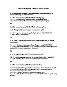
Table 3 Proposed diagnostic criteria for neuromyelitis optica (NMO) Definite NMO Optic neuritis Acute myelitis At least two of three supportive criteria 1. Contiguous spinal cord MRI lesion extending over ⱖ3 vertebral segments 2. Brain MRI not meeting diagnostic criteria for multiple sclerosis 3. NMO-IgG seropositive status
cluded one or both of CSF abnormalities or severe weakness, variables that were used to support NMO diagnosis in the 1999 diagnostic criteria. Although we confirmed that these features are more suggestive of NMO than MS, the best model (one of the three supportive criteria plus either severe, fixed, post-attack motor weakness or CSF pleocytosis ⬎50 WBC/mm3 or neutrophils ⬎5/mm3) achieved only 87% sensitivity and 85% specificity.
Discussion. We propose revised NMO diagnostic criteria that remove the absolute restriction on CNS involvement beyond the optic nerves and spinal cord and emphasize the specificity of longitudinally extensive spinal cord lesions and NMO-IgG seropositivity (table 3). Although brain MRI findings are generally either negative or nonspecific in NMO, brain lesions do not preclude the diagnosis.9,24 CSF pleocytosis or neutrophilia and the occurrence of severe, fixed, attack-related motor weakness were also validated as characteristic features of NMO but with less diagnostic power. A history of systemic autoimmunity or presence of non-organ-specific autoantibodies was common in both NMO and MS but did not distinguish them. Our results provide quantitative data to support clinical NMO diagnostic criteria and are the first to incorporate the NMO-IgG biomarker. Derivation or revision of valid diagnostic criteria in the absence of a pathologic or quantitative reference standard poses a difficult challenge. Ideally, the reference standard and the diagnostic test are evaluated in every subject independently and in blinded fashion. In our study, although we used 1999 Mayo Clinic NMO criteria as a guideline, we did not formally apply them to establish the reference standard. Therefore, we believe that our approach of evaluating each individual criterion and then constructing new combinations with optimal diagnostic properties is valid. The database allows maintenance of independence among the reference standard diagnosis, individual criteria, and the NMO-IgG result. Our results highlight the difficulties inherent in using arbitrary and subjective clinical criteria for diagnostic purposes. The tradition of excluding NMO as a diagnostic possibility in a patient who has experienced any extra-optic-spinal neurologic symptoms is no longer valid. Continued use of this arbitrary requirement will undoubtedly provide a pure cohort but precludes a valid and complete assessment of the spectrum of NMO. The concept of pure NMO should 1488 NEUROLOGY 66
May (2 of 2) 2006
be abandoned. Our data demonstrate that a wide variety of neurologic symptoms may precede or accompany NMO and may or may not be associated with an identifiable CNS lesion. The revisions we propose improve the diagnostic properties of NMO criteria. It is imperative, however, that individual components be ascertained appropriately. Brain MRI results at disease onset must be reviewed if follow-up scans reveal lesions that meet MS criteria. We used older (Paty) MRI diagnostic criteria for MS18 rather than those in current use25 for purposes of consistency. However, because newer criteria are designed to enhance specificity for MS, failure to meet the more sensitive Paty criteria26 should more likely yield a true negative result. The spinal cord MRI manifestation of a longitudinally extensive lesion is the single most useful diagnostic test but is also subject to timing issues, since a lengthy T2-weighted lesion may not have developed fully in the first few days after clinical symptom onset or it may have contracted or resolved with time. Some degree of redundancy and flexibility in the diagnostic criteria, such as the minimum requirement of only two of three supportive criteria, is therefore most practical for clinical use and we have demonstrated equivalent diagnostic properties with this model. The onset brain MRI and the initial spinal cord MRI are available after the presentation of the first myelitis event. Since diagnosis requires only two of three supportive criteria, access to NMO-IgG testing is not necessary to use this system. Some of the difficulties noted above may be eliminated if additional biomarkers can be identified for NMO. The NMO-IgG autoantibody was 76% sensitive and 94% specific for a final clinical diagnosis of NMO. This is a powerful and clinically meaningful result since this cohort represents patients with optic-spinal disease, not other typical forms of MS, and the determination of whether a patient has NMO or MS may be difficult. The autoantigen to which NMO-IgG binds was recently shown to be aquaporin-4,14 the principal water channel involved in fluid homeostasis in the CNS.27 The involvement of aquaporin-4 in the pathogenesis of NMO has not yet been investigated. However, the specificity of the antibody as a marker for NMO and its immunoreactive sites in the spinal cord (abluminal surface of blood vessels and astrocytic foot processes),28 where pathology occurs in NMO,13 is consistent with it being a primary effector of disease rather than a secondary or nonspecific phenomenon. We derived our data from a group of patients who had already experienced both optic neuritis and acute myelitis. However, the biomarker NMO-IgG is proving to be predictive of NMO development after a first event of longitudinally extensive idiopathic acute transverse myelitis.16 Thus our newly proposed criteria will likely require further revision to include disorders that represent inaugural symptoms of NMO or limited NMO variants, including recurrent myelitis associated with negative brain MRI, recur-
rent isolated optic neuritis, or isolated optic neuritis or myelitis presentations associated with NMO-IgG seropositivity. Use of these criteria, and future refinements that allow earlier diagnosis, is also of therapeutic importance. Although existing reports include only small open-label experience and no randomized controlled trials, the generally accepted approach for attack prevention in NMO is immunosuppression using therapies that reduce serum autoantibody levels6,29,30 rather than immunomodulation with currently approved MS therapies.31 We believe that the revised diagnostic criteria we propose represent an important advance in NMO research and clinical practice. The criteria for definite NMO diagnosis are simple, practical, and have excellent diagnostic accuracy. They discriminate NMO from MS beginning with optic neuritis and myelitis, a scenario in which NMO is a reasonable initial diagnostic consideration. Further validation and refinement of these diagnostic criteria, application to individuals of different ethnic and racial backgrounds in different countries and clinical settings, and continued evaluation of NMO-IgG and future biomarkers are necessary next steps in advancing the diagnosis and reducing the morbidity and mortality of this often devastating disorder.
References 1. O’Riordan JI, Gallagher HL, Thompson AJ, et al. Clinical, CSF, and MRI findings in Devic’s neuromyelitis optica. J Neurol Neurosurg Psychiatry 1996;60:382–387. 2. Cree BA, Goodin DS, Hauser SL. Neuromyelitis optica. Semin Neurol 2002;22:105–122. 3. Mandler RN, Davis LE, Jeffery DR, Kornfeld M. Devic’s neuromyelitis optica: a clinicopathological study of 8 patients. Ann Neurol 1993;34: 162–168. 4. de Seze J. Neuromyelitis optica. Arch Neurol 2003;60:1336–1338. 5. Wingerchuk DM, Hogancamp WF, O’Brien PC, Weinshenker BG. The clinical course of neuromyelitis optica (Devic’s syndrome). Neurology 1999;53:1107–1114. 6. Wingerchuk DM, Weinshenker BG. Neuromyelitis optica. Curr Treat Options Neurol 2005;7:173–182. 7. Zaffaroni M. Cerebrospinal fluid findings in Devic’s neuromyelitis optica. Neurol Sci 2004;25 Suppl 4:S368–370. 8. Ghezzi A, Bergamaschi R, Martinelli V, et al. Clinical characteristics, course and prognosis of relapsing Devic’s Neuromyelitis Optica. J Neurol 2004;251:47–52. 9. de Seze J, Lebrun C, Stojkovic T, Ferriby D, Chatel M, Vermersch P. Is Devic’s neuromyelitis optica a separate disease? A comparative study with multiple sclerosis. Mult Scler 2003;9:521–525.
10. Papais-Alvarenga RM, Miranda-Santos CM, Puccioni-Sohler M, et al. Optic neuromyelitis syndrome in Brazilian patients. J Neurol Neurosurg Psychiatry 2002;73:429–435. 11. Poppe AY, Lapierre Y, Melancon D, et al. Neuromyelitis optica with hypothalamic involvement. Mult Scler 2005;11:617–621. 12. Weinshenker BG. Neuromyelitis optica: what it is and what it might be. Lancet 2003;361:889–890. 13. Lucchinetti CF, Mandler RN, McGavern D, et al. A role for humoral mechanisms in the pathogenesis of Devic’s neuromyelitis optica. Brain 2002;125:1450–1461. 14. Lennon VA, Kryzer TJ, Pittock SJ, Verkman AS, Hinson SR. IgG marker of optic-spinal multiple sclerosis binds to the aquaporin-4 water channel. J Exp Med 2005;202:473–477. 15. Lennon VA, Wingerchuk DM, Kryzer TJ, et al. A serum autoantibody marker of neuromyelitis optica: distinction from multiple sclerosis. Lancet 2004;364:2106–2112. 16. Weinshenker BG, Wingerchuk DM, Vukusic S, et al. Neuromyelitis optica IgG predicts relapse after longitudinally extensive transverse myelitis. Ann Neurol 2006;59:566 –569. 17. Yu Z, Kryzer TJ, Griesmann GE, Kim K, Benarroch EE, Lennon VA. CRMP-5 neuronal autoantibody: marker of lung cancer and thymomarelated autoimmunity. Ann Neurol 2001;49:146–154. 18. Paty DW, Oger JJ, Kastrukoff LF, et al. MRI in the diagnosis of MS: a prospective study with comparison of clinical evaluation, evoked potentials, oligoclonal banding, and CT. Neurology 1988;38:180–185. 19. Deeks JJ, Altman DG. Diagnostic tests 4: likelihood ratios. BMJ 2004; 329:168–169. 20. Sackett DL, Haynes RB, Guyatt GH, Tugwell P. Clinical epidemiology: a basic science for clinical medicine. 2nd ed. Boston, MA: Little, Brown; 1991. 21. Beck JR, Shultz EK. The use of relative operating characteristic (ROC) curves in test performance evaluation. Arch Pathol Lab Med 1986;110: 13–20. 22. Centor RM, Schwartz JS. An evaluation of methods for estimating the area under the receiver operating characteristic (ROC) curve. Med Decis Making 1985;5:149–156. 23. Hosmer DW, Lemeshow S. Applied logistic regression. 2nd ed. New York: John Wiley & Sons; 2000. 24. Pittock SJ, Lennon VA, Krecke K, Wingerchuk DM, Lucchinetti CF, Weinshenker BG. Brain MRI abnormalities in neuromyelitis optica. Arch Neurol 2006;63:390 –396. 25. McDonald WI, Compston A, Edan G, et al. Recommended diagnostic criteria for multiple sclerosis: guidelines from the International Panel on the diagnosis of multiple sclerosis. Ann Neurol 2001;50:121–127. 26. Tintore M, Rovira A, Brieva L, et al. Isolated demyelinating syndromes: comparison of CSF oligoclonal bands and different MR imaging criteria to predict conversion to CDMS. Mult Scler 2001;7:359–363. 27. Agre P, Kozono D. Aquaporin water channels: molecular mechanisms for human diseases. FEBS Lett 2003;555:72–78. 28. Nielsen S, Nagelhus EA, Amiry-Moghaddam M, Bourque C, Agre P, Ottersen OP. Specialized membrane domains for water transport in glial cells: high-resolution immunogold cytochemistry of aquaporin-4 in rat brain. J Neurosci 1997;17:171–180. 29. Mandler RN, Ahmed W, Dencoff JE. Devic’s neuromyelitis optica: a prospective study of seven patients treated with prednisone and azathioprine. Neurology 1998;51:1219–1220. 30. Cree BA, Lamb S, Morgan K, Chen A, Waubant E, Genain C. An open label study of the effects of rituximab in neuromyelitis optica. Neurology 2005;64:1270–1272. 31. Goodin DS, Frohman EM, Garmany GP Jr., et al. Disease modifying therapies in multiple sclerosis: report of the Therapeutics and Technology Assessment Subcommittee of the American Academy of Neurology and the MS Council for Clinical Practice Guidelines. Neurology 2002; 58:169–178.
2006 ANNUAL MEETING PROGRAMS AVAILABLE The 2006 Syllabi on CD-ROM includes more than 170 Educational Programs from the AAN Annual Meeting in San Diego. If you missed the meeting, this is a convenient way to catch up on the latest science and education. Make sure you visit the AAN Annual Meeting Online Library, a centralized location featuring more than 220 hours of content, including six Plenary sessions and 58 programs available in various multimedia formats, including Web casts and MP3. For more information, visit www.aan.com/amprograms.
May (2 of 2) 2006
NEUROLOGY 66
1489