The New England Journal of Medicine
Review Article
Medical Progress
AORTIC STENOSIS Recognition
V ALVULAR H EART D ISEASE BLASE A. CARABELLO, M.D., FRED A. CRAWFORD, JR., M.D.
AND
T
HE past 15 years have brought a remarkable improvement in the clinical outcome of patients with valvular heart disease. It is impossible to attribute the change to any single advance in the field. However, it is likely that more effective noninvasive monitoring of ventricular function, improvement in prosthetic valves, advances in valvereconstruction techniques, and the development of useful guidelines for choosing the proper timing of surgical intervention have all worked in concert to improve prognosis. Moreover, advances in minimally invasive surgical techniques may make valve procedures more easily tolerated by the patient.1 All valvular heart diseases place a hemodynamic burden on the left or right ventricle, or on both ventricles, which is initially tolerated as the cardiovascular system compensates for the overload. However, hemodynamic overload eventually leads to muscle dysfunction and congestive heart failure, and sometimes sudden death. Two major questions must be answered in the management of every case of valvular heart disease: Is the valvular disease severe enough to cause morbidity or mortality for which mechanical intervention would be beneficial? And if the answer to this question is yes, what are the best medical therapy and the best time for surgical intervention to minimize or eliminate morbidity and mortality? We will discuss these questions for the four major acquired left-sided valvular lesions: aortic stenosis, mitral stenosis, nonischemic mitral regurgitation, and aortic regurgitation.
From the Cardiology Division, Department of Medicine (B.A.C.), the Gazes Cardiac Research Institute (B.A.C.), and the Division of Cardiothoracic Surgery, Department of Surgery (F.A.C.), Medical University of South Carolina; and the Ralph H. Johnson Department of Veterans Affairs (B.A.C.) — both in Charleston, S.C. Address reprint requests to Dr. Carabello at the Cardiology Division, Medical University of South Carolina, 171 Ashley Ave., Charleston, SC 29425-2221. ©1997, Massachusetts Medical Society.
32 �
July 3 , 1 9 9 7
Acquired aortic stenosis is usually an idiopathic disease resulting from degeneration and calcification of the aortic leaflets.2,3 Stenosis is more likely to occur in persons born with bicuspid aortic valves than in those with normal tricuspid valves, and it develops earlier, in the fourth and fifth decades of life, in such persons. When the disease is acquired in previously normal tricuspid aortic valves, stenosis develops in the sixth, seventh, and eighth decades. Why some persons are afflicted by this disease whereas others are spared is unknown, but it shares some of the pathologic features of coronary atherosclerosis and is associated with some of the same risk factors, such as hypertension and hypercholesterolemia.4 The classic symptoms of aortic stenosis are angina, syncope, and the symptoms of congestive heart failure. Angina develops in aortic stenosis in part because of reduced coronary flow reserve and in part because of increased myocardial oxygen demand caused by high afterload.5-7 The origin of exertional syncope in aortic stenosis remains controversial. According to one theory, an exercise-induced decrease in total peripheral resistance is uncompensated because cardiac output is restricted by the stenotic valve.8 Another possible mechanism is the precipitation of a vasodepressor response.9 Heart failure in aortic stenosis can be caused by diastolic dysfunction, systolic dysfunction, or both. Diastolic dysfunction results from increased left-ventricular-wall thickness and increased collagen content.10 Systolic dysfunction results from excess afterload, decreased contractility, or a combination of these factors.7 The most common sign of aortic stenosis is a systolic ejection murmur radiating to the neck. The murmur is usually heard best in the aortic area. It often disappears over the sternum and then reappears in the apical area, mimicking mitral regurgitation (Gallivardin’s phenomenon). In mild aortic stenosis, the murmur usually peaks early in systole, it is often associated with a thrill, and the carotid upstrokes are well preserved. As the severity of stenosis increases, the murmur peaks progressively later in systole and may become softer as cardiac output diminishes. The carotid upstrokes classically become diminished in amplitude and delayed in time (parvus et tardus). The second heart sound may become single as the aortic closing component is lost, or S2 may become paradoxically split because of delay in left ventricular emptying.
MED ICA L PROGR ES S
Assessment of Severity
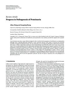
Although a reasonable estimate of the severity of aortic stenosis can be made during physical examination, echocardiography with Doppler examination of the aortic valve now provides a more accurate assessment of the transvalvular gradient and the area of the aortic valve. Since flow is the product of crosssectional area and bloodstream velocity, as the bloodstream reaches a narrowing (Fig. 1), velocity must increase for flow to remain constant.11 This increase in velocity, detected by the Doppler technique, can be translated into a gradient (four times the velocity squared) that accurately mirrors the gradient assessed by direct pressure measurement,12 or the velocity can be used in the continuity equation to estimate aortic-valve area (Fig. 1). Echocardiography is also useful in assessing the extent of left ventricular hypertrophy and in estimating left ventricular ejection performance. Cardiac Catheterization and Coronary Angiography
The severity of aortic stenosis can usually be gauged accurately by noninvasive techniques. However, because patients with aortic stenosis are often elderly and therefore at risk for coronary disease, coronary angiography is usually performed before valve replacement, especially in patients with angina.13,14 Although initial attempts to detect coronary artery disease in patients with aortic stenosis by noninvasive techniques appear fruitful, further study will be required before this practice is adopted as routine.15,16 In cases where the severity of aortic stenosis cannot be determined by noninvasive testing, invasive measurement of the pressure gradient and determination of cardiac output are performed to derive data from which to calculate the area of the aortic-valve orifice.17 Timing of Surgery
Except for prophylaxis against endocarditis, there is no proved medical therapy for aortic stenosis. The only effective relief of this mechanical obstruction to blood flow is aortic-valve replacement.18 Although the noninvasive assessment of the severity of aortic stenosis is quite accurate and thus useful in determining the timing of surgery, it is not severity alone that determines the timing of aortic-valve replacement. Rather, the decision to replace the aortic valve is based on the presence of the classic symptoms of aortic stenosis (noted above) along with a severely stenotic valve. As shown in Figure 2, the survival of patients with aortic stenosis is nearly normal until the onset of symptoms, when a precipitous decrease in survival occurs.19 The prognostic importance of symptoms in an early study has been corroborated by more recent
V1
A2
V2
A1 � V 1 � A 2 � V2 Figure 1. Principles of the Use of Doppler Ultrasonography and the Continuity Equation in Estimating Aortic-Valve Area. For blood flow (A1 � V1) to remain constant when it reaches a stenosis (A2), velocity must increase to V2. Doppler examination of the stenosis detects the increase in velocity, which can be used to calculate the aortic-valve gradient or to solve the continuity equation for A2. A denotes area, and V velocity.
Onset of severe symptoms
100 Latent period
Survival (%)
Echocardiography
A1
Angina Syncope Failure
80 60
0
40 20
6 2 4 Average survival (yr)
Average age at death
0 40
50
60
70
80
Age (yr) Figure 2. Natural History of Aortic Stenosis. There is a long latent period of increasing obstruction and myocardial overload, during which the asymptomatic patient has a normal life span. However, once angina, syncope, or heart failure develops, survival is greatly reduced. If the aortic valve is not replaced, approximately 50 percent of patients will be dead within five years after angina develops, 50 percent will be dead within three years after syncope develops, and 50 percent will be dead within only two years after heart failure develops. Adapted from Ross and Braunwald,19 with the permission of the publisher.
studies in which Doppler echocardiography established the severity of stenosis in initially asymptomatic patients.20,21 About 75 percent of patients with symptomatic aortic stenosis will be dead three years after the onset of symptoms unless the aortic valve is replaced.22 Typically, a gradient of more than 50 mm Hg or a valve area of less than 0.8 cm2 indicates critical stenosis that is capable of causing symptoms and death. However, there are exceptions to these rules. Some patients with larger valve areas or smaller gradients have symptoms that are clearly due to aortic stenosis. Such patients will benefit from aortic-valve replacement. Age
It should be emphasized that even though most patients with aortic stenosis are elderly, the prognosis with surgery, even in an octogenarian, is excellent Vol ume 337
Numbe r 1
�
33
The New England Journal of Medicine
in the absence of coexisting illnesses.23-25 Thus, age is not a contraindication to surgery in such patients. Furthermore, the age-corrected survival after aorticvalve replacement in patients older than 65 is not different from that in the general population.26 Balloon Aortic Valvotomy
Balloon aortic valvotomy for adult acquired aortic stenosis is useful only for palliation of the disease. Although the procedure was initially greeted with enthusiasm, interest in balloon aortic valvotomy has waned in recent years. The rate of serious complications, including death, stroke, aortic rupture, aortic regurgitation, and vascular injury, exceeds 10 percent.27 Furthermore, the mortality after this procedure is 60 percent at 18 months, a rate similar to that in an untreated population.28 The event-free survival at two years is only 20 percent, and many patients thought to be candidates only for balloon valvotomy eventually have a good outcome with aortic-valve replacement.28,29 Although the procedure is not lifesaving, it may be useful in alleviating symptoms in patients who are clearly not candidates for aortic-valve replacement because of other medical problems. Some practitioners have successfully used balloon valvotomy as a bridge to aortic-valve replacement in very sick patients. However, no controlled data are available to prove this approach superior to direct aortic-valve replacement. Stress Testing
Although the presence or absence of symptoms is the key factor in the management of aortic stenosis, in some patients who have vague symptoms or for whom a reliable history is difficult to obtain, exercise testing may be useful to establish the symptomatic state more objectively. However, this procedure is attended by increased risk in patients with aortic stenosis, and it is ill-advised to perform exercise testing in patients who are symptomatic.30 The test can be performed safely in asymptomatic patients or those with vague symptoms if great caution is used and a physician is present.31-33 In such patients, exercise testing may provide additional information on which to base clinical decision making.
with a low ejection fraction and a low transvalvular gradient. Studies of this subgroup of patients have found high operative mortality and persistence of symptoms after surgery in many cases.34,37,38 However, it is clear that other patients in this group improve after surgery.38 The criteria for deciding which patients in this group should have aortic-valve replacement continue to evolve. Initial studies indicate that patients in this group may have a favorable surgical outcome if both the cardiac output and the transvalvular gradient are increased by either inotropic stimulation or the administration of nitroprusside.39,40 Patients in whom inotropic stimulation augments cardiac output but not the transvalvular gradient form a group with milder stenosis that was not the primary cause of left ventricular dysfunction. Such patients are unlikely to benefit from valve replacement. Women with Aortic Stenosis
Recently, differences in left ventricular geometry between men and women with aortic stenosis have been recognized.41-43 Women with aortic stenosis are likely to have thicker ventricular walls, which reduces wall stress (which, according to the Laplace equation, is calculated as the product of the left ventricular pressure and the radius, divided by two times the wall thickness), and higher ejection fractions. Preoperative recognition of these differences is important, because postoperative management of low cardiac output requires volume expansion rather than the use of pressor agents.44 In summary, aortic stenosis is a disease of aging that is likely to become more prevalent as the proportion of older people in our population increases. Once stenosis has been identified during physical examination, its severity can be accurately quantified by Doppler echocardiography. Asymptomatic patients with aortic stenosis are followed medically until the onset of one of the classic symptoms of aortic stenosis, at which time the aortic valve is replaced. Aortic-valve replacement is successful in most elderly patients and even in many patients with advanced heart failure. MITRAL STENOSIS
Patients with Congestive Heart Failure and Reduced Systolic Performance
Recognition and Assessment of Severity
For most patients with advanced congestive heart failure, even those with a marked reduction in the ejection fraction, aortic-valve replacement provides remarkable relief of symptoms and improved ejection performance, because relieving the obstruction to outflow reduces left ventricular afterload and might also eventually lead to the restoration of contractile function.34-36 However, a persistently problematic group of patients with aortic stenosis is the group
Mitral stenosis is a sequela of rheumatic heart disease that primarily affects women. Unfortunately, a reliable history of rheumatic fever is often difficult to obtain and thus cannot usually be used as a guide to the likely presence or absence of this disease. In developed countries, the steady decline in the incidence of rheumatic fever has reduced the incidence of mitral stenosis. Both rheumatic fever and mitral stenosis remain common in developing nations.
34 �
July 3 , 1 9 9 7
MED ICA L PROGR ES S
Patients with mitral stenosis usually have symptoms typical of left-sided heart failure: dyspnea on exertion, orthopnea, and paroxysmal nocturnal dyspnea. Less frequently, they have hemoptysis, hoarseness, and symptoms of right-sided heart failure; these symptoms are somewhat more specific for mitral stenosis. Often the patient remains asymptomatic until she becomes pregnant or has atrial fibrillation, when dyspnea and orthopnea are noted. The symptoms of mitral stenosis stem from increased left atrial pressure and reduced cardiac output, primarily caused by mechanical obstruction of filling of the left ventricle. Although the symptoms are those of left ventricular failure, contractility of the left ventricle is usually normal in mitral stenosis.45 However, in some cases, the left ventricular ejection fraction is reduced because of excessive afterload secondary to a reflexive increase in systemic vascular resistance. Since it is the right ventricle that ultimately bears the burden of propelling blood through the mitral valve, right ventricular function is compromised first by the afterload imposed on it by high left atrial pressure and then by the development of secondary pulmonary vasoconstriction. During physical examination, mitral stenosis is suspected because of the presence of the classic diastolic rumble that follows an opening snap. S1 is characteristically loud, because the mitral valve is held open by the transmitral gradient until the force of ventricular systole closes the valve. The presence of a loud P2, right ventricular lift, elevated neck veins, ascites, and edema indicates that pulmonary hypertension producing right ventricular overload has developed. This is an ominous sign in the progression of the disease, because pulmonary hypertension increases the risk associated with surgery.46 Echocardiography is the premier noninvasive diagnostic tool for assessing the severity of mitral stenosis and for judging the applicability of balloon mitral valvotomy. Echocardiography can usually permit an accurate planimeteric calculation of valve area47 and can also be used to assess the severity of stenosis by measuring the decay of the transvalvular gradient or the “pressure half-time,” an empirical measurement.48,49 The latter determination is based on the principle that as the severity of stenosis worsens, it takes progressively longer for the transmitral flow velocity to decay. By empirically dividing the constant of 220 by the pressure half-time, one can make an approximation of valve area.48 Therapy and Timing of Intervention
For the asymptomatic patient in sinus rhythm, prophylaxis against endocarditis is the only medical therapy indicated. When mild symptoms develop, diuretics are usually effective in lowering left atrial pressure and reducing symptoms. If atrial fibrillation develops, rate control with digoxin, a beta-blocker,
or a calcium-channel blocker is crucial, since a rapid heart rate further impairs left ventricular filling, simultaneously reducing cardiac output and increasing left atrial pressure. Anticoagulant therapy is required, since there is a high risk of embolism in patients with chronic atrial fibrillation and mitral stenosis. If the symptoms are more than mild, or if there is evidence that pulmonary hypertension is beginning to develop, mechanical relief of the mitral stenosis is indicated, since further delay worsens the prognosis.50 In many cases, balloon valvotomy provides excellent mechanical relief that usually results in prolonged benefit, unlike valvotomy in aortic stenosis.51 The presence of heavy valvular calcification, severe subvalvular distortion, or more than mild mitral regurgitation militates against the use of balloon valvotomy.52 In such cases, open commissurotomy, valve reconstruction, or mitral-valve replacement improves survival and reduces symptoms. Still unresolved is the proper treatment of patients with mitral stenosis who are asymptomatic except for the presence of atrial fibrillation. This unwanted arrhythmia is associated with extensive morbidity and mortality.53 It is often hoped that correction of mitral stenosis before the atrial fibrillation has become prolonged (and thus more likely to be permanent) will allow the reestablishment of sinus rhythm after valvotomy or surgery. However, there is currently no conclusive evidence that this management strategy is successful. Indeed, some now advocate a combination of the appropriate mitral-valve procedure and the Cox maze procedure in appropriate patients to ensure the maintenance of sinus rhythm postoperatively.54-56 NONISCHEMIC MITRAL REGURGITATION Recognition and Assessment of Severity
The usual causes of mitral regurgitation are infective endocarditis, myxomatous degeneration of the mitral valve (including the mitral valve prolapse syndrome), collagen vascular disease, spontaneous rupture of the chordae tendineae, and rheumatic fever. Figure 3 depicts the pathophysiologic stages of mitral regurgitation, progressing from acute mitral regurgitation to chronic compensated mitral regurgitation and to chronic decompensated mitral regurgitation.57 Chronic mitral regurgitation is compensated by the development of eccentric cardiac hypertrophy, and cardiac enlargement should therefore be manifest on physical examination. A holosystolic apical murmur heard on physical examination alerts the examiner that mitral regurgitation is present. An S3 suggests that the disease is severe. However, an S3 heard in mitral regurgitation does not necessarily indicate the presence of congestive heart failure, since in this situation the sound is caused by rapid filling Vol ume 337
Numbe r 1
�
35
The New England Journal of Medicine
A
B
Normal 100 ml
LA, 10 mm Hg
Acute mitral regurgitation
70 ml
95 ml
LA, 15 mm Hg EDV, 170 ml
95 ml
LA, EDV, 25 mm Hg 240 ml
PRE- AFTERLOAD LOAD ESS
mm
kdyn/ cm 2
EF
RF FSV
90
N
0.67 0.00 100
AMR
60
N
0.82 0.50
2.25
STAGE
SL
ESS
mm
kdyn/ cm 2
2.25
60
CCMR 2.19
90
ml
Normal 2.07
70
EDV, 260 ml 85 ml ESV, 110 ml
PRE- AFTERLOAD LOAD CF
65 ml
Chronic decompensated mitral regurgitation
ESV, 50 ml
ESV, 30 ml
SL
ESV, 50 ml
ESV, 30 ml
95 ml
70 ml
STAGE
EDV, 240 ml
Chronic compensated mitral regurgitation
Acute mitral regurgitation LA, 25 mm Hg
LA, 15 mm Hg
EDV, 170 ml 70 ml
ESV, 50 ml
C
95 ml
70 ml
LA, EDV, 25 mm Hg 150 ml
Chronic compensated mitral regurgitation
AMR
PRE- AFTERLOAD LOAD CF
SL
ESS
ml
mm
kdyn/ cm 2
N
0.82 0.50 70
CCMR 2.19
90
N
0.79 0.50 95
CDMR 2.19
120
EF
RF FSV
STAGE
CF
EF
RF FSV ml
N
0.79 0.50 95 0.58 0.57 65
Figure 3. Pathophysiologic Stages of Mitral Regurgitation. Panel A shows the transition from normal physiology to acute mitral regurgitation (AMR). The volume overload of acute mitral regurgitation increases preload sarcomere length (SL) so that end-diastolic volume (EDV) increases from 150 to 170 ml. The presence of a new pathway for the ejection of blood into the left atrium (LA) reduces afterload, described as end-systolic stress (ESS), and therefore end-systolic volume (ESV) is reduced from 50 to 30 ml. The ejection fraction (EF) increases acutely, but because 50 percent of the total stroke volume is regurgitated into the left atrium, resulting in a regurgitant fraction (RF) of 0.50, forward stroke volume (FSV) is reduced from 100 to 70 ml. The increased volume in the left atrium raises pressure there from normal to 25 mm Hg. Panel B shows the transition from acute mitral regurgitation to chronic compensated mitral regurgitation (CCMR). The development of eccentric hypertrophy has increased end-diastolic volume from 170 to 240 ml. The now larger ventricle has an increase in afterload because the radius applied in the Laplace equation for stress has increased. This in turn increases end-systolic volume to normal. The presence of eccentric hypertrophy, however, allows for an increase in total stroke volume as well as forward stroke volume. Enlargement of the left atrium allows the volume overload there to be accommodated at a lower filling pressure (15 mm Hg). The ejection fraction is supernormal. Panel C shows the transition to chronic decompensated mitral regurgitation (CDMR). The now weakened ventricle can no longer contract well, and end-systolic volume therefore increases from 50 to 110 ml. Forward stroke volume is reduced, and cardiac dilatation leads to an increased regurgitant fraction. However, the still favorable loading conditions permit the ejection fraction to remain normal (0.58). CF denotes contractile function; N, normal; and the downward arrow, depressed. Reproduced from Carabello,57 with the permission of the publisher.
of the left ventricle by the large volume of blood stored in the left atrium in diastole. Echocardiography confirms enlargement of the chamber, and colorflow examination of the mitral valve establishes the pattern of disturbed flow caused by regurgitation across the mitral valve. Although a variety of methods of quantifying the severity of regurgitation have been used, none have met with universal success. Currently, echocardiography provides only a semiquantitative estimate of the severity of mitral regurgitation. Left ventriculography 36 �
July 3 , 1 9 9 7
performed during cardiac catheterization provides an additional but also imperfect estimate of the severity of mitral regurgitation. However, catheterization is used only when surgery is being contemplated, and it is not suitable for longitudinal follow-up. Timing of Surgery
Unlike the stenotic lesions, regurgitant lesions may progress insidiously, causing left ventricular damage before symptoms have developed.58 Thus, although the presence of symptoms in chronic mitral regurgi-
MED ICA L PROGR ES S
tation usually indicates disordered physiology and the need for valve surgery, surgery should also be performed if asymptomatic left ventricular dysfunction has begun to develop. The loading conditions in mitral regurgitation are favorable to left ventricular ejection; preload is increased whereas afterload is normal or occasionally decreased, and thus the lesion itself facilitates left ventricular emptying.57 Therefore, in the presence of normal muscle function, the ejection fraction should be supernormal in the patient with mitral regurgitation.59 Once the ejection fraction falls below 60 percent, the prognosis worsens.60 Left ventricular performance can also be gauged in mitral regurgitation by assessing the diameter to which the left ventricle can contract at the end of systole. End-systolic dimension is less dependent on preload than is ejection fraction and can be used as another measure of left ventricular contractile function.61 When the end-systolic dimension exceeds 45 mm, the prognosis worsens.62-64 Thus, patients should be referred for surgery if more than mild symptoms develop, or if the ejection fraction falls toward 60 percent or the end-systolic dimension approaches 45 mm, even in the absence of symptoms. Hochreiter et al.65 demonstrated a worsened prognosis if right ventricular function is reduced, emphasizing the prognostic role of pulmonary hypertension in this disease. Patients with a right ventricular ejection fraction of less than 30 percent are at especially high risk. Importance of the Mitral-Valve Apparatus
Although the importance of the mitral-valve apparatus was described decades ago,66 its role in sustaining left ventricular function has become almost universally recognized only recently. Mitral-valve repair has a lower operative mortality and a better late outcome67 than mitral-valve replacement. Thus, mitral-valve repair rather than replacement should be performed whenever possible.68-70 Even when the mitral valve must be replaced because of extensive degeneration of the valve, an attempt should be made to conserve the chordal structures and their connections. In the past, when standard replacement of the mitral valve involved destruction of the chordal apparatus, the ejection fraction almost always fell after the operation. Currently, however, the ejection fraction is usually maintained at its preoperative level when the chordal apparatus is preserved in either repair or replacement of the mitral valve.69,71,72 Repair rather than replacement also obviates the need for anticoagulant therapy in patients in sinus rhythm and avoids possible failure of the prosthetic valve. With these improved surgical techniques, postoperative survival after well-timed mitral-valve surgery now approaches that of the general population, as it does for patients with aortic or mitral stenosis.60 It
should be noted, however, that whereas in aortic stenosis, age alone is not a contraindication to surgery, in mitral stenosis patients more than 75 years of age have a worse prognosis after surgery than younger patients, especially if mitral-valve replacement rather than repair has been performed or if coronary disease is present.73 Medical Therapy
Although vasodilators are successfully used to increase forward output and decrease left ventricular filling pressure in patients with acute mitral regurgitation, there is currently no apparent benefit to longterm use, especially in asymptomatic patients.74 Although such benefit might be possible, no long-term, large studies have demonstrated that the use of vasodilators safely reduces or delays the need for surgery or improves outcome. AORTIC REGURGITATION
Aortic regurgitation results from disease of either the aortic leaflets or the aortic root that distorts the leaflets to prevent their coaptation. Common causes of leaflet abnormalities that result in aortic regurgitation include infective endocarditis and rheumatic fever. Aortic-root causes of aortic regurgitation include annuloaortic ectasia (idiopathic root dilatation associated with hypertension and aging), Marfan’s syndrome, aortic dissection, collagen vascular disease, and syphilis. In chronic aortic regurgitation, left ventricular enlargement produces a large total stroke volume that is entirely ejected into the aorta. In contrast, in mitral regurgitation the regurgitant volume enters the left atrium. Increased stroke volume increases pulse pressure, causing systolic hypertension, which imposes increased afterload on the left ventricle. Indeed, afterload can be as high in aortic regurgitation as it is in aortic stenosis.75,76 Recognition and Assessment of Severity
The large total stroke volume in aortic regurgitation increases pulse pressure, which produces a myriad of clinical signs. Although the typical diastolic blowing murmur heard along the left sternal border is the usual sign of aortic regurgitation, the peripheral signs of a hyperdynamic circulation often indicate that the disease is severe. A partial list of these signs includes Quincke’s pulse (systolic plethora and diastolic blanching in the nail bed when gentle pressure is placed on it), Corrigan’s pulse (a bounding, full carotid pulse with a rapid downstroke), Musset’s sign (head bobbing), and Hill’s sign (systolic blood pressure in the leg at least 30 mm Hg higher than that in the arm). In addition to the typical murmur of aortic insufficiency, a diastolic rumble (Austin Flint murmur) may also be heard over the cardiac apex. Although Vol ume 337
Numbe r 1
�
37
The New England Journal of Medicine
its origin is debatable, the Austin Flint murmur is probably produced as the aortic jet impinges on the mitral valve, causing it to vibrate; also, simultaneous diastolic filling of the left ventricle from the left atrium and aorta tends to close the mitral valve in diastole, producing physiologic stenosis. Once aortic regurgitation is suspected on physical examination, echocardiography with Doppler examination of the aortic valve can help estimate its severity. Aortography during catheterization helps confirm the severity of the disease. Therapy Surgical Correction
As with mitral regurgitation, symptoms may not appear until left ventricular dysfunction in aortic insufficiency is well advanced. The symptoms are usually those of left-sided heart failure (dyspnea, orthopnea, and fatigue). Angina may also occur in patients with aortic insufficiency without coronary disease, but less frequently than in patients with aortic stenosis.13 Although aortic insufficiency should be corrected when more than mild symptoms develop, there is compelling evidence that aortic regurgitation should be corrected before the onset of permanent left ventricular damage, even in asymptomatic patients.77-81 As noted above, aortic insufficiency increases left ventricular afterload, in part because the high stroke volume produces a wide pulse pressure and systolic hypertension. After aortic-valve replacement, afterload is reduced and ejection fraction improves. Thus, it is not surprising that patients with aortic insufficiency can have a greater decrease in ejection performance and a larger end-systolic dimension than patients with mitral insufficiency, while still having a good postoperative outcome. In general, the “55 rule” has been useful in gauging the timing of surgery for this disease.77-81 Aortic-valve surgery should be performed before the ejection fraction falls below 55 percent or the endsystolic dimension exceeds 55 mm. The markers for the timing of surgery in mitral regurgitation and aortic insufficiency are shown in Table 1. Although replacement of the aortic valve with a tissue or mechanical prosthesis has been the definitive therapy
TABLE 1. ECHOCARDIOGRAPHIC PREDICTORS OF GOOD OUTCOME IN AORTIC AND MITRAL REGURGITATION. TYPE OF REGURGITATION
END-DIASTOLIC DIMENSION
EJECTION FRACTION
SHORTENING FRACTION
Aortic
�55
�0.55
�0.27
Mitral
�45
�0.60
�0.32
38 �
July 3 , 1 9 9 7
for severe aortic regurgitation, experience with the pulmonary autograft (Ross procedure) and aorticvalve reconstruction is rapidly increasing.82-85 Medical Therapy
Because aortic regurgitation represents a state of excess afterload, it could be anticipated that reduction of afterload with vasodilators would improve left ventricular performance while simultaneously decreasing the amount of aortic regurgitation, thus reducing or delaying the need for surgery. The most compelling evidence supporting this concept is from a study showing that the use of nifedipine in asymptomatic patients with severe aortic regurgitation and normal left ventricular function can delay the need for surgery by two to three years.86 It is likely that other vasodilators will also be efficacious in safely forestalling surgery.87 Acute Aortic Insufficiency Assessment of Severity and Timing of Surgery
Acute severe aortic insufficiency is usually a surgical emergency. The large regurgitant volume suddenly entering the left ventricle, before adaptation to the volume load has developed, increases left ventricular filling pressure, causing acute pulmonary congestion. Severe regurgitation impairs forward cardiac output, thus reducing organ perfusion. Reduced output, in concert with elevated left ventricular filling pressure, probably reduces coronary blood flow, possibly potentiating myocardial ischemia and further left ventricular deterioration. A fact of diagnostic importance is that the large left ventricular stroke volume present in compensated chronic aortic insufficiency is absent in acute aortic insufficiency, because left ventricular enlargement has not yet occurred. Therefore, many of the signs of severe aortic regurgitation discussed above are absent, and the diagnosis is easy to miss.88 Important clues during physical examination include the diastolic blowing murmur of aortic insufficiency and a soft first heart sound. A soft first heart sound occurs because rapid ventricular filling due to aortic insufficiency closes the mitral valve before the onset of systole, and thus S1 is constituted only by the closure sound of the tricuspid valve. Preclosure of the mitral valve, suspected on physical examination and confirmed by echocardiography, is an ominous development, usually indicating the need for urgent surgery.89 Because acute aortic insufficiency is usually caused by infective endocarditis, there is always concern about aortic-valve replacement in the presence of infection. However, in most cases the risk of sudden death from cardiac causes outweighs the relatively small risk (less than 10 percent) of prosthetic-valve infection.90 Most consider the aortic homograft the
MED ICA L PROGR ES S
valve of choice in this situation. Thus, aortic-valve replacement should be contemplated in any patient with acute aortic insufficiency who has evidence of even mild congestive heart failure or mitral-valve preclosure. CORONARY ARTERY DISEASE
The presence of coronary disease in patients with either mitral or aortic valve disease worsens the long-term prognosis.73,91,92 Although the operative risk may not be increased,93 the long-term prognosis in combined coronary and valvular heart disease is not as good as that in valvular disease alone, even when coronary bypass surgery is performed at the time of valve replacement. This is presumably a result of the progressive nature of coronary disease. Ischemic mitral regurgitation carries the worst prognosis: operative mortality is 10 to 20 percent, and longterm survival is substantially lower than with nonischemic mitral regurgitation.94,95 An unresolved issue is the approach to the aortic valve in patients who have mild-to-moderate aortic stenosis and who are undergoing coronary bypass grafting. Although some centers report that patients who undergo later reoperation because of progression to severe aortic stenosis do not have an increased risk of morbidity and mortality,96 others suggest a very high mortality rate.97 Because progression to severe aortic stenosis may occur rapidly,98 it has been suggested that serious consideration should be given to elective valve replacement at the time of the initial bypass operation.97 SUMMARY
The prognosis for patients with valvular heart disease has improved substantially over the past 15 years. A better understanding of the proper timing of surgery is one of the key reasons. In general, surgery for stenotic valvular disease can be delayed until symptoms appear. Conversely, in regurgitant valvular heart disease, prognostically important left ventricular dysfunction may develop in the absence of symptoms, and thus valve surgery for some asymptomatic patients is entirely appropriate. It is likely that in the future there will be progress toward increasing conservation of the patient’s native valve. This will be beneficial because even modern prosthetic valves have inherent risks.99 Acquired aortic stenosis will often continue to require prosthetic aortic-valve replacement. However, valvular disease will increasingly be treated by procedures that conserve native valves. These include pulmonary autografts for aortic stenosis, balloon commissurotomy for mitral stenosis, mitral-valve repair for mitral regurgitation, and aortic-valve repair for aortic regurgitation. These procedures will make surgery more attractive by eliminating the risks associated with prostheses. Thus, continuing advances in noninva-
sive assessment of the aortic and mitral valves, appropriate timing of referral for surgery, improved surgical techniques for valve replacement and reconstruction, and very recent advances in less invasive surgical approaches should combine to improve the outlook for patients with valvular heart disease. REFERENCES 1. Cosgrove DM III, Sabik JF. Minimally invasive approach for aortic valve operations. Ann Thorac Surg 1996;62:596-7. 2. Passik CS, Ackermann DM, Pluth JR, Edwards WD. Temporal changes in the causes of aortic stenosis: a surgical pathologic study of 646 cases. Mayo Clin Proc 1987;62:119-23. 3. Waller B, Howard J, Fess S. Pathology of aortic valve stenosis and pure aortic regurgitation: a clinical morphologic assessment. Clin Cardiol 1994; 17:85-92. 4. Otto CM, Kuusisto J, Reichenbach DD, Gown AM, O’Brien KD. Characterization of the early lesion of ‘degenerative’ valvular aortic stenosis: histological and immunohistochemical studies. Circulation 1994;90:844-53. 5. Marcus ML, Doty DB, Hiratzka LF, Wright CB, Eastham CL. Decreased coronary reserve: a mechanism for angina pectoris in patients with aortic stenosis and normal coronary arteries. N Engl J Med 1982;307: 1362-7. 6. Strauer B-E. Ventricular function and coronary hemodynamics in hypertensive heart disease. Am J Cardiol 1979;44:999-1006. 7. Huber D, Grimm J, Koch R, Krayenbuehl HP. Determinants of ejection performance in aortic stenosis. Circulation 1981;64:126-34. 8. Schwartz LS, Goldfischer J, Sprague GJ, Schwartz SP. Syncope and sudden death in aortic stenosis. Am J Cardiol 1969;23:647-58. 9. Richards AM, Nicholls MG, Ikram H, Hamilton EJ, Richards RD. Syncope in aortic valvular stenosis. Lancet 1984;2:1113-6. 10. Hess OM, Ritter M, Schneider J, Grimm J, Turina M, Krayenbuehl HP. Diastolic stiffness and myocardial structure in aortic valve disease before and after valve replacement. Circulation 1984;69:855-65. 11. Carabello BA. Aortic stenosis. In: Crawford MH, ed. Current diagnosis & treatment in cardiology. Norwalk, Conn.: Appleton & Lange, 1995: 87-98. 12. Currie PJ, Seward JB, Reeder GS, et al. Continuous-wave Doppler echocardiographic assessment of severity of calcific aortic stenosis: a simultaneous Doppler-catheter correlative study in 100 adult patients. Circulation 1985;71:1162-9. 13. Alexopoulos D, Kolovou G, Kyriakidis M, et al. Angina and coronary artery disease in patients with aortic valve disease. Angiology 1993;44:70711. 14. Lund O, Nielsen TT, Pilegaard HK, Magnussen K, Knudsen MA. The influence of coronary artery disease and bypass grafting on early and late survival after valve replacement for aortic stenosis. J Thorac Cardiovasc Surg 1990;100:327-37. 15. Kupari M, Virtanen KS, Turto H, et al. Exclusion of coronary artery disease by exercise thallium-201 tomography in patients with aortic valve stenosis. Am J Cardiol 1992;70:635-40. 16. Kettunen R, Huikuri HV, Heikkilä J, Takkunen JT. Preoperative diagnosis of coronary artery disease in patients with valvular heart disease using technetium-99m isonitrile tomographic imaging together with high-dose dipyridamole and handgrip exercise. Am J Cardiol 1992;69: 1442-5. 17. Gorlin R, Gorlin SG. Hydraulic formula for calculation of the area of the stenotic mitral valve, other cardiac valves, and central circulatory shunts. Am Heart J 1951;41:1-29. 18. Schwarz F, Baumann P, Manthey J, et al. The effect of aortic valve replacement on survival. Circulation 1982;66:1105-10. 19. Ross J Jr, Braunwald E. Aortic stenosis. Circulation 1968;38:Suppl V: V-61–V-67. 20. Kelly TA, Rothbart RM, Cooper CM, Kaiser DL, Smucker ML, Gibson RS. Comparison of outcome of asymptomatic to symptomatic patients older than 20 years of age with valvular aortic stenosis. Am J Cardiol 1988; 61:123-30. 21. Pellikka PA, Nishimura RA, Bailey KR, Tajik AJ. The natural history of adults with asymptomatic, hemodynamically significant aortic stenosis. J Am Coll Cardiol 1990;15:1012-7. 22. O’Keefe JH Jr, Vlietstra RE, Bailey KR, Holmes DR Jr. Natural history of candidates for balloon aortic valvuloplasty. Mayo Clin Proc 1987;62: 986-91. 23. Levinson JR, Akins CW, Buckley MJ, et al. Octogenarians with aortic stenosis: outcome after aortic valve replacement. Circulation 1989;80: Suppl I:I-49–I-56.
Vol ume 337
Numbe r 1
�
39
The New England Journal of Medicine
24. Elayda MA, Hall RJ, Reul RM, et al. Aortic valve replacement in patients 80 years and older: operative risks and long-term results. Circulation 1993;88:Suppl II:II-11–II-16. 25. Culliford AT, Galloway AC, Colvin SB, et al. Aortic valve replacement for aortic stenosis in persons aged 80 years and over. Am J Cardiol 1991; 67:1256-60. 26. Lindblom D, Lindblom U, Qvist J, Lundström H. Long-term relative survival rates after heart valve replacement. J Am Coll Cardiol 1990;15: 566-73. 27. Berman AD, McKay RG, Grossman W. Balloon valvuloplasty. In: Baim DS, Grossman W, eds. Cardiac catheterization, angiography, and intervention. 5th ed. Baltimore: Williams & Wilkins, 1996:659-87. 28. Otto CM, Mickel MC, Kennedy JW, et al. Three-year outcome after balloon aortic valvuloplasty: insights into prognosis of valvular aortic stenosis. Circulation 1994;89:642-50. 29. Lieberman EB, Bashore TM, Hermiller JB, et al. Balloon aortic valvuloplasty in adults: failure of procedure to improve long-term survival. J Am Coll Cardiol 1995;26:1522-8. 30. Schlant RC, Friesinger GC II, Leonard JJ. Clinical competence in exercise testing: a statement for physicians from the ACP/ACC/AHA Task Force on Clinical Privileges in Cardiology. J Am Coll Cardiol 1990;16: 1061-5. 31. Linderholm H, Osterman G, Teien D. Detection of coronary artery disease by means of exercise ECG in patients with aortic stenosis. Acta Med Scand 1985;218:181-8. 32. Clyne CA, Arrighi JA, Maron BJ, Dilsizian V, Bonow RO, Cannon RO III. Systemic and left ventricular responses to exercise stress in asymptomatic patients with valvular aortic stenosis. Am J Cardiol 1991;68:146976. 33. Otto CM, Pearlman AS, Kraft CD, Miyake-Hull CY, Burwash IG, Gardner CJ. Physiologic changes with maximal exercise in asymptomatic valvular aortic stenosis assessed by Doppler echocardiography. J Am Coll Cardiol 1992;20:1160-7. 34. Carabello BA, Green LH, Grossman W, Cohn LH, Koster JK, Collins JJ Jr. Hemodynamic determinants of prognosis of aortic valve replacement in critical aortic stenosis and advanced congestive heart failure. Circulation 1980;62:42-8. 35. Smith N, McAnulty JH, Rahimtoola SH. Severe aortic stenosis with impaired left ventricular function and clinical heart failure: results of valve replacement. Circulation 1978;58:255-64. 36. St John Sutton M, Plappert T, Spiegel A, et al. Early postoperative changes in left ventricular chamber size, architecture, and function in aortic stenosis and aortic regurgitation and their relation to intraoperative changes in afterload: a prospective two-dimensional echocardiographic study. Circulation 1987;76:77-89. 37. Lund O. Preoperative risk evaluation and stratification of long-term survival after valve replacement for aortic stenosis: reasons for earlier operative intervention. Circulation 1990;82:124-39. 38. Brogan WC III, Grayburn PA, Lange RA, Hillis LD. Prognosis after valve replacement in patients with severe aortic stenosis and a low transvalvular pressure gradient. J Am Coll Cardiol 1993;21:1657-60. 39. Cannon JD Jr, Zile MR, Crawford FA Jr, Carabello BA. Aortic valve resistance as an adjunct to the Gorlin formula in assessing the severity of aortic stenosis in symptomatic patients. J Am Coll Cardiol 1992;20:151723. 40. deFilippi CR, Willett DL, Brickner ME, et al. Usefulness of dobutamine echocardiography in distinguishing severe from nonsevere valvular aortic stenosis in patients with depressed left ventricular function and low transvalvular gradients. Am J Cardiol 1995;75:191-4. 41. Carroll JD, Carroll EP, Feldman T, et al. Sex-associated differences in left ventricular function in aortic stenosis of the elderly. Circulation 1992; 86:1099-107. 42. Aurigemma GP, Silver KH, McLaughlin M, Orsinelli D, Sweeney AM, Gaasch WH. Gender influences the pattern of left ventricular hypertrophy in elderly patients with aortic stenosis. Circulation 1992;86:Suppl I:I-538. abstract. 43. Douglas PS, Otto CM, Mickel MC, Labovitz A, Reid CL, Davis KB. Gender differences in left ventricle geometry and function in patients undergoing balloon dilatation of the aortic valve for isolated aortic stenosis: NHLBI Balloon Valvuloplasty Registry. Br Heart J 1995;73:548-54. 44. Morris JJ, Schaff HV, Mullany CJ, Morris PB, Frye RL, Orszulak TA. Gender differences in left ventricular functional response to aortic valve replacement. Circulation 1994;90:Suppl II:II-183–II-189. 45. Gash AK, Carabello BA, Kent RL, Frazier JA, Spann JF. Left ventricular performance in patients with coexistent mitral stenosis and aortic insufficiency. J Am Coll Cardiol 1984;3:703-11. 46. Ward C, Hancock BW. Extreme pulmonary hypertension caused by mitral valve disease: natural history and results of surgery. Br Heart J 1975; 37:74-8.
40 �
July 3 , 1 9 9 7
47. Martin RP, Rakowski H, Kleiman JH, Beaver W, London E, Popp RL. Reliability and reproducibility of two dimensional echocardiograph measurement of the stenotic mitral valve orifice area. Am J Cardiol 1979;43: 560-8. 48. Hatle L, Brubakk A, Tromsdal A, Angelsen B. Noninvasive assessment of pressure drop in mitral stenosis by Doppler ultrasound. Br Heart J 1978; 40:131-40. 49. Assey ME, Usher BW, Carabello BA, Spann JF Jr. The patient with valvular heart disease. In: Pepine CJ, Hill JA, Lambert CR, eds. Diagnostic and therapeutic cardiac catheterization. 3rd ed. Baltimore: Williams & Wilkins (in press). 50. Carabello BA. Timing of surgery for mitral and aortic stenosis. Cardiol Clin 1991;9:229-38. 51. Reyes VP, Raju BS, Wynne J, et al. Percutaneous balloon valvuloplasty compared with open surgical commissurotomy for mitral stenosis. N Engl J Med 1994;331:961-7. 52. Wilkins GT, Weyman AE, Abascal VM, Block PC, Palacios IF. Percutaneous balloon dilatation of the mitral valve: an analysis of echocardiographic variables related to outcome and the mechanism of dilatation. Br Heart J 1988;60:299-308. 53. Petersen P, Godtfredsen J. Risk factors for stroke in chronic atrial fibrillation. Eur Heart J 1988;9:291-4. 54. Kobayashi J, Kosakai Y, Isobe F, et al. Rationale of the Cox maze procedure for atrial fibrillation during redo mitral valve operations. J Thorac Cardiovasc Surg 1996;112:1216-22. 55. Kawaguchi AT, Kosakai Y, Sasako Y, Eishi K, Nakano K, Kawashima Y. Risks and benefits of combined maze procedure for atrial fibrillation associated with organic heart disease. J Am Coll Cardiol 1996;28:985-90. 56. Sandoval N, Velasco VM, Orjuela H, et al. Concomitant mitral valve or atrial septal defect surgery and the modified Cox-maze procedure. Am J Cardiol 1996;77:591-6. 57. Carabello BA. Mitral regurgitation: basic pathophysiologic principles. Mod Concepts Cardiovasc Dis 1988;57:53-8. 58. Carabello BA, Nolan SP, McGuire LB. Assessment of preoperative left ventricular function in patients with mitral regurgitation: value of the end-systolic wall stress-end-systolic volume ratio. Circulation 1981;64: 1212-7. 59. Wisenbaugh T. Does normal pump function belie muscle dysfunction in patients with chronic severe mitral regurgitation? Circulation 1988;77: 515-25. 60. Enriquez-Sarano M, Tajik AJ, Schaff HV, Orszulak TA, Bailey KR, Frye RL. Echocardiographic prediction of survival after surgical correction of organic mitral regurgitation. Circulation 1994;90:830-7. 61. Carabello BA. Clinical assessment of systolic dysfunction. ACC Curr J Rev 1994;3:25-9. 62. Zile MR, Gaasch WH, Carroll JD, Levine HJ. Chronic mitral regurgitation: predictive value of preoperative echocardiographic indexes of left ventricular function and wall stress. J Am Coll Cardiol 1984;3:23542. 63. Wisenbaugh T, Skudicky D, Sareli P. Prediction of outcome after valve replacement for rheumatic mitral regurgitation in the era of chordal preservation. Circulation 1994;89:191-7. 64. Crawford MH, Souchek J, Oprian CA, et al. Determinants of survival and left ventricular performance after mitral valve replacement: Department of Veterans Affairs Cooperative Study on Valvular Heart Disease. Circulation 1990;81:1173-81. 65. Hochreiter C, Niles N, Devereux RB, Kligfield P, Borer JS. Mitral regurgitation: relationship of noninvasive descriptors of right and left ventricular performance to clinical and hemodynamic findings and to prognosis in medically and surgically treated patients. Circulation 1986;73:900-12. 66. Rushmer RF. Initial phase of ventricular systole: asynchronous contraction. Am J Physiol 1956;184:188-94. 67. Horskotte D, Schulte HD, Bircks W, Strauer BE. The effect of chordal preservation on late outcome after mitral valve replacement: a randomized study. J Heart Valve Dis 1993;2:150-8. 68. Hansen DE, Sarris GE, Niczyporuk MA, Derby GC, Cahill PD, Miller DC. Physiologic role of the mitral apparatus in left ventricular regional mechanics, contraction synergy, and global systolic performance. J Thorac Cardiovasc Surg 1989;97:521-33. 69. Goldman ME, Mora F, Guarino T, Fuster V, Mindich BP. Mitral valvuloplasty is superior to valve replacement for preservation of left ventricular function: an intraoperative two-dimensional echocardiographic study. J Am Coll Cardiol 1987;10:568-75. 70. Carabello BA. The mitral valve apparatus: is there still room to doubt the importance of its preservation? J Heart Valve Dis 1993;2:250-2. 71. David TE, Burns RJ, Bacchus CM, Druck MN. Mitral valve replacement for mitral regurgitation with and without preservation of chordae tendineae. J Thorac Cardiovasc Surg 1984;88:718-25. 72. Rozich JD, Carabello BA, Usher BW, Kratz JM, Bell AE, Zile MR. Mi-
MED ICA L PROGR ES S
tral valve replacement with and without chordal preservation in patients with chronic mitral regurgitation: mechanisms for differences in postoperative ejection performance. Circulation 1992;86:1718-26. 73. Enriquez-Sarano M, Schaff HV, Orszulak TA, Tajik AJ, Bailey KR, Frye RL. Valve repair improves the outcome of surgery for mitral regurgitation: a multivariate analysis. Circulation 1995;91:1022-8. 74. Wisenbaugh T, Sinovich V, Dullabh A, Sareli P. Six month pilot study of captopril for mildly symptomatic, severe isolated mitral and isolated aortic regurgitation. J Heart Valve Dis 1994;3:197-204. 75. Wisenbaugh T, Spann JF, Carabello BA. Differences in myocardial performance and load between patients with similar amounts of chronic aortic versus chronic mitral regurgitation. J Am Coll Cardiol 1984;3:916-23. 76. Taniguchi K, Nakano S, Kawashima Y, et al. Left ventricular ejection performance, wall stress, and contractile state in aortic regurgitation before and after aortic valve replacement. Circulation 1990;82:798-807. 77. Henry WL, Bonow RO, Borer JS, et al. Observations on the optimum time for operative intervention for aortic regurgitation. I. Evaluation of the results of aortic valve replacement in symptomatic patients. Circulation 1980;61:471-83. 78. Bonow RO, Lakatos E, Maron BJ, Epstein SE. Serial long-term assessment of the natural history of asymptomatic patients with chronic aortic regurgitation and normal left ventricular systolic function. Circulation 1991;84:1625-35. 79. Gaasch WH. Chronic aortic regurgitation: echocardiographic indices of function and prognosis. Primary Cardiol 1987;13(3):104-16. 80. Carabello BA, Usher BW, Hendrix GH, Assey ME, Crawford FA, Leman RB. Predictors of outcome for aortic valve replacement in patients with aortic regurgitation and left ventricular dysfunction: a change in the measuring stick. J Am Coll Cardiol 1987;10:991-7. 81. Zile MR. Chronic aortic and mitral regurgitation: choosing the optimal time for surgical correction. Cardiol Clin 1991;9:239-53. 82. David TE. Aortic valve repair in patients with Marfan syndrome and ascending aorta aneurysms due to degenerative disease. J Card Surg 1994; 9:Suppl:182-7. 83. Cosgrove DM, Rosenkranz ER, Hendren WG, Bartlett JC, Stewart WJ. Valvuloplasty for aortic insufficiency. J Thorac Cardiovasc Surg 1991; 102:571-6. 84. Duran CM. Present status of reconstructive surgery for aortic valve disease. J Card Surg 1993;8:443-52. 85. Fraser CD Jr, Wang N, Mee RBB, et al. Repair of insufficient bicuspid aortic valves. Ann Thorac Surg 1994;58:386-90. 86. Scognamiglio R, Rahimtoola SH, Fasoli G, Nistri S, Dalla Volta S. Ni-
fedipine in asymptomatic patients with severe aortic regurgitation and normal left ventricular function. N Engl J Med 1994;331:689-94. 87. Schön HR. Hemodynamic and morphologic changes after long-term angiotensin converting enzyme inhibition in patients with chronic valvular regurgitation. J Hypertens 1994;12:Suppl 4:S95-S104. 88. Mann T, McLaurin L, Grossman W, Craige E. Assessing the hemodynamic severity of acute aortic regurgitation due to infective endocarditis. N Engl J Med 1975;293:108-13. 89. Sareli P, Klein HO, Schamroth CL, et al. Contribution of echocardiography and immediate surgery to the management of severe aortic regurgitation from active infective endocarditis. Am J Cardiol 1986;57:413-8. 90. al Jubair K, al Fagih MR, Ashmeg A, Belhaj M, Sawyer W. Cardiac operations during active endocarditis. J Thorac Cardiovasc Surg 1992;104: 487-90. 91. Lytle BW, Cosgrove DM, Gill CC, et al. Aortic valve replacement combined with myocardial revascularization: late results and determinants of risk for 471 in-hospital survivors. J Thorac Cardiovasc Surg 1988;95: 402-14. 92. Lytle BW, Cosgrove DM, Taylor PC, et al. Primary isolated aortic valve replacement: early and late results. J Thorac Cardiovasc Surg 1989;97:67594. 93. Cohn LH, Couper GS, Kinchla NM, Collins JJ Jr. Decreased operative risk of surgical treatment of mitral regurgitation with or without coronary artery disease. J Am Coll Cardiol 1990;16:1575-8. 94. Connolly MW, Gelbfish JS, Jacobowitz IJ, et al. Surgical results for mitral regurgitation from coronary artery disease. J Thorac Cardiovasc Surg 1986;91:379-88. 95. Akins CW, Hilgenberg AD, Buckley MJ, et al. Mitral valve reconstruction versus replacement for degenerative or ischemic mitral regurgitation. Ann Thorac Surg 1994;58:668-75. 96. Hoff SJ, Merrill WH, Stewart JR, Bender HW Jr. Safety of remote aortic valve replacement after prior coronary artery bypass grafting. Ann Thorac Surg 1996;61:1689-92. 97. Odell JA, Mullany CJ, Schaff HV, Orszulak TA, Daly RC, Morris JJ. Aortic valve replacement after previous coronary artery bypass grafting. Ann Thorac Surg 1996;62:1424-30. 98. Otto CM, Pearlman AS, Gardner CL. Hemodynamic progression of aortic stenosis in adults assessed by Doppler echocardiography. J Am Coll Cardiol 1989;13:545-50. 99. Kratz JM, Crawford FA Jr, Sade RM, Crumbley AJ, Stroud MR. St. Jude prosthesis for aortic and mitral valve replacement: a ten-year experience. Ann Thorac Surg 1993;56:462-8.
MASSACHUSETTS MEDICAL SOCIETY REGISTRY ON CONTINUING MEDICAL EDUCATION To obtain information about continuing medical education courses in New England, call between 9 a.m. and 12 noon, Monday through Friday, (617) 893-4610, or in Massachusetts, 1-800-322-2303, ext. 1342.
Vol ume 337
Numbe r 1
�
41