Special Article Review Article: Management of Bone Loss in Revision Knee Arthroplasty Sarit Hongvilai MD*, Aree Tanavalee MD* * Department of Orthopaedics, Faculty of Medicine, Chulalongkorn University, Bangkok, Thailand The number of total knee arthroplasty (TKA) has been increasing during the past few years, as this procedure is reliable and efficacious for patients who suffered from late stage knee osteoarthritis. According to higher number this procedure on varies patient’s age, there has been increasing number of younger patients. Thus, there is potential to increase incidence of revision TKA in the future. In revision TKA, one of the major problems to deal with is the bone loss, which may affect the prosthesis placement, the alignment of the limb and prosthesis longevity. Bone loss in revision TKA varies according to the degree of severity. Management options are based on the severity and the principle of bone reconstruction, which range from bone cement, autogenous graft, allograft, metal augment, and mega prosthesis. Recently, new alloys with high porosity have been introduced with satisfactory short-tem results. In this review article, the authors summarized the scientific evidences of current treatment options and outcomes of bone loss according to the degree of severity. Keywords: Bone loss, Revision, Total knee arthroplasty, TKA, Porous, Augment
J Med Assoc Thai 2012; 95 (Suppl. 10): S230-S237 Full text. e-Journal: http://jmat.mat.or.th Currently, there has been increasing number of primary total knee arthroplasty (TKA) in both elderly and younger patients(1). Furthermore, with the advance of medical technology and knowledge, the average life expectancy of human beings has been longer than that of the past decade. Thus, the incidence of revision knee arthroplasty tends to increase rapidly in the near future. Among several problems related to revision TKA, bone defect is one of the most challenging problems to deal with in this particular procedure regardless of infection(2). In fact, bone loss occurs on both proximal tibia and distal femur, which potentially affects the stability of the knee reconstruction in revision surgery. To maintain the proper limb alignment and stable prosthetic fixation, the sufficient bone stock and stable bone implant interface must be accomplished. Regarding bone loss management in revision TKA, several surgical options have been proposed with reports of clinical results. The present review article collects surgical options of bone loss management and clinical outcomes including the contemporary Correspondence to: Tanavalee A, Department of Orthopaedics, Faculty of Medicine, Chulalongkorn University, 1873 Rama IV Road, Bangkok 10330, Thailand. Phone: 0-2256-4212, Fax: 0-2256-4625 Email:
[email protected]
S230
technology for severe bone loss with early results. Classification of bone loss The severity of bone loss should be well evaluated preoperatively and intraoperatively. The standard radiographic study, including anteroposterior and lateral view of the knee, may not sufficient to properly evaluated the severity of the bone loss(3-5). Additional investigations, such as radiographic study in oblique view or computerized tomography (CT) scan, are useful for preoperative evaluation of bone loss and preparation of revision total knee system and augmentation for the surgery. Several classifications of bone loss in revision TKA were proposed. According to Clatworthy and Gross(6), they simply classified bone defects as contained central forms, and uncontained peripheral forms with or without metaphyseal bone involvement, which is easy to remember. However, this classification does not provide specific management method. The most popular classification is the Anderson Orthopedic Research Institute (AORI) classification, which was described by Engh(7). This classification specifically defined bone loss according to the site and the severity with specific management method. According to AORI classification of bone loss, the bone loss site is defined as the femur (F) and the tibia (T), independently, and the severity of bone is graded from type 1 to 3 (Table 1).
J Med Assoc Thai Vol. 95 Suppl. 10 2012
Table 1. The AORI Classification(7) Type 1
Type 2
Type 3
Metaphysis
Intact, minor defect (contained)
Damaged, required augmentation 2A: one condyle or plateau 2B: both condyles or plateaus (uncontained)
Deficient (uncontained)
Collateral ligament
Intact
Intact
Usually detach
Options for management of bone loss Following the emerging of failure of primary TKA, variation of bone loss in revision surgery caused many investigators to propose several surgical options for management of this problem. In general, the key to select an appropriate surgical option for individual patient is based on two main factors. The first factor is the patient-related factor, such as age, body mass index, activity level and life expectancy. The second factor is the knee-related factor, such as the location, the size, and the character of bone loss (contained or noncontained), as well as the ligament stability. Currently, several short- to long-term reports on outcomes of different surgical options for management of bone loss in revision TKA have been reported in the literature(8). Each surgical option and its clinical outcomes are discussed as the following: 1. Polymethylmethacrylate (PMMA) with or without reinforcing screws Using the PMMA or bone cement to fill the bone defect is the surgical option which is commonly used in both primary and revision TKA. This surgical option is simple without special preparation of the total knee systems or surgical equipments. According to the biomechanical property, the PMMA complex has an inferior load transfer for shear force compared with compression force. Thus, it is rational that applying the bone cement for filling the mild contained bone defect provides more superior outcome than that of the uncontained defect. In primary TKA, as the uncontained bone defect is the most common bone lesion, Ritter et al(9) proposed the use of PMMA with reinforcing screws in order to minimize the shearing force. Although, combined using screws and bone cement for filling the uncontained bone defect showed slightly improvement of the construction(10,11), Ritter(9) reported on 57 patients with no radiolucency between cement-bone interface in a mean 3-year follow-up. Lotke et al(12) reported satisfactory mid-term results in a series
J Med Assoc Thai Vol. 95 Suppl. 10 2012
of 59 patients with the mean 7-year follow-up. In this series, 42 patients developed nonprogressive radiolucent lines, while one patient had component failure and had undergo revision TKA. Lotke concluded that the bone cement should be used when the bone defects are small and affect less than 50% of tibial plateau. 2. Bone graft Bone grafting, regardless of autogenous graft or allograft, can restore the bone defect and increase residual bone stock(13). Although autogenous bone graft provides inexpensive cost and simple obtaining method, the amount needed for reconstruction in revision surgery may not be adequate in moderate to severe bone loss. Thus, in revision TKA, allograft may be the preferred choice(14). Both autogenous bone graft and allograft are commonly used in structural and morsellised preparation. a) Autograft The most common applicable form of autogenous bone graft is the morsellized form, which is suitable for a small contained bone defect, as this form of graft provides less morbidity at the donor site. In the study of Watanabe(15) on 30 patients with at an average 7 years of follow-up, the grafted bone united and formed good continuity with the tibial bone in all except one knees with 96% success rate. Similarly, Scuderi et al(13) reported a good result in filling a small contained bone defect with autogenous graft. Regarding clinical outcomes, Ahmed et aI(16) reported a comparable postoperative American Knee Society Score (AKSS) between 18 patients who were treated uncontained tibial defect with autogenous bone graft and 132 patients who had no bone loss. b) Allograft The structural allograft has more advantage on restoring a large uncontained bone defect(17,18).
S231
Similar satisfactory mid- to long-term reports on structural allograft have been reported. Engh et al(19) showed no graft collapse or aseptic loosening associated with using structural allograft in severe tibial bone defect at a mean 8-year follow-up. Clatworthy et al(20) reported 75% success rate in using structural allograft for uncontained bone defect and 72% of allograft survivorship at ten years. Richards and associates(21) demonstrated that the clinical outcomes of patients having femoral head structural allograft (FHSA) for the management of massive bone defects during revision TKA had better clinical outcomes than those who did not have. Similarly, Bezwada(22) and Ghazavi(23) reported a favorable outcome using the structural allograft in massive bone defect. Regarding reports on morsellised allograft, Lotke(24) reported a series of impacted morsellised allograft for intact cortical rim which had good result with no mechanical failures. The radiographic study showed that there was well graft incorporation and remodeling. Additionally, there were studies(25-27) reported good clinical outcomes of using morsellised allograft graft for filling the bone defect and the pressfit cementless long-stemmed in revision TKA. However, disadvantages of allograft were concerned, including grafts resorbtion(28), graft fracture due to improper weight bearing, increased risk of infection and disease transmission(17,29,30). 3. Modular component (metal augment) Following the study of Brooks et al(10) who demonstrated a comparable biomechanical study between a metal wedge and a custom-made component, the modular metal augmentation has become popular, as most contemporary total knee systems are more versatile which allow the surgeon to add the modular augment to the femoral or tibial component regardless of primary or revision knee system. Thus, this option provides the surgical ease to reconstruct the uncontained bone loss. As the metal augment does not restore bone stock, it may be appropriate option for elderly patients. There were evidences which proved the good efficacy of metal augments in treatment of uncontained bone defect. Brand et al(31) reported no failures and no loosening of tibial components on the use of modular metal wedges to augment tibial bone stock deficiency in 22 knees with average 37-month follow-up. Similarly, studies from the Mayo Clinic reported favorable midterm outcome using the metal augment in tibial deficiency(31,32).
S232
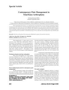
4. Porous trabecular metal and metaphyseal cone Recently, a new biomaterial, the porous trabecular metal, has been introduced and become widely used in complex revision knee arthroplasty. Biomechanically, the porous metal, made from tantalum or titanium, has good biocompatibility. Regarding trabecular tantalum metal, it has approximately 400micron porous diameter, high volumetric porosity (7080%), low modulus of elasticity (3MPa), excellent corrosion resistance, and high coefficient of friction(32). The porous tantalum is also available in several shapes which can be used in both hip and knee (Fig. 1). With specific property of high volumetric porosity, it enhances and fastens the process of bone ingrowth. The major advantage of the porous trabecular metal is that can be used in moderate to severe bone
Fig. 1
A and B) demonstrating anteroposterior and lateral radiographs of loosening TKA with type 2B femoral bone loss (F2B), C and D) demonstrating the use of tantalum metaphyseal cone for distal femoral reconstruction during revision TKA
J Med Assoc Thai Vol. 95 Suppl. 10 2012
loss in order to avoid the possible complications from massive structural bone graft, which seems to be a future solution for management of bone loss in revision TKA. However, this new device does not restore bone stock which the future problem regarding bone loss in re-revision surgery may be in concern. In a series of 16 patients, who were treated tibial bone defect with porous tantalum, with an average 31-month follow-up, the reconstructions had good function with no reoperations in 14 cases. In addition, the radiographic evaluation demonstrated stable osteointegration into the porous tantalum(33). The metaphyseal cone is another porous metal augment which has been recommended to use in a significant metaphyseal bone loss in order to achieve structural and biomechanical restoration. It was designed to solve the problems related to bulk allograft reconstruction. According to its advantage on osteointegration property, the fixation of the metaphyseal cone to the host bone is cementless. However, the fixation of the metaphyseal cone and the prosthesis with intramedullary stem is cemented. Regarding the surgical technique, the extension of metaphyseal bone defect is evaluated. The trial the metaphyseal cone is inserted and a fine tuning inner bone shape is made for maximum contact surface between the host bone and the metaphyseal cone using a high-speed burr. With proper size of metaphyseal cone, impact it into the tibial or the femur to seat in the stable position. The noncontact surface at the periphery area should be filled up with morsellized bone graft. Then, the prosthesis with intramedullary stem is inserted and cemented with or without additional metal augment. Even though the reports are still in the shortterm clinical outcomes, a series of Howard et al(34) demonstrated 24 revision TKAs with tantalum cone were followed at an average of 35 months, the average Knee Society clinical score improved from 55 points, preoperatively, to 81 points, postoperatively. Similarly, Meneghini et al(35) reported good short-term results of using porous tantalum metaphyseal cone for severe bone loss in 15 revision TKAs at an average of 34 month-follow-up without evidence of loosening or migration. Lachiewicz et al(36) reported a retrospective review of 27 revision knee arthroplasties with tantalum metaphyseal cone at a mean follow-up of 39 months, the mean Knee Society pain score improved from 40 points, preoperatively, to 79 points, postoperatively. However, the mid- to long-term results of using this new biomaterial are needed to confirm its efficacy.
J Med Assoc Thai Vol. 95 Suppl. 10 2012
5. Mega-prostheses, Custom-made prostheses, Rotating-hinge prostheses In a large bone defect, custom-made or rotating-hinge prostheses may be required. Bistolfi et al(37) and Utting et al(38) reported acceptable outcomes of custom-made or rotating-hinge prosthesis in revision total knee arthroplasty with severe ligament instability and bone loss. . Deehan et al(39) reviewed a series of 72 salvaged knee procedures using a Kinematic rotating hinge prosthesis and reported the survival analysis of best-case 10-year implant survival of 90%. while Pour et al(40) reported the rate of prosthetic survival was 79.6% at one year and 68.2% at five years in 44 revision TKA with rotating hinge prostheses. Barrack at al(41) and Hossain at al(42) reported the comparable outcomes to condylar revision knee prostheses, it is much more expensive than that of standard prosthesis. In addition, it takes a certain time to manufacture. Thus, it should be considered as an option when other type of knee systems is not feasible. Additionally, Pour (40) suggested that it should be used in an elderly patients. Selection of option for management of bone loss Although there are several successful options for management of bone loss in revision TKA, each treatment option should be considered based on the characteristic of bone loss, surgeon’s expertise and the availability of bone graft and complex options of TKA system at the surgeon’s institution. In many revision TKA scenarios, a preliminary preoperative evaluation for bone loss enhances the surgeon to prepare for bone graft or more complex options of TKA system. Then, a final evaluation of bone loss is made intraoperatively after removal of the prosthesis. Using a thin saw blade for breaking the cement-prosthesis interface and remove the bone cement with direct visualization may minimize bone loss. With the use of structural allograft, the surgeon should aware of proper weight bearing before the graft incorporates. Qiu and associates(43) summarized a useful treatment options as shown in Table 2. In addition, the authors summarized the overall results, advantages and disadvantages of each treatment option related to the time of follow-up in Table 3 and Table 4. Conclusion Bone loss is a common problem found in revision TKA which several options for management have been proposed. Regarding mild to moderate bone loss, treatment options have been well developed. However, the treatment options for massive bone loss
S233
Table 2. Summarized treatment options for management of bone loss(43) Defect
Treatment options
Contained 10 mm Mild uncontained 50% femoral condyle or tibial plateau, intact ligament Severe uncontained Lateral ligament involved
Cement Cement + Screw Impaction morselised allografting, structural allografting Cement Cement + Screw Impaction allograft bone, metal augment Metal augment, structural allografting, Modular prosthesis
Metal augment, structural allografting, Megaprosthesis, tantalum augment
Table 3. Result of each treatment options Study Cement with or without screw Ritter MA(9) Lotke et al(12) Morselized bone graft (impaction) Bradley GW(27) Lotke et al(24) Ahmed et al(16) Hanna et al(26) Structural allograft Ghazavi et al(23) Clatworthy et al(20) Engh and Ammeen(19) Bauman et al(17) Modular component (metal augment) Brand et al(31) Patel et al(44) Porous trabecular metal and metaphyseal cone Meneghini et al(35) Long and Scuderi(33) Howard et al(34) Lachiewicz et al(36) Mega-prostheses, Custom-made prostheses, Rotating-hinge prostheses Pour et al(40) Bistolfi et al(37)
S234
Year
Mean Follow-up
N
Outcome
1986 1991
36 months 85.2 months
57 59
No revision No revision
2000 2006 2008 2011
33 months 45.6 months 120 months 87.6 months
19 42 11 56
Revision 1 case (5.26%) Revision 6 cases (14%) No revision Revision 5 cases (9%)
1997 2001 2007 2009
50 months 96.9 months 60 months 90 months
28 29 49 70
Revision 7 cases (25%) Revision 12 cases (23%) Revision 4 cases (8.16%) Revision 16 cases (22.8%)
1989 2004
37 months 84 months
20 79
No revision Revision 6 cases (7.59%)
2008 2009 2011 2012
34 months 31 months 35 months 39.3 months
15 16 24 27
No revision Revision 2 cases (12.5%) No revision Revision 4 cases (14.8%)
2007 2012
50.4 months 60.3 months
44 26
Revision 8 cases (18.18%) Revision 2 cases (7.69%)
J Med Assoc Thai Vol. 95 Suppl. 10 2012
Table 4. Advantages and disadvantages of each treatment option Management options
Advantage
Disadvantage
Cement with or without screw
Simple method
Risk of thermal necrosis Radiolucent Only used in small defect
Morselized bone graft (impaction)
Osteoinduction Osteoconduction Potentially provide bone stock
Not recommend in uncontained defect Graft resorption Graft collapse
Structural allograft
Physiologic material Manage large bone defect Potentially provide bone stock
Disease transmission Graft resorption Fracture Risk of infection
Modular component (metal augment)
Various shapes and sizes Biomechanical support
Stress-shielding Fretting and corrosion Potential bone loss in the long-term follow-up
Porous trabecular metal and metaphyseal cone
Biocompatibility Induce osteointegration Manage large bone defect
No long-term study Expensive Potential bone loss in the long-term follow-up?
Mega-prostheses, Custom-made prostheses, Rotating-hinge prostheses
Manage severe bone defect
Expensive
are still debatable. The new porous metal augments have become alternative option besides the structural allograft with a good short-term outcome.
5.
Potential conflicts of interest None. References 1. Memtsoudis SG, Della Valle AG, Besculides MC, Gaber L, Laskin R. Trends in demographics, comorbidity profiles, in-hospital complications and mortality associated with primary knee arthroplasty. J Arthroplasty 2009; 24: 518-27. 2. Lombardi AV, Berend KR, Adams JB. Management of bone loss in revision TKA: it’s a changing world. Orthopedics 2010; 33: 662. 3. Nadaud MC, Fehring TK, Fehring K. Underestimation of osteolysis in posterior stabilized total knee arthroplasty. J Arthroplasty 2004; 19: 110-5. 4. Zotti MG, Campbell DG, Woodman R. Detection of periprosthetic osteolysis around total knee
J Med Assoc Thai Vol. 95 Suppl. 10 2012
6.
7.
8.
9.
10.
arthroplasties an in vitro study. J Arthroplasty 2012; 27: 317-22. Reish TG, Clarke HD, Scuderi GR, Math KR, Scott WN. Use of multi-detector computed tomography for the detection of periprosthetic osteolysis in total knee arthroplasty. J Knee Surg 2006; 19: 25964. Clatworthy MG, A.E. Management of bony defects in revision total knee arthroplasty. JJ Callaghan AR, HE Rubash, PT, Simonian TW, (Eds.), editors2003. Engh GA, Ammeen DJ. Classification and preoperative radiographic evaluation: knee. Orthop Clin North Am 1998; 29: 205-17. Whittaker JP, Dharmarajan R, Toms AD. The management of bone loss in revision total knee replacement. J Bone Joint Surg Br 2008; 90: 981-7. Ritter MA. Screw and cement fixation of large defects in total knee arthroplasty. J Arthroplasty 1986; 1: 125-9. Brooks PJ, Walker PS, Scott RD. Tibial component fixation in deficient tibial bone stock. Clin Orthop
S235
Relat Res 1984; 302-8. 11. Saha S PS. Mechanical properties of bone cement: a review. J Biomed Mater Res B Appl Biomater. 2009;89(2)(May):558-74. 12. Lotke PA, Wong RY, Ecker ML. The use of methylmethacrylate in primary total knee replacements with large tibial defects. Clin Orthop Relat Res 1991; 288-94. 13. Dorr LD, Ranawat CS, Sculco TA, McKaskill B, Orisek BS. Bone graft for tibial defects in total knee arthroplasty. Clin Orthop Relat Res 1986; 153-65. 14. Engh GA, Parks NL. The management of bone defects in revision total knee arthroplasty. Instr Course Lect 1997; 46: 227-36. 15. Watanabe W, Sato K, Itoi E. Autologous bone grafting without screw fixation for tibial defects in total knee arthroplasty. J Orthop Sci 2001; 6: 481-6. 16. Ahmed I, Logan M, Alipour F, Dashti H, Hadden WA. Autogenous bone grafting of uncontained bony defects of tibia during total knee arthroplasty a 10-year follow up. J Arthroplasty 2008; 23: 74450. 17. Bauman RD, Lewallen DG, Hanssen AD. Limitations of structural allograft in revision total knee arthroplasty. Clin Orthop Relat Res 2009; 467: 81824. 18. Tsahakis PJ, Beaver WB, Brick GW. Technique and results of allograft reconstruction in revision total knee arthroplasty. Clin Orthop Relat Res 1994; 8694. 19. Engh GA, Ammeen DJ. Use of structural allograft in revision total knee arthroplasty in knees with severe tibial bone loss. J Bone Joint Surg Am 2007; 89: 2640-7. 20. Clatworthy MG, Ballance J, Brick GW, Chandler HP, Gross AE. The use of structural allograft for uncontained defects in revision total knee arthroplasty. A minimum five-year review. J Bone Joint Surg Am 2001; 83-A: 404-11. 21. Richards CJ, Garbuz DS, Pugh L, Masri BA. Revision total knee arthroplasty: clinical outcome comparison with and without the use of femoral head structural allograft. J Arthroplasty 2011; 26: 1299-304. 22. Bezwada HP, Shah AR, Zambito K, Cerynik DL, Johanson NA. Distal femoral allograft reconstruction for massive osteolytic bone loss in revision total knee arthroplasty. J Arthroplasty 2006; 21: 242-8. 23. Ghazavi MT, Stockley I, Yee G, Davis A, Gross AE. Reconstruction of massive bone defects with
S236
24.
25.
26.
27.
28.
29.
30.
31.
32.
33.
34.
35.
36.
37.
allograft in revision total knee arthroplasty. J Bone Joint Surg Am 1997; 79: 17-25. Lotke PA, Carolan GF, Puri N. Impaction grafting for bone defects in revision total knee arthroplasty. Clin Orthop Relat Res 2006; 446: 99-103. Completo A, Simoes JA, Fonseca F. Revision total knee arthroplasty: the influence of femoral stems in load sharing and stability. Knee 2009; 16: 275-9. Hanna SA, Aston WJ, de Roeck NJ, Gough-Palmer A, Powles DP. Cementless revision TKA with bone grafting of osseous defects restores bone stock with a low revision rate at 4 to 10 years. Clin Orthop Relat Res 2011; 469: 3164-71. Bradley GW. Revision total knee arthroplasty by impaction bone grafting. Clin Orthop Relat Res 2000; (371): 113-8. Backstein D, Safir O, Gross A. Management of bone loss: structural grafts in revision total knee arthroplasty. Clin Orthop Relat Res 2006; 446: 10412. Delloye C, Cornu O, Druez V, Barbier O. Bone allografts: What they can offer and what they cannot. J Bone Joint Surg Br 2007; 89: 574-9. Dennis DA. The structural allograft composite in revision total knee arthroplasty. J Arthroplasty 2002; 17: 90-3. Brand MG, Daley RJ, Ewald FC, Scott RD. Tibial tray augmentation with modular metal wedges for tibial bone stock deficiency. Clin Orthop Relat Res 1989; (248): 71-9. Levine BR, Sporer S, Poggie RA, Della Valle CJ, Jacobs JJ. Experimental and clinical performance of porous tantalum in orthopedic surgery. Biomaterials 2006; 27: 4671-81. Long WJ, Scuderi GR. Porous tantalum cones for large metaphyseal tibial defects in revision total knee arthroplasty: a minimum 2-year follow-up. J Arthroplasty 2009; 24: 1086-92. Howard JL, Kudera J, Lewallen DG, Hanssen AD. Early results of the use of tantalum femoral cones for revision total knee arthroplasty. J Bone Joint Surg Am 2011; 93: 478-84. Meneghini RM, Lewallen DG, Hanssen AD. Use of porous tantalum metaphyseal cones for severe tibial bone loss during revision total knee replacement. J Bone Joint Surg Am 2008; 90: 78-84. Lachiewicz PF, Bolognesi MP, Henderson RA, Soileau ES, Vail TP. Can tantalum cones provide fixation in complex revision knee arthroplasty? Clin Orthop Relat Res 2012; 470: 199-204. Bistolfi A, Massazza G, Rosso F, Crova M.
J Med Assoc Thai Vol. 95 Suppl. 10 2012
38.
39.
40.
41.
Rotating-hinge total knee for revision total knee arthroplasty. Orthopedics 2012; 35: e325-30. Utting MR, Newman JH. Customised hinged knee replacements as a salvage procedure for failed total knee arthroplasty. Knee 2004; 11: 475-9. Deehan DJ, Murray J, Birdsall PD, Holland JP, Pinder IM. The role of the rotating hinge prosthesis in the salvage arthroplasty setting. J Arthroplasty 2008; 23: 683-8. Pour AE, Parvizi J, Slenker N, Purtill JJ, Sharkey PF. Rotating hinged total knee replacement: use with caution. J Bone Joint Surg Am 2007; 89: 1735-41. Barrack RL, Lyons TR, Ingraham RQ, Johnson JC. The use of a modular rotating hinge component in
salvage revision total knee arthroplasty. J Arthroplasty 2000; 15: 858-66. 42. Hossain F, Patel S, Haddad FS. Midterm assessment of causes and results of revision total knee arthroplasty. Clin Orthop Relat Res 2010; 468: 1221-8. 43. Qiu YY, Yan CH, Chiu KY, Ng FY. Review article: Treatments for bone loss in revision total knee arthroplasty. J Orthop Surg (Hong Kong ) 2012; 20: 78-86. 44. Patel JV, Masonis JL, Guerin J, Bourne RB, Rorabeck CH. The fate of augments to treat type2 bone defects in revision knee arthroplasty. J Bone Joint Surg Br 2004; 86: 195-9.
บทความฟื้นฟูวิชาการ: การรักษาภาวะการสูญเสียกระดูกในการผ่าตัดเปลี่ยนข้อเข่าเทียมซ้ำ ศริษฏ์ หงษ์วไิ ล, อารี ตนาวลี การผ่าตัดเปลี่ยนข้อเข่าเทียมมีจำนวนเพิ่มขึ้นอย่างรวดเร็วในช่วงหลายปีมานี้ เนื่องจากเป็นการผ่าตัดรักษา ภาวะข้ อ เข่ า เสื ่ อ มในระยะท้ า ยที ่ ไ ด้ ผ ลดี แ ละมี ป ระสิ ท ธิ ภ าพ ทั ้ ง นี ้ มี ค วามหลากหลายอายุ ข องผู ้ ป ่ ว ยที ่ ไ ด้ ร ั บ การผ่าตัดมากขึ้น จึงมีผู้ป่วยที่อายุน้อยจำนวนเพิ่มขึ้น ดังนั้น โอกาสที่ผู้ป่วยจะได้รับการผ่าตัดเปลี่ยนข้อเข่าเทียมซ้ำ (revision total knee arthroplasty) ในครัง้ ถัดไปจึงมากขึน้ ปัญหาใหญ่ทส่ี ำคัญปัญหาหนึง่ ในการผ่าตัดซ้ำ คือการรักษา ภาวะการสูญเสียกระดูก (bone loss) ซึ่งมีผลกระทบต่อตำแหน่งที่ดี และความยืนยาวของการใช้งานในการใส่ ข้อเทียมข้อใหม่ ภาวะการสูญเสียกระดูกมีความรุนแรงหลายระดับ ซึ่งแต่ละระดับมีแนวทางการรักษาแตกต่างกันไป ตั้งแต่การเสริมด้วยสารยึดกระดูก (bone cement) การปลูกกระดูกซึ่งนำมาจากผู้ป่วยเอง (autogenous graft) หรือจากผูอ้ น่ื (allograft) การใช้โลหะเพือ่ เป็นตัวหนุน (metal augment) การใช้ขอ้ เทียมทีพ่ เิ ศษเฉพาะ จนถึงการใช้โลหะ ชนิดใหม่ที่คุณสมบัติเป็นรูพรุน (porous material)l ซึ่งให้ผลการรักษาระยะสั้นเป็นที่พอใจ บทความฟื้นฟูวิชาการนี้ ผู้นิพนธ์ได้รวบรวมแนวทางและผลการรักษาภาวะการสูญเสียกระดูกแต่ละระดับตา แนวทางหลักฐานทางวิชาการ ทีม่ อี ยูใ่ นปัจจุบนั J Med Assoc Thai Vol. 95 Suppl. 10 2012
S237