Arch Iranian Med 2003; 6 (2): 127– 140
REVIEW ARTICLE GASTROESOPHAGEAL REFLUX DISEASE: THE NEW EPIDEMIC •
Reza Malekzadeh MD , Siavosh Nasseri-Moghaddam MD, Masoud Sotoudeh MD Digestive Disease Research Center, Tehran University of Medical Sciences, Tehran, Iran
Introduction
G
astroesophageal reflux disease (GERD), although known for a long time, has come to clinical attention only during the past 15 years. Currently, it is one of the most common gastrointestinal problems in the west 1 – 4 and apparently on increase in Iran 5 – 8 and other eastern countries as well.9 Therefore, it is timely to review this topic and update our knowledge on various aspects including the epidemiology, pathophysiology, symptomatology, diagnosis, and treatment of GERD, and its future trends (Table 1). Epidemiology Epidemiologic studies of GERD in the west date back to the late 1970s. Initial reports indicated a prevalence of 7 – 15% in the community.10 Later studies using better instruments for detecting GERD11 reported weekly GERD symptoms in 20% of the population and occasional GERD symptoms in about 40% of the population in the USA. 1 A recent meta-analysis of different studies has confirmed these figures.2 Studies from East and South-East Asia have shown much lower prevalence rates12 and it is generally believed that the disease is not as frequent in the east, 13 although more recent reports have challenged this concept. 9, 14 Unfortunately, no true population-based studies have been performed in Iran yet, but available data from clinical and epidemiologic studies point to an increasing burden.5 – 8 As there is no gold standard for diagnosis of GERD, except for careful history taking, epidemiologic studies may be challenging. A widely accepted instrument for populationbased studies is the Mayo questionnaire,11 which has been used in several studies both in the USA •Correspondence: R. Malekzadeh, MD, Digestive Disease Research Center, Shariati Hospital, Kargar-e-Shomali Street, Tehran 14114, Iran. Fax: +98-21-802 6481, E-mail:
[email protected].
and in other countries. An attempt to validate this questionnaire showed that the translation of this instrument is neither valid nor reliable for an Iranian population.15 Currently, we are validating a modified Mayo questionnaire, and if it proves to be useful, a population-based study can be expected. Otherwise, we need to construct a locally valid and reliable questionnaire for this purpose. What is GERD and what are the risk factors? Almost everybody has some degree of reflux of gastric contents into the esophagus. This physiologic gastroesophageal reflux (GER) does not usually cause any symptoms and is not associated with physical damage to the esophagus. On the other hand, some people suffer from pathologic reflux disease (GERD). By definition, all patients exposed to the risk of physical Table 1. GERD: key points. • GERD is a chronic and recurrent disease. • GERD can seriously affect the quality of life. • There is no gold standard for diagnosis of GERD. • Currently a careful history with consideration of extra-esophageal and atypical GERD symptoms is the best way to diagnose GERD. • Normal endoscopy and/or 24-hour pH-metry do not rule out presence of GERD. • 50 – 70% of patients with GERD have a normal endoscopy (nonerosive reflux disease, NERD). • NERD is not a mild form of GERD, but a separate entity with little chance of progression to GERD. • Treatment of NERD should be as aggressive as GERD. • The GERD patient who has not responded adequately to medical therapy is not an appropriate candidate for surgery. • Adequate (and probably long-term) medical acid suppression is the mainstay of treatment of these patients. • GERD is increasing in Iran.
Archives of Iranian Medicine, Vol 6, No 2, April 2003 127
Gastroesophageal Reflux Disease: The New Epidemic
Table 2. Risk factors for GERD. Obesity Fatty meals Heavy meals Spicy food Cigarette smoking Tight fitting garments
Emotional stress Rapid eating behavior Coffee, tea Pregnancy Medication Reclining after eating
complications of GER or those who experience clinically significant impairment of their quality of life due to GE reflux after adequate reassurance of the benign nature of their symptoms, have GERD.16 Some occasional pyrosis or heartburn may be experienced by many people. By convention, clinical GERD is diagnosed when a patient suffers two or more episodes of pyrosis, heartburn or other GERD symptoms in a week 17 or the symptoms seriously affect the quality of life. Known risk factors for GERD are summarized in Table 2. Among these, obesity and fatty or heavy meals appear to be the most important ones. Natural history and complications of GERD GERD is a chronic and recurrent disease.17 It has been shown that more than 90% of cases of erosive GERD and more than 75% of cases of nonerosive GERD (nonerosive reflux disease, NERD) recur after discontinuation of effective medical therapy.17 It is also well documented that a significant minority (up to 30%) of patients who undergo an initially successful antireflux surgery develop recurrent GERD symptoms within 10 years after surgery, requiring further medical treatment. Therefore, GERD should be considered a chronic and recurrent disease. Current evidence also supports the central role of GERD in development of specialized intestinal metaplasia in the distal esophagus (Barrett’s esophagus), which by itself increases the risk of distal esophageal adenocarcinoma 30 to 100-folds. 4 A red velvetty mucosa lining more than 2 – 3 cm of the distal esophagus instead of the usual pale esophageal mucosa extending to the gastroesophageal junction has traditionally been used to define Barrett’s esophagus.18, 19 More recently, it has been shown that the histological type of the metaplastic mucosa (fundal, oxynto-cardiac, or specialized intestinal) is more important than the length of the columnar-lined mucosa. 20 – 22 Only specialized intestinal metaplasia, containing goblet cells (Figure 1) is considered Barrett’s esophagus; the other histological types do not appear to increase the risk of malignancy. Therefore, the 128 Archives of Iranian Medicine, Vol 6, No 2, April 2003
Figure 1. Lower esophageal biopsy showing specialized intestinal metaplasia with goblet cells of Barrett’s esophagus (H&E stain).
diagnosis of Barrett’s esophagus can only be made after careful examination of adequate biopsy samples of apparently metaplastic mucosa from the distal esophagus.20 – 22 Hence the terms short- and long-segment Barrett’s esophagus (SSBE and LSBE) (Figure 2), referring to respective less and more than 2 cm of columnar-lined mucosa in the distal esophagus respectively, have been coined. 22 However, the pathophysiologic differences between, and natural histories of SSBE and LSBE remain to be determined. 22, 23 It is generally believed that SSBE and LSBE are different entities and that the chances of SSBE transforming to LSBE are minimal. Recent data indicate that the chance of developing specialized columnar gastric
Figure 2. Long-segment Barrett’s esophagus (LSBE)—long segment columnar metaplasia in the lower esophagus.
R. Malekzadeh, S. Nasseri-Moghaddam, M. Sotoudeh
Figure 3. Sliding endoscopy.
hiatal
hernia
(SHH)
on
metaplasia increases significantly with longer duration of and more severe GERD symptoms.18 Specialized columnar metaplasia and its dysplastic changes have been reported in apparently normal gastroesophageal junctions without any evidence of visible Barrett’s esophagus.24, 25 Although this makes the issue a little more complicated, looking for such changes in an apparently normal gastroesophageal junction is not recommended. Other complications of GERD include erosive distal esophagitis, esophageal strictures and ulcers, and significant impairment of quality of life. 26 – 28 Some reports indicate that the quality of life in patients with severe GERD may be even worse than that in patients with chronic ischemic heart disease.29, 30 In practice, this impaired quality of life may be the most bothersome aspect of the patient’s complaints and should be addressed carefully. Thus, GERD should be considered a pathophysiologically heterogeneous group of diseases (erosive GERD, NERD, and Barrett’s esophagus) that, despite having relatively similar clinical manifestations, follow different natural courses and may respond differently to treatment. These are hot issues actively being investigated. It is generally accepted that there is no direct correlation between GERD symptoms, endoscopic, and histological findings in the mucosa of the distal esophagus. What is the underlying mechanism? The underlying mechanism of GERD is not yet fully understood, but various factors contribute to the generation of GERD. The most important known mechanisms include the following: 1. Transient lower esophageal relaxation
2.
3.
(tLESR) is currently considered a major pathophysiologic cause of GERD,31, 32 especially in milder forms of endoscopic esophagitis. Lower esophageal relaxation is a physiological action during swallowing. tLESR has several characteristics: it is not associated with swallowing, it has a longer duration, and it is not associated with esophageal peristalsis.31 tLESR is considered a physiological response to gastric fundal distension, to rid the stomach of excessive swallowed gas (as part of the belching reflex). Increased tLESR frequency exposes the esophagus to acid and other gastric and duodenal contents, which if not cleared normally, leads to GERD. Despite good reports relating tLESR to GERD,33 it is wellestablished that most episodes of tLESR are not associated with GERD symptoms and not all people with GERD have tLESR.34 Hiatal hernia is a second contributing factor. Usually, the lower esophageal sphincter (LES) is located at the level of the diaphragmatic opening (hiatus) through which the esophagus traverses from the chest to the abdomen. Therefore, the diaphragmatic crura add to the contractile force of the LES. In some people, the LES is located in the chest instead of the level of the diaphragmatic hiatus, hence losing diaphragmatic crural support during contraction. Meanwhile, this abnormal location of the LES gives rise to a pouch of stomach just beneath the LES and above the diaphragm, which acts as a reservoir that can contain gastric contents to be regurgitated into the esophageal lumen. This is called a sliding hiatal hernia (SHH), and contributes (Figure 3) to development of GERD.35 However, many people have SHH without experiencing GERD symptoms or its complications and vice versa. 35 – 37 A new concept is that, even in the absence of a hiatal hernia, there is a small area around the gastric cardia that maintains a low pH among patients with GERD. This acidic milieu then contributes to the symptoms and consequences of GERD. Hypotensive LES, defined as a resting LES pressure of less than 4 to 10 mmHg, is another contributing factor. Although uncommon, it seems to be especially important in the more severe forms of
Archives of Iranian Medicine, Vol 6, No 2, April 2003 129
Gastroesophageal Reflux Disease: The New Epidemic
4.
5.
6.
erosive endoscopic esophagitis, i.e. grades C and D (Los Angeles Classification) (Table 3). This is usually associated with hiatal hernias and the two (hypotensive LES and hiatal hernia) act in concert to cause severe GERD. Newer experimental therapeutic options, such as baclofen, have this mechanism as their main target of action. Impaired esophageal acid clearance is another contributing factor.38 Acid, regurgitated into the esophagus, is usually cleared with esophageal peristalsis and the bicarbonate secreted in the saliva. If either of these mechanisms is impaired, GERD supervenes.38 – 40 Esophageal hypersensitivity has also been reported among GERD patients.41 Patients with esophageal hypersensitivity may have no evidence of pathologic reflux on 24-hour pH monitoring or endoscopy but still suffer from significant GERD symptoms.41, 42 This is a very important aspect and should be considered especially in patients scheduled for antireflux surgery, as they may respond poorly to surgery. Esophageal hypersensitivity is best detected by Bernstein’s test (see below). Duodenogastroesophageal reflux (DGER) has attracted more attention recently. Bile and other duodenal contents may be present
Table 3. Savary-Miller and Los Angeles Classifications of endoscopic GERD. Savary-Miller Classification Grade 1: One or more supravestibular reddish spots, with or without exudates. Grade 2: Erosive and exudative lesions in the distal esophagus that may be confluent but not circumferential. Grade 3: Circumferential erosions in the distal esophagus covered by hemorrhagic and pseudomembranous exudates. Grade 4: Presence of chronic complications such as deep ulcers, stenosis, or scarring with Barrett’s metaplasia. Los Angeles Classification Grade A: One or more mucosal breaks ≤ 5 mm in length. Grade B: At least one mucosal break > 5 mm long, but not continuous between the tops of adjacent mucosal folds. Grade C: At least one mucosal break which is continuous between adjacent mucosal folds, but not circumferential (< 75% of periphery). Grade D: Mucosal breaks that involve at least threequarters of the luminal circumference. Note: Ulcers, strictures, Barrett’s metaplasia, and other findings are reported as an adjunct to each grade.
130 Archives of Iranian Medicine, Vol 6, No 2, April 2003
8.
in the esophagus of patients with GERD.31, 32 Different duodenal contents have different effects on the esophageal mucosa. For instance, conjugated bile acids and pepsin are more injurious to the esophageal mucosa at acidic pH, while unconjugated bile acids and trypsin are more harmful at pH 5 – 8. 32 Hydrochloric acid inhibits the damaging effects of trypsin and unconjugated bile acids, whereas conjugated bile acids decrease the damaging effect of pepsin at acidic pH.32, 43 Therefore, alkaline reflux esophagitis is caused by both unconjugated bile acids and trypsin at neutral pH.32, 44 – 46 Recent studies of patients with and without complications of Barrett’s esophagus have found increased reflux of bile and acid into the lower esophagus of both groups as compared with controls.47 More importantly, reflux of acid paralleled DGER, and both were significantly higher in patients with complicated Barrett’s esophagus than in those with uncomplicated Barrett’s esophagus.47 These studies support earlier findings in animals that suggest a possible synergy between acid and DGER in the development of esophagitis and Barrett’s esophagus. DGER in the absence of acid reflux is a rare event (7%) in patients without prior gastric surgery. 32, 48 DGER without excessive acid reflux can cause reflux symptoms but does not usually produce esophagitis.32, 48 Therefore, reflux of duodenal contents and gastric acid put the patient at increased risk of esophageal injury, especially complicated Barrett’s esophagus (double reflux, double jeopardy).49 Initially, DGER was considered synonymous with alkaline reflux (vs acid reflux), but it is welldocumented that the two terms are not synonymous, because other potentially harmful substances are present in bile. 32 Hence, the preferred term is “bile reflux”. Helicobacter pylori has been the focus of numerous investigations in the field of gastroenterology. There is unequivocal evidence that H. pylori has a pathogenic role in peptic ulcer disease, gastric adenocarcinoma, and mucosa-associated lymphoid tissue lymphoma.50 – 57 The literature on the relationship between H. pylori and GERD is controversial. Although H. pylori does not affect LES pressure or
R. Malekzadeh, S. Nasseri-Moghaddam, M. Sotoudeh Table 4. Other symptoms of GERD. Dysphagia Chest pain Globus sensation Water brash Chronic cough Aspiration pneumonitis Sleep apnea Nausea
or related conditions Chronic hoarseness Bitter mouth and sore throat Halitosis Laryngitis Sinusitis Hiccups Asthma
tLESR, its effect on esophageal mucosal hypersensitivity is still not known.58 Currently, it should be considered that H. pylori has no direct pathogenetic role in GERD, but it affects the clinical outcome in some instances. Therefore, there is no consensus to eradicate or not eradicate H. pylori in GERD and the issue is under intense investigation. 8. Other mechanisms that potentially contribute to GERD pathogenesis include impaired salivary gland secretions, impaired gastric emptying, and abnormal esophageal motility. Despite all these proposed mechanisms, the exact pathophysiology of GERD is as yet poorly understood. Considering that GERD is a group of heterogeneous diseases with some common symptomatology, it is conceivable that various mechanisms could be responsible for them, with no one explanation capable of explaining the whole range of diseases (one size does not fit all). Symptomatology and diagnosis There are a variety of diagnostic modalities for GERD. These include symptom analysis, symptom score, 24-hour ambulatory pH monitoring, Bernstein test, and upper gastrointestinal endoscopy. None of these are the gold standard, but the most reliable is currently careful symptom evaluation.59, 60 Therefore, the diagnosis and study of GERD may be problematic. Major GERD symptoms include acid regurgitation and heartburn. 59, 60 Heartburn may be defined differently by different patients; therefore, it should be addressed carefully and through words that are comprehensible to the patient. 59, 60 By heartburn, we mean feeling a burning sensation just below the sternum. Acid regurgitation, another major GERD symptom, refers to the feeling of a sour, bitter fluid running up to the mouth from the abdomen. Other symptoms and related conditions are shown in Table 4. Interestingly, more than 50%
of patients with non-cardiac chest pain and more than 50% of patients with chronic cough, for whom no demonstrable etiology is found, actually suffer from GERD and may respond to prolonged and aggressive treatment for GERD.61 Antireflux surgery has even been used for long-term control of symptoms in these patients. Although chest pain is a relatively common GERD symptom, patients presenting with chest pain should always be evaluated for cardiac causes of chest pain. Reports on the association of asthma and GERD are numerous, but the association is one of the most controversial.62, 63 Overall, it seems that in patients with severe asthma, GERD is relatively common and asthma symptoms may be better controlled by concomitant treatment of GERD, especially among children.64 Minor, extraesophageal, and atypical GERD symptoms such as nausea or waterbrash may be the only presenting symptoms of GERD and therefore should be specifically addressed by the evaluating physician. It should be kept in mind that these atypical or extraesophageal symptoms may be very difficult to control even in the absence of endoscopic esophagitis. For 24-hour pH monitoring, a thin pH-sensitive catheter is passed into the lower esophagus and connected to a portable electronic pH-meter. The device is held in place for 24 hours and the patient is instructed to do his/her routines and just push specific buttons for events such as eating, reclining, and having symptoms. The beginning and the end of each event is recorded and correlated with the recorded pH. The data are then analyzed by specific computer software and several indices including a total score (DeMeester score) are calculated. This information can be interpreted in several ways. Twenty-four-hour pH monitoring is about 80–85% sensitive and specific for diagnosing GERD.16 There are several reasons for normal pH readings while the patient has GERD, including a hypersensitive esophageal mucosa, bile reflux, proximal reflux, esophageal motor abnormalities, and inadequacy of the scores and calculations.64 Therefore, this method is reserved to make the diagnosis of GERD in patients whose diagnosis is controversial after careful history taking and endoscopy. It is also indicated in patients who are candidates for antireflux surgery.65 A device similar to that used for 24-hour pH monitoring, the Bilitec 2000® (Synectics Medical AB, Stockholm, Sweden), has been developed to measure bile reflux and its applications are increasing. In this instance, the Archives of Iranian Medicine, Vol 6, No 2, April 2003 131
Gastroesophageal Reflux Disease: The New Epidemic
Table 5. Generally recommended indications for upper gastrointestinal endoscopy in GERD. •
Any suspicion of GERD complications or GERDrelated malignancy
•
Presence of alarming signs including significant weight loss, anemia, upper gastrointestinal bleeding or melena
•
Recent change in character of symptoms
•
Age of onset after 40 years
•
Long-term GERD symptoms (more than 5 years)
•
Family history of GERD-related cancer
•
Suspicion of concomitant upper GI lesions
•
Before antireflux surgery
•
Equivocal diagnosis
•
Patient reassurance
catheter is sensitive to electromagnetic wavelengths in the range of 470 nm (the range of bile) instead of pH.66, 67 It is still difficult and cumbersome to use this device and at best it identifies up to 70% of patients with bile reflux. 67 The other diagnostic modality for GERD is upper gastrointestinal endoscopy (UGIE). Generally recommended indications for UGIE are shown in Table 5. Signs of GERD on UGIE include distal esophageal erosions (the most frequent), ulcers in the lower esophagus, esophageal stricture, and distal esophageal gastric metaplasia (Barrett’s esophagus).68 Currently, it is widely accepted that distal esophageal redness without discrete mucosal breaks should not be considered evidence of endoscopic GERD.68 Endoscopic GERD can be diagnosed if definite mucosal breaks are observed in the distal esophagus or if strictures, ulcers, and columnar metaplasia are noted. The two most widely used classification systems for endoscopic GERD (the Savary-Miller and the Los Angeles Classifications) are shown in Table 3. It is recommended that the endoscopist describes what he/she sees exactly and then decides about the degree of esophagitis and notes which system he/she has used. Detailed descriptions should not be omitted in favor of recording the classification. Although finding endoscopic signs of GERD is quite specific,16 these signs can be seen in 30–50% of GERD cases at best. Most GERD patients do not have any endoscopically visible damage to their mucosa. These patients have NERD.69, 70 Some data show that symptoms in NERD patients may be even more difficult to control than those in
132 Archives of Iranian Medicine, Vol 6, No 2, April 2003
patients with erosive GERD.69 Hence, it is generally a false belief that patients with GERD symptoms but normal endoscopy have a milder form of disease. Longitudinal studies have shown that most NERD patients never develop endoscopically visible esophageal mucosal damage, yet they have a recurrent and sometimes difficult-to-control condition. A significant portion of NERD patients present with extraesophageal symptoms and even a normal 24-hour ambulatory pH-metry. Therefore, it is widely believed that symptoms and endoscopic and pathologic findings are not directly correlated in GERD patients.70 It is extremely important to bear in mind that not seeing endoscopic evidence of GERD does not rule out GERD. Potent proton pump inhibitors (PPIs) such as omeprazole have been used for both diagnosis and treatment of GERD.71, 72 In the so-called omeprazole test (O-test), omeprazole at a dose of 20 – 40 mg/day is prescribed orally for 7 – 14 days. If the patient’s symptoms are totally controlled, the test is interpreted as positive and GERD is the basis of the patient’s symptoms. 71, 72 If used in conjunction with a detailed history and careful follow-up, the O-test is useful and costeffective for diagnosis of GERD.72, 73 Biopsies from the lower esophagus are also helpful in diagnosing GERD. Pathologic findings suggestive of GERD are shown in Table 6. None of these findings are specific to GERD and a constellation of clinical and pathologic findings is needed to make the correct diagnosis. Taking biopsies from appropriate sites is very important for correct interpretation and yield of the pathology report.70 Usually, biopsies are taken only if there is an ulcer, a stricture, columnar metaplasia, lumps, or other lesions raising the suspicion of more serious conditions such as dysplasia or malignancy.70 Some investigators have noted columnar metaplasia and even dysplasia in an apparently completely normal gastroesophageal junction,5, 23 but routine biopsies of this area are not generally recommended. 5, 23 If the biopsy is taken merely to diagnose GERD and none of the above lesions are present, then it should be taken from about 3 cm above the Z-line, because minor changes suggestive of GERD are present lower than that even in people without GERD. Other, rarely used, methods to diagnose GERD include barium esophagography, which is neither sensitive nor specific. It is mostly useful for highgrade esophagitis and detection of some of the
R. Malekzadeh, S. Nasseri-Moghaddam, M. Sotoudeh
complications of GERD,16, 74 The Bernstein test (dripping 0.1 normal hydrochloric acid over the distal esophagus at a rate of 6 – 8 mL/min and looking for reproduction of symptoms not reproduced with saline infusion), although sensitive, is cumbersome and has a low specificity.75 The intraluminal impedance technique is a relatively new way to diagnose GERD but is still considered investigational and is not in general clinical use.76 Transabdominal ultrasonography has been used to diagnose GERD among newborns, with varying results. Overall, taking a detailed history with adequate attention to atypical and extraesophageal manifestations of GERD is the best way to make the diagnosis. Sometimes, it may be necessary to rule out life-threatening conditions (e.g. coronary artery disease) before securing the diagnosis of GERD. Treatment The goals of treating GERD are two-fold: 1) relieving the patient’s symptoms and improving his/her quality of life, and 2) minimizing or eliminating the physical damage to the esophagus caused by GERD, which can lead to potentially serious long-term consequences such as esophageal adenocarcinoma. There are four categories of treatments for GERD: lifestyle modification and dietary changes, medical therapy, surgery, and endoscopic interventions. A) Lifestyle modification and dietary changes Risk factors for developing GERD are shown in Table 2. Correcting these risk factors, i.e. losing weight; avoiding weight gain; avoiding fatty, heavy, and high-calorie food; quitting smoking; avoiding reclining at least 2 to 3 hours after meals; avoiding activities involving bending forward after meals; engaging in regular exercise that does not increase intraabdominal pressure; and Table 6. Histologic findings suggestive of GERD on lower esophageal biopsy. •
Proliferation of the basal cell layer
•
Elongation of papillae
•
Infiltration of neutrophils and eosinophils in lamina propria
•
Dilated vascular channels in papillae of the lamina propria
•
Distended, pale squamous ("balloon") cells
avoiding tight clothing may help in decreasing GER episodes and symptoms. Also, elevating the head of the bed by 20 – 30° by placing blocks beneath the bed may help. These measures alone can improve GERD symptoms in up to 20% of cases at best, although if not observed, they can potentiate GERD symptoms. Therefore, they are used as an adjunct to other modalities. 77 They should be used alone only in the mildest forms of disease. B) Medical therapy All acid-reducing agents have been used in treatment of GERD, albeit with variable success. Antacids that directly neutralize acid have been used at high doses. Although they are effective in alleviating acute GERD symptoms, they maintain a symptom-free state in only about 20% of cases and are therefore not suitable choices for long-term use. Various histamine H2 blockers, which proved to be excellent for peptic ulcer treatment in the pre-H. pylori era, were effective in about 50 – 60% of GERD cases, even at higher doses than usual.77 Prokinetic agents such as cisapride (Prepulsid ®), which is no longer available in North America because of concern about its cardiac sideeffects, has the same efficacy as H2 blockers.78 Cisapride improves gastric emptying and increases LES pressure. Metoclopramide is of limited use in GERD, and there is only evidence for some benefit in diabetic patients with GERD.78, 79 H2 blockers may be started at the usual dosage used to treat peptic ulcer disease, but it is usually necessary to increase the dosage to control patients’ symptoms.78 Sucralfate, a coating agent, can be used in some circumstances, especially in postgastric surgery patients, but it has limited value in the overall management of GERD. PPIs, such as omeprazole, rabeprazole, pantoprazole, lansoprazole, and esomeprazole, have proven to be the most effective medical treatments for GERD.80, 81 These drugs have revolutionized both GERD management and our clinical understanding of this disease. At adequate dosage, they control GERD symptoms in at least 85 – 90% of cases.77, 80, 81 They have been shown Archives of Iranian Medicine, Vol 6, No 2, April 2003 133
Gastroesophageal Reflux Disease: The New Epidemic
to be safe even at higher doses for extended periods of time (up to 11 years).82 – 84 The problem with PPIs is the so-called nocturnal acid break-through. Most patients maintained on PPIs have nocturnal acid break-through, defined as esophageal pH less than 4 for more than an hour during the night.85 This may or may not be associated with symptoms that awaken the patient in the middle of the night. Increasing the dosage of conventional PPIs has not controlled this phenomenon, because of the difference in mechanisms of acid secretion during the day and night (fed and unfed states). Adding an H2 blocker at bedtime to the regimen of these patients has proved to be very useful for controlling these nocturnal symptoms.85 This is the only instance that combining PPIs and H2 blockers has been supported by scientific evidence. Newer studies propose that the newer generation of PPIs (e.g. esomeprazole, and pantoprazole) can overcome the problem of nocturnal acid break-through without the need for an H2 blocker, but further studies are expected. Interestingly, PPIs also decrease DGER and its symptoms. This probably happens through decreased gastric volume.32 Acid-reducing agents do not directly affect the underlying mechanism of reflux. Therefore, a great deal of research is under way to find an agent that adequately increases LES pressure. One of the proposed medications in this newer category is baclofen, a gamma-aminobutyric acid type B (GABA-B) receptor agonist, which in animal and human studies has been shown to increase LES pressure.86 Despite this, the clinical benefit has been marginal and conflicting so far. Therefore, its routine use for this indication is not currently suggested. The following are useful tips for medical management of GERD: 1. Recommend general measures and lifestyle modification to all patients. 2. H2 blockers may be the starting agent in patients with NERD or patients with milder symptoms. It may be necessary to prescribe them at double the usual dosage. There is no preference between different agents. The best time for taking these agents is between dinner and bedtime,87 although split 134 Archives of Iranian Medicine, Vol 6, No 2, April 2003
3.
4.
5.
6.
7.
doses can be used if higher doses are prescribed. They can be used on a continuous or intermittent basis. PPIs are the most potent antireflux medication and the choice for erosive reflux disease and patients with extraesophageal symptoms. The best time to take PPIs is before breakfast and before dinner. As their effect is cumulative, it is better to use them continuously (6 – 24 months) rather than intermittently. Adding an H2 blocker to the firstgeneration PPIs controls nocturnal acid break-through in most patients. It is not yet proven whether medical treatment at the lowest effective dose should be advised for very prolonged periods of time (years) or proposed on an as-needed basis (courses of 3 – 6 months with each recurrence), although there are reports that consider the latter approach to be more costeffective.88 With continuation of therapy, higher doses of the same medication may be necessary to control symptoms. Careful evaluation and reevaluation whenever necessary (any suspicion of complications or malignancy) should be the cornerstone of long-term management of GERD.
C) Surgery Before the advent of PPIs, surgery was considered the only effective long-term treatment for GERD. Surgery was a last resort used to treat patients with refractory symptoms. Over the past decade, with more extensive use of PPIs and the introduction of effective laparoscopic methods for antireflux operations, the debate has grown and the role of surgery has changed dramatically. 81 There are various methods for antireflux surgery, such as Nissen’s fundoplication, Toupet’s procedure, floppy Nissen, and NissenRosetti. Currently, with potent PPIs available, there is a general consensus that patients who respond poorly to medical therapy will most probably not respond to antireflux surgery either. Therefore, surgery is generally preserved for patients who respond fully to medical treatment but
R. Malekzadeh, S. Nasseri-Moghaddam, M. Sotoudeh
relapse upon discontinuation of therapy and need PPIs in the long-term.89, 90 Surgery may still be offered to selected patients with refractory symptoms, but they should be warned of the relatively high chances of not responding to surgery before they decide. 90 Surgery has also been advocated for those who have extraesophageal symptoms of GERD, with variable outcomes.91 Overall, the best results are expected for patients with typical GERD symptoms on presentation (heartburn, and acid regurgitation), endoscopic findings suggestive of GERD before surgery, 24-hour pH monitoring suggestive of GERD, and excellent clinical response to medical therapy either with H 2 blockers or PPIs.89 There are reports indicating that the psychological status of the patient is also a determinant of the outcome of antireflux surgery. Hence, surgery should be advised with caution for patients with depression, serious anxiety, or other psychiatric conditions mandating psychiatric attention. If cases are selected carefully, antireflux surgery is effective in controlling patients’ symptoms in about 90% of instances.89, 90 Reports of long-term outcomes of surgery show variable results, but between 15 and 30% of patients will experience recurrence of symptoms within 10 years and need continuous or frequent use of appropriate medications and specific modifications of lifestyle and diet,89 – 91 although patient satisfaction is acceptable.90, 91 It is important to discuss all these issues with patients before referring them to a surgeon. In addition, the surgeon’s experience in antireflux surgery, either laparoscopic or open, is an important determinant of the outcome of surgery. 92 Various surgical methods are used to treat GERD. All these can be performed either laparoscopically or as open procedures. Experience with Nissen’s fundoplication (either laparoscopic or open) has so far been the largest, but there is no convincing evidence that any of the procedures has definite superiority to the others. During Nissen’s fundoplication, the gastric fundus is wrapped around the LES, hence potentiating its resistance to reflux of gastric and duodenal contents. The choice of surgery in obese people
may be somewhat different. As obesity is a risk factor for development of GERD, these patients may often need surgery to control their symptoms. Usual antireflux surgical procedures may not be as effective among this population. Roux-en-Y gastric bypass or transverse gastric ligation have been claimed to be a better choice in morbidly obese patients requiring antireflux surgery. Patients with GERD and esophageal dysmotility have been the focus of extensive research.95 Recent data suggest that they do not perform less well than patients without esophageal dysmotility, and the motility problems may resolve after surgery. 93 Complications of laparoscopic surgery include transient dysphagia, inability to burp, and some local pain. These are usually transient, but on occasion may become troublesome, mandating revision of surgery. Morbidity is much higher with open antireflux surgery. D) Endoscopic methods Over the past few years, endoscopic modalities to potentiate LES contractility and resistance have been proposed as alternatives to surgery for GERD.39, 94 – 98 Various endoscopic suturing devices have been developed and are being used in clinical trials.39, 94 – 96 Although interim results are encouraging, their role in longterm control of GERD symptoms and complications is largely unknown, therefore they are not yet recommended outside investigational protocols. The other device developed for endoscopic treatment of GERD is the “Stretta procedure”,97, 98 which delivers highfrequency ultrasound waves to the LES, increasing its contractility. Results so far have been encouraging. This device has been approved by the FDA for this indication and it may be in common clinical use in the coming years, although investigations are still ongoing. There are also methods in which biopolymers are injected into the LES to potentiate its resistance. Endoscopic methods, if proven to be effective in the long-term, would be promising options for treatment of this chronic and recurrent disease. Archives of Iranian Medicine, Vol 6, No 2, April 2003 135
Gastroesophageal Reflux Disease: The New Epidemic
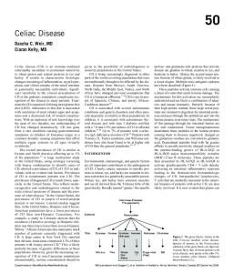
100% 80%
No Endoscopic GERD Endoscopic GERD
60% 40% 20% 0% 1994
1996
1998
Year
Figure 4. Increasing prevalence of endoscopic GERD among dyspeptic Iranians (n = 4545) undergoing upper gastrointestinal endoscopy between 1994 and 1999 in Tehran.
GERD in Iran Several studies from East and South-East Asia indicate a lower prevalence of GERD and its complications in this area, although recent reports have shown an increased prevalence. There are no well-defined population-based studies in Iran. A retrospective study of endoscopies at a single referral gastrointestinal center in Tehran between 1994 and 1999 showed that endoscopic GERD (esophagitis, Barrett’s metaplasia, and peptic stricture) in patients referred because of dyspepsia increased from less than 20% in 1994 to more than 70% in 1999 (Figure 4).6 In another prospective study in the same referral center in Tehran performed on 343 dyspeptic patients, 77% of patients had endoscopic evidence of GERD.5 Although patients with a body mass index (BMI) of more than 25 kg/m2 were more likely to experience acid regurgitation, there was no correlation between increasing BMI and endoscopic esophagitis or other GERD symptoms. The same study showed a higher prevalence of globus sensation and chronic cough among patients with erosive esophagitis seen on endoscopy. Therefore, these extra-esophageal symptoms should be addressed carefully in a patient being evaluated for dyspepsia and GERD. Epigastric burning sensation and bloating sensed most prominently in the epigastrium were associated with the presence of erosive GERD. Various studies, including our own, have shown that nausea, especially when it happens in the morning and is associated with morning anorexia, may be the only symptom of GERD and should alert the physician to its presence. This is especially true in younger patients. In this prospective study, H. pylori was not more common among GERD 136 Archives of Iranian Medicine, Vol 6, No 2, April 2003
patients, but it was significantly more common among patients with dysplasia at the gastroesophageal junction. 5 In another study in 3,328 healthy blood donors, 3.9% reported daily GERD symptoms, 5.2% reported weekly GERD symptoms, and 5.9% reported monthly GERD symptoms. This gives a prevalence of around 14%, which is comparable to western reports. In a population-based endoscopic screening program in Ardabil Province performed for detection of asymptomatic cancerous and precancerous lesions, 36.7% of the 504 individuals screened had endoscopic esophagitis. 8 About 60% of the population studied in Ardabil had a BMI of more than 25 kg/m2 . Endoscopic esophagitis counts for about half of patients with GERD at most. Therefore, these data show that GERD is rapidly increasing in Iran. It is currently the most common endoscopic finding in patients referred for endoscopy. The authors’ personal experiences also imply that GERD is probably the most common cause of referral to a gastroenterologist in Tehran. A population-based study is awaited and needs validation of questionnaires detecting GERD in a systematic manner. 15 Why are we facing such an increase in a once uncommon problem? Lifestyle and dietary habit changes are important factors. According to official national data, each Iranian citizen consumes about 42 liters of carbonated beverages per year. These drinks contain considerable amounts of sucrose (28 g/300 mL) in addition to the pressurized gas. This may contribute to the increased prevalence of GERD. The same report indicates that about 40% of Iranians consume more food than they need and the average Iranian consumes 40% more carbohydrate and 30% more fat than needed. Added to these is the increased interest of the community in a more westernized diet containing junk and fast food (sandwiches, pizza, potato chips, and snacks) that are fatty and spicy themselves and are usually served with fatty and spicy sauces. Rapid eating (which is associated with consumption of junk food), sedentary lifestyle due to lack of adequate exercise and decreased walking, and stressful life conditions should be added to this constellation. Therefore, it is important to advise patients and their families about these aspects of the disease and counsel them to modify these risk factors as far as possible. Otherwise, we should expect a continuing rise in the prevalence of GERD and its complications,
R. Malekzadeh, S. Nasseri-Moghaddam, M. Sotoudeh
including esophageal adeno-carcinoma, in our community. Summary GERD is increasing in prevalence and is the most commonly reported upper gastrointestinal disease in the West and probably in Iran. It has various clinical presentations, the most typical and common being heartburn and acid regurgitation. Extraesophageal and atypical symptoms such as nausea should be carefully addressed by the physician, as they may be the sole presenting symptoms of GERD. GERD may cause important complications including a significant decrease in quality of life, erosive esophagitis, esophageal ulcers and strictures, specialized columnar metaplasia (Barrett’s esophagus), dysplasia, and adenocarcinoma. The pathophysiology of GERD is not well understood, but tLESR, hypotensive LES, SHH, hypersensitive esophagus, and DGER are among the most widely accepted mechanisms. Current knowledge indicates that probably different pathophysiologic mechanisms are responsible for different subgroups of disease (i.e. erosive GERD, NERD, and Barrett’s esophagus). GERD, regardless of presence or absence of esophageal erosions, is a chronic and recurrent disease that seriously affects quality of life, mandating long-term medical attention. It is important to discuss this issue with the patient in an appropriate manner. The nonerosive form should be taken as seriously as the erosive form and be treated appropriately and aggressively. Various methods for treatment are available including lifestyle modifications, H2 blockers or PPIs, and surgical and endoscopic treatments. It is generally recommended to start with general measures plus H2 blockers or PPIs until the patient’s symptoms are controlled and then maintain them on the lowest effective dose, either continuously or intermittently. Surgery is usually reserved for patients who respond adequately to medical therapy but suffer from frequent relapses and are either reluctant to take their medication (long-term) or are too young to receive potent acidreducing agents for an undefined period of time. Careful patient selection and discussion of postsurgery outcomes are important before sending patients to surgery. A large minority of these patients may need maintenance antireflux medication within 10 years after surgery, despite an initially successful operation. Endoscopic treatments are on the runway and await more
clinical studies. With better understanding of the pathophysiologic mechanisms of GERD, better management plans will be feasible. Data from our own country indicate that GERD is increasing in various parts of the country with different ethnic and socioeconomic backgrounds and we should be alert to this chronic-recurrent disease. Prompt diagnosis and adequate maintenance therapy are the cornerstones of appropriate management of these patients. Dietary and lifestyle changes contribute significantly to this new epidemic and should be addressed properly in each counseling occasion with patients and their families.
References 1
2
3
4
5
6
7
8
9
10
11
12 13
14
Locke GR, Talley NJ, Fett SL, et al. Prevalence and clinical spectrum of gastroesophageal reflux: a population-based study in Olmsted County, Minnesota. Gastroenterology. 1997; 112: 1448 – 56. Fass R. Epidemiology and pathophysiology of symptomatic gastroesophageal reflux disease. Am J Gastroenterol. 2003; 98 (3 Suppl): 2 – 7. Hollenz M, Stolte M, Labenz J. Prevalence of gastrooesophageal reflux disease in general practice [in German]. Dtsch Med Wochenschr. 2002; 127: 1007 – 12. Shaheen N, Ransohoff DF. Gastroesophageal reflux, Barrett esophagus, and esophageal cancer: scientific review. JAMA. 2002; 287: 1972 – 81. Nasseri-Moghaddam S, Malekzadeh R, Sotoudeh M, et al. Lower esophagus in dyspeptic Iranian patients. J Gastroenterol Hepatol. 2003; 18: 315 – 21. Sotoudehmanesh R, Nasseri-Moghaddam S, Shirazian N, et al. Prevalence of endoscopic gastroesophageal reflux disease in a 6-year period. Endoscopy. 2000; 32: 33. Pourshams A, Malekzadeh R, Azimi K, et al. Relative frequency of functional gastrointestinal diseases among Iranian blood donors. Digest J. 2001; 31: 27. Saidi F, Malekzadeh M, Sotoudeh M, et al. Endoscopic esophageal cancer survey in the western part of the Caspian Littoral. Dis Esophagus. 2002; 15: 214 – 8. Yeh C, Hsu CT, Ho AS, et al. Erosive esophagitis and Barrett’s esophagus in Taiwan: a higher frequency than expected. Dig Dis Sci. 1997; 42: 702 – 6. Nebel OT, Fornes MF, Castell DO. Symptomatic gastroesophageal reflux: incidence and precipitating factors. Am J Dig Dis. 1976; 21: 953 – 6. Locke GR, Talley NJ, Weaver AL, et al. A new questionnaire for gatsroesophageal reflux disease. Mayo Clin Proc. 1994; 69: 539 – 47. Makuuchi H. Clinical study of esophageal hiatal hernia. Jpn J Gastroenterol. 1985; 79: 1557 – 67. Kang JY, Tay HH, Yap I, et al. Low frequency of endoscopic esophagitis in Asian patients. J Clin Gastroenterol. 1993; 16: 70 – 3. Chang CS, Poon SK, Lien HC, et al. The incidence of reflux esophagitis among the Chinese. Am J
Archives of Iranian Medicine, Vol 6, No 2, April 2003 137
Gastroesophageal Reflux Disease: The New Epidemic
Gastroenterol. 1997; 92: 668 – 71. 15 Habibi R, Rafaat-Zand K, Nasseri-Moghaddam S. Assessment of Validity, Reliability and Feasibility of the Mayo GERD Questionnaire in Tehran, Iran [Doctorate Thesis]. Tehran: Tehran University of Medical Sciences; 2000. 16 Dent J, Brun J, Fendrick AM, et al. An evidencebased appraisal of reflux disease management. The Genva Workshop Report. Gut. 1999; 44: 1 – 16. 17 Carlsson R, Dent J, Watts R, et al. Gastroesophageal reflux disease in primary care: an international study of different treatment strategies with omeprazole. Eur J Gastroenterol Hepatol. 1998; 10: 119 – 24. 18 Lagergren J, Bergstrom R, Lindgren A, et al. Symptomatic gastroesophageal reflux as a risk factor for esophageal adenocarcinoma. N Engl J Med. 1999; 340: 825 – 31. 19 Weston AP, Krmpotich P, Makdisi WF, et al. Short segment Barrett's esophagus: clinical and histological features, associated endoscopic findings, and association with gastric intestinal metaplasia. Am J Gastroenterol. 1996; 91: 981– 6. 20 Spechler SJ. The columnar-lined esophagus: a riddle wrapped in a mystery inside an enigma. Gut. 1997; 41: 710 – 1. 21 Spechler SJ. The columnar-lined esophagus. History, terminology, and clinical issues. Gastroenterol Clin North Am. 1997; 26:455 – 66. 22 Spechler SJ. Short and ultrashort Barrett's esophagus—what does it mean? Semin Gastrointest Dis. 1997; 8: 59 – 67. 23 Hirota WK, Loughney TM, Lazas DJ, et. Specialized intestinal metaplasia, dysplasia , and cancer of the esophagus and esophagogastric junction: prevalence and clinical data. Gastroenterology. 1999; 116: 277 – 85. 24 Trudgill NJ, Suvarna SK, Kapur KC, et al. Intestinal metaplasia at the squamocolumnar junction in patients attending for diagnostic gastroscopy. Gut. 1997; 41: 585 – 9. 25 Spechler SJ, Zeroogian JM, Antoniolli DA, et al. Prevalence of metaplasia at the gastrooesophageal junction. Lancet. 1994; 344: 1533 – 6. 26 Glise H, Hallerback B, Johansson B. Quality of life assessments in the evaluation of gastroesophageal reflux and peptic ulcer disease before, during and after treatment. Scand J Gastroenterol Suppl. 1995; 208: 133 – 5. 27 Revicki DA, Wood M, Maton PN, et al. The impact of gastroesophageal reflux disease on health-related quality of life. Am J Med. 1998; 104: 252 – 8. 28 Glise H, Hallerback B, Wiklund I. Quality of life: a reflection of symptoms and concerns. Scand J Gastroenterol Suppl. 1996; 221: 14 – 7. 29 Dimenas E, Carlsson G, Glise H, et al. Relevance of norm values as part of the documentation of quality of life instruments for use in upper gastrointestinal disease. Scand J Gastroenterol Suppl. 1996; 221: 8 – 13. 30 Lind T, Havilund T, Carlsson R, et al. Heartburn without esophagitis: efficacy of omeprazole therapy and features of therapeutic response. Scand J Gastroenterol. 1997; 32: 974 – 9. 31 Orlando RC. Overview of the mechanisms of
138 Archives of Iranian Medicine, Vol 6, No 2, April 2003
32
33
34
35
36
37
38
39
40
41
42
43
44
45
46
47
48
gastroesophageal reflux. Am J Med. 2001; 111 (Suppl 8A): 174 – 7. Vaezi MF, Richter JE. Duodenogastroesophageal reflux and methods to monitor nonacidic reflux. Am J Med. 2001; 111(Suppl 8A): 160 – 8. Mittal RK, Holloway RH, Penagini R, et al. Transient lower esophageal sphincter relaxation. Gastroenterology. 1995; 109: 601 – 10. Kahrilas PJ, Gupta RR. Mechanisms of acid reflux associated with cigarette smoking. Gut. 1990; 31: 4 – 10. Sloan S, Rademaker AW, Kahrilas PJ. Determinants of gastroesophageal junction incompetence: hiatal hernia, lower esophageal sphincter, or both? Ann Intern Med. 1992; 117: 977 – 82. Jones MP, Sloan SS, Rabine JC, et al. Hiatal hernia size is the dominant determinant of esophagitis presence and severity in gastroesophageal reflux disease. Am J Gastroenterol. 2001; 96: 1711 – 7. Kahrilas PJ, Shi G, Manka M, et al. Increased frequency of transient lower esophageal sphincter relaxation induced by gastric distension in reflux patients with hiatal hernia. Gastroenterology. 2000; 118: 688 – 95. Ho SC, Chang CS, Wu CY, et al. Ineffective esophageal motility is a primary motility disorder in gastroesophageal reflux disease. Dig Dis Sci. 2002; 47: 652 – 6. Dent J. Recent views on the pathogenesis of gastrooesophageal reflux disease. Baillieres Clin Gastroenterol. 1987; 1: 727 – 45. Korsten MA, Rosman AS, Fishbein S, et al. Chronic xerostomia increases esophageal acid exposure and is associated with esophageal injury. Am J Med. 1991; 90:701 – 6. Rodriguez-Stanley S, Robinson M, Earnest DL, et al. Esophageal hypersensitivity may be a major cause of heartburn. Am J Gastroenterol. 1999; 94: 628 – 31. Trimble KC, Douglas S, Pryde A, et al. Clinical characteristics and natural history of symptomatic but not excess gastroesophageal reflux. Dig Dis Sci. 1995; 40: 1098 – 104. Osugi H, Higashino M, Kaseno S, et al. Ambulatory intraesophageal bilirubin monitoring in Japanese patients with gastroesophageal reflux. J Gastroenterol. 2002; 37: 697 – 702. Kivilaakso E, Fromm D, Silen W. Effect of bile salts and related compounds on isolated esophageal mucosa. Surgery. 1980; 87: 280 – 5. Vaezi MF, Richter JE. Role of acid and duodenogastroesophageal reflux in gastroesophageal reflux disease. Gatroenterology. 1996; 111: 1192 – 9. Stipa F, Stein HJ, Feussner H, et al. Assessment of nonacid esophageal reflux: comparison between longterm reflux aspirartion test and fiberoptic bilirubin monitoring. Dis Esophagus. 1997; 10: 24 – 8. Champion G, Richter JE, Vaezi MF, et al. Duodenogastroesophageal reflux : relationship to pH and importance in Barrett’s esophagus. Gastroenterology. 1994; 107: 747 – 54. Sears RJ, Champion GL, Richter JE. Characteristics of partial gastrectomy patients with esophageal symptoms of duodenogastric reflux. Am J Gastroenterol. 1995; 90: 211 – 5.
R. Malekzadeh, S. Nasseri-Moghaddam, M. Sotoudeh
49 Gutschow CA, Schroder W, Holscher AH. Barrett’s esophagus: what is the poison-alkaline, biliary or acidic reflux? Dis Esophagus. 2002; 15: 5 – 9. 50 Tytgat G, Langenberg W, Rauws E, et al. Campylobacter-like organism (CLO) in the human stomach. Gastroenterology. 1985; 88: 1620. 51 Sipponen P, Varis K, Fraki O, et al. Cumulative 10year risk of symptomatic duodenal and gastric ulcer in patients with or without chronic gastritis: a clinical follow-up study of 454 outpatients. Scand J Gastroenterol. 1990; 25: 966 – 73. 52 Nomura A, Stemmermann GN, Chyou PH, et al. Helicobacter pylori infection and the risk for duodenal and gastric ulceration. Ann Intern Med. 1994; 120: 977 – 81. 53 Leoci C, Lerardi E, Chiloiro M, et al. Incidence and risk factors of duodenal ulcer. A retrospective cohort study. J Clin Gastroenterol. 1995; 20: 104 – 9. 54 Laine L, Hopkins RJ, Girardi LS, et al. Has the impact of Helicobacter pylori therapy on ulcer recurrence in the United States been overstated? A meta-analysis of rigorously designed trials. Am J Gastroenterol. 1998; 93: 1409 – 3. 55 Parsonnet J, Vandersteen D, Goates J, et al. Helicobacter pylori infection in intestinal- and diffuse-type gastric adenocarcinomas. J Natl Cancer Inst. 1991; 83: 640 – 3. 56 The EUROGAST Study Group. An international association between Helicobacter pylori and gastric cancer. Lancet. 1993; 341: 1359 – 62. 57 Wotherspoon AC, Ortiz-Hidalgo C, Falzon M, et al. Helicobacter pylori-associated gastritis and primary B-cell gastric lymphoma. Lancet. 1991; 338: 1175 – 6. 58 Parsonnet J, Hansen S, Rodriguez L, et al. Helicobacter pylori infection and gastric lymphoma. N Engl J Med. 1994; 330: 1267 – 71. 59 Shirota T, Kusano M, Kawamura O, et al. Helicobacter pylori infection correlates with severity of reflux esophagitis: with manometry findings. J Gastroenterol. 1999; 34: 553 – 9. 60 Dent J. Gastrooesophageal reflux disease. Digestion. 1998; 59: 433 – 45. 61 Dent J. Definitions of reflux disease and its separation from dyspepsia. Gut. 2002; 50 (Suppl 4): 17 – 20. 62 Castell D, Katz P. The acid suppression test for unexplained chest pain. Gastroenterology. 1998; 115: 222 – 4. 63 Field SK, Underwood M, Brant R, et al. Prevalence of gastroesophageal reflux symptoms in asthma. Chest. 1996; 109: 316 – 22. 64 Harding SM, Richter JE. The role of gastroesophageal reflux in chronic cough and asthma. Chest. 1997; 111: 1389 – 402. 65 Harding SM, Richter JE, Guzzo MR, et al. Asthma and gastroesophageal reflux: acid suppressive therapy improves asthma outcome. Am J Med. 1996; 100: 395 – 405. 66 Waring JP, Fackler WK. Ambulatory esophageal pH monitoring: proper probe placement and normal values. Aliment Pharmacol Ther. 2001; 15: 1155 – 62. 67 Bechi P, Pucciani F, Baldini F, et al. Long-term ambulatory enterogastric reflux monitoring: validation of a new fiberoptic technique. Dig Dis Sci.
1993; 38: 1297 – 306. 68 Vaezi MF, Lacamera RG, Richter JE. Validation studies of Bilitec 2000: an ambulatory duodenogastric reflux monitoring system. Am J Physiol. 1994; 267: 1050 – 7. 69 Lundell LR, Dent J, Bennett JR, et al. Endoscopic assessment of oesophagitis: clinical and functional correlates and further validation of the Los Angeles Classification. Gut. 1999; 45: 172 – 80. 70 Fass R, Fennerty B, Vakil N. Nonerosive reflux disease—current concepts and dilemmas. Am J Gastroenterol. 2001; 96: 303 – 14. 71 Tew S, Jamieson TS, Pilwsky I, et al. The illness behavior of patients with gastroesophageal reflux disease with and without endoscopic esophagitis. Dis Esophagus. 1997; 10: 9 – 15. 72 Fass R, Fennerty MB, Ofman JJ, et al. The clinical and economic value of a short course of omeprazole in patients with noncardiac chest pain. Gastroenterology. 1998; 115: 42 – 9. 73 Fass R, Ofman JJ, Gralnek IM, et al. Clinical and economic assessment of the omeprazole test in patients with symptoms suggestive of gastroesophageal reflux disease. Arch Intern Med. 1999; 159: 2161 – 8. 74 Fennerty MB. Strategies for diagnosis and short and long-term treatment: respondent overview. Am J Gastroenterol. 2001; 96: 27 – 8. 75 Madsen E, Aksglaede K, Jacobsen NO, et al. Gastrooesophageal reflux demonstrated by radiography. A supplement to 24-hour pH monitoring. Acta Radiol. 2001; 42: 521 – 5. 76 Howard PJ, Maher A, Pryde L, et al. Symptomatic gastroesophageal reflux, abnormal esophageal acid exposure, and mucosal acid sensitivity are three separate, though related, aspects of gastroesophageal reflux disease. Gut. 1991; 32: 128 – 32. 77 Wenzl TG. Investigating esophageal reflux with the intraluminal impedance technique. J Pediatr Gastroenterol Nutr. 2002; 34: 121 – 8. 78 Silverstein MD, Petterson T, Talley NJ. Initial endoscopy or empirical therapy with or without testing for Helicobacter pylori for dyspepsia: a decision analysis. Gastroenterology. 1996; 110: 72 –83. 79 Geldof H, Hazelgoff B, Otten MH. Two different doses of cisapride in the treatment of reflux esophagitis: a double-blind comparison with ranitidine. Aliment Pharmacol Ther. 1993; 7: 409 –15. 80 Pope CE 2nd. Acid-reflux disorders. N Engl J Med. 1994; 331: 656 – 60. 81 Zagari M, Villa KP, Freston JW. Proton pump inhibitors versus H2-receptor antagonists for the treatment of erosive gastroesophageal reflux disease: a cost-cooperative study. Am J Managed Care. 1995; 1: 247 – 55. 82 Dent J. Management of reflux disease. Gut. 2002; 50 (Suppl 4): 67 – 71. 83 Klinkenberg-Knol EC, Festen H, Jansen J, et al. Long-term treatment with omeprazole for refractory reflux esophagitis: efficacy and safety. Ann Intern Med. 1994; 121: 161 – 7. 84 Klinkenberg-Knol EC. Eleven years’ experience of
Archives of Iranian Medicine, Vol 6, No 2, April 2003 139
Gastroesophageal Reflux Disease: The New Epidemic
85
86
87
88
89
90
91
continuous maitenance treatment with omeprazole in GERD patients. Gastroenterology. 1998; 114: 180. Klinkenberg-Knol EC. Long-term omeprazole treatment in resistant gastroesophageal reflux disease: efficacy, safety, and influence on gastric mucosa. Gastroenterology. 2000; 118: 661 – 9. Katz PO, Anderson C, Khoury R, et al. Gastroesophageal reflux associated with nocturnal gastric acid breakthrough on proton pump inhibitors. Aliment Pharmacol Ther. 1998; 12: 1231 – 4. Lidums I, Lehmann A, Checklin H, et al. Control of transient lower esophageal sphincter relaxations and reflux by the GABA(B) agonist baclofen in normal subjects. Gastroenterology. 2000; 118: 7 – 13. Wolfe MM, Sachs G. Acid suppression: optimizing therapy for gastroduodenal ulcer healing, gastroesophageal reflux disease, and stress-related erosive syndrome. Gatsroenterology. 2000; 118 (Suppl 1): 9 – 31. Bardhan KD, Müller-Lissner S, Bigard MS, et al. Symptomatic gastrooesophageal reflux disease: double blinded controlled study of intermittent treatment with omeprazole or ranitidine. The European Study Group. BMJ. 1999; 318: 502 – 7. Lundell L, Miettinen P, Myrvold HE, et al. Long-term management of gastrooesophageal reflux disease with omeprazole or open antireflux surgery: results of a prospective, randomized clinical trial. The Nordic GORD Study Group. Eur J Gastroenterol Hepatol. 2000; 12: 879 – 87. Allgood PC, Bachmann M. Medical or surgical treatment for chronic gastroesophageal reflux? A systematic review of published evidence of effectiveness. Eur J Surg. 2000; 166: 713 – 21.
140 Archives of Iranian Medicine, Vol 6, No 2, April 2003
92 Spechler SJ, Lee E, Ahnen D, et al. Long-term outcome of medical and surgical therapies for gastroesophageal reflux disease: follow-up of a randomized controlled trial. JAMA. 2001; 285: 2331 – 8. 93 Watson DI, Jamieson GG, Baigrie RJ, et al. Laparoscopic surgery for gastrooesophageal reflux: beyond the learning curve. Br J Surg. 1996; 83: 1284 – 7. 94 Lehman GA, Dunne DP, Hieston K, et al. Suturing plication of cardia with endocinch devide: effect of supplemental cautery. A human prospective randomized trial. Gastrointest Endosc. 2002; 55: AB260. 95 Filipi C, Lehman G, Rothstein RI, et al. Transoral endoscopic sututring for gastroesophageal reflux disease: a multicenter trial. Gastrointest Endosc. 2001; 53: 416. 96 Chuttani R, Kozarek R, Sachdev R, et al. A novel endoscopic full-thickness plicator for the treatment of GERD: early clinical results (abstract). Endoscopy. 2001; 33S: 2200. 97 Chuttani R, Sud R, Sachdev G, et al. Endoscopic fullthickness plicator for GERD: final results of human pilot study (abstract). Gastrointest Endosc. 2002; 55: AB258. 98 Triadafilopoulos G, Dibaise JK, Nostrant TT, et al. Radiofrequency energy delivery to the gastroesophageal junction for the treatment of GERD. Gastrointest Endosc. 2001; 53: 407. 99 Triadafilopoulos G, Dibaise JK, Nostrant TT, et al. The Stretta procedure for the treatment of GERD: 6and 12-month follow-up of the US open label trial. Gastrointest Endosc. 2002; 55: 149.