BEHAVIOR Wilhelm et al.MODIFICATION / RESPIRATORY/ September DYSREGULATION 2001
Respiration is a complex physiological system affecting a variety of physical processes that can act as a critical link between mind and body. This review discusses the evidence for dysregulated breathing playing a role in three clinical syndromes: panic disorder, functional cardiac disorder, and chronic pain. Recent technological advances allowing the ambulatory assessment of endtidal partial pressure of CO2 (PCO2) and respiratory patterns have opened up new avenues for investigation and treatment of these disorders. The latest evidence from laboratories indicates that subtle disturbances of breathing, such as tidal volume instability and sighing, contribute to the chronic hypocapnia often found in panic patients. Hypocapnia is also common in functional cardiac and chronic pain disorders, and studies indicate that it mediates some of their symptomatology. Consistent with the role of respiratory dysregulation in these disorders, initial evidence indicates efficacy of respiration-focused treatment.
Respiratory Dysregulation in Anxiety, Functional Cardiac, and Pain Disorders Assessment, Phenomenology, and Treatment FRANK H. WILHELM Stanford University
RICHARD GEVIRTZ California School of Professional Psychology—San Diego
WALTON T. ROTH Department of Veterans Affairs Health Care System
Respiration is a physiological function situated strategically at the interface of mind and body. It is capable of operating automatically, its usual mode, but can be brought under voluntary control at least briefly. In contrast to autonomic functions such as heart rate or blood pressure, respiratory changes are perceivable if attention is focused on them. AUTHORS’ NOTE: Preparation of this article was supported by National Institutes of Health Grant MH56094 and the Department of Veterans Affairs. BEHAVIOR MODIFICATION, Vol. 25 No. 4, September 2001 513-545 © 2001 Sage Publications
513
514
BEHAVIOR MODIFICATION / September 2001
Respiration is essential for the maintenance of life, with every organ system depending on proper gas exchange. Its regulation is subject to complex homeostatic mechanisms, and derangement of regulation can have severe health consequences. The centrality of breathing in human emotion, health, and consciousness is obvious from references to it in intellectual domains as wide ranging as literature, philosophy, religion, and physiology. Given these facts, it is surprising that respiration had not been widely recognized in the field of psychophysiology until recently. In response to the lack of emphasis on respiration in psychological science, the International Society for the Advancement of Respiratory Psychophysiology was formed in 1984 (Timmons & Ley, 1994). Meanwhile, advances in pulmonary physiology and a better understanding of the central and peripheral processes involved in breathing regulation (Dempsey & Pack, 1995) have made it clear that homeostasis in the human body is greatly influenced by respiratory behaviors and especially by changes in arterial carbon dioxide (CO2) concentration. Building on this knowledge, clinical psychophysiologists have recognized that interactions between emotional states, respiratory behaviors (such as fast and deep breathing, strained breathing, sighing, yawning, or inhibited breathing), and physiological changes at the level of chemical blood composition and autonomic nervous system regulation play a role in disorders such as hypertension (Anderson, Dhokalia, Parsons, & Bagrov, 1996), hyperventilation syndrome (Folgering, 1999b), panic disorder (PD), functional cardiac disorder (FCD), and chronic pain syndrome (which are reviewed below). Furthermore, dysregulated respiratory behavior may play a role in the quality of performance both in the workplace and in sports (Forster & Pan, 1988; Schleifer & Ley, 1994). We will first discuss the basic respiratory physiology processes, which are crucial for understanding the impact dysregulated breathing can have on the body. Then, we will present common measurement methods and review the data on respiratory abnormalities in anxiety, functional cardiac, and pain disorders, with a focus on anxiety, where most work has been done. Finally, we will discuss implications for treatment. As the reader will see, we are in the early stages of gathering a scientific database in this fascinating mind-body research area.
Wilhelm et al. / RESPIRATORY DYSREGULATION
515
BACKGROUND PHYSIOLOGY OF BREATHING
The respiratory system functions basically to supply oxygen (O2) to the organs of the body to use in the chemical combustion of fuel (carbohydrates) and to rid the body of the gas CO2, resulting from this metabolism. Although CO2 is often thought to be only a waste product, it plays a crucial role in maintaining acid-base balance within the internal milieu of the body. During inspiration, a contraction of the diaphragm and intercostal muscles increases the volume of the thoracic cavity, and air is drawn into the lung. It passes through the oral and nasal passages, the trachea, bronchi, and bronchioles, where it is cleaned, warmed, and moistened, before it finally enters the alveoli where the gas exchange with the body takes place. The alveoli are extremely small sacs that spread out 2 to produce an enormous surface area (50 to 100 m in adults) and are surrounded by pulmonary capillaries. The tissues are only about ½ micron thick and allow passive diffusion of O2 and CO2 through them. Because inhaled room air typically consists of about 21% O2 and only 0.05% CO2 (the rest is mainly nitrogen), the O2 in the alveoli has a very high partial pressure (PO2) of about 105 mm Hg. The result is a tendency to diffuse through the walls of the alveoli and capillaries into the blood. At the same time, the alveolar CO2 at the end of inspiration normally has a relatively low partial pressure (PCO2) of about 40 mm Hg, which results in diffusion of the CO2 from venous blood (where it has a higher PCO2 of about 45 mm Hg) into the alveoli. Thus, the exhaled breath is enriched in CO2, having a partial pressure of about 40 mm Hg. The O2 is absorbed in the blood mainly by the hemoglobin (an iron containing protein) of the red blood cells and transported to the target organs. The amount of O2 carried depends on factors such as the amount of O2 available in the inhaled air, the temperature, and the blood’s pH, normally 7.4. The relationship between PO2 and the percentage of hemoglobin saturation (the amount of O2 already absorbed by the hemoglobin) is depicted in the hemoglobin-oxygen dissociation curve (see Figure 1). The x-axis represents the differing levels of
516
BEHAVIOR MODIFICATION / September 2001
Figure 1. Hemoglobin-oxygen dissociation curve and Bohr effect (see text). Hyperventilation causes a left-shift of the curve and a stronger affinity of oxygen (O2 ) to hemoglobin. Under certain conditions this can result in a reduced release of O2 to tissues.
PO 2. At normal blood pH, the curve is quite steep between 0 and 40 mm Hg PO2 and almost flat above 60 mm Hg PO2. The y-axis labeled hemoglobin saturation indicates how bound or stuck the oxygen is to the hemoglobin. The shape of the curve indicates that hemoglobin has a strong affinity to O2, which assures a stable O2 supply to the organs, even under difficult conditions (e.g., during physical exertion), when O2-demand is high, or at high altitudes, when much less O2 is available in the inhaled air. On the other hand, if overbound under certain conditions, the oxygen cannot dissociate and nourish target organs. When the arterial blood leaves the lungs, it normally has an O2 saturation of 97.5%. The CO2 is dissolved in the blood as carbonic acid. Any change in the concentration of the dissolved CO2 leads to a change in the blood pH, which shifts the hemoglobin-oxygen dissociation curve (see Figure 1) to the left (reduction in PCO2 and rise in pH) or to the right (rise in PCO2 and reduction in pH). This is called the Bohr effect, which can be seen as an ingenious invention of natural evolution because it plays a significant role in enhancing oxygenation of the blood in the lungs and then again in enhancing the release of oxygen from the blood to the tissue cells. As the blood passes through the lungs, CO2 diffuses from the blood into the alveoli. This reduces the blood PCO2 and increases its pH (due to a decrease in its carbonic acid content), which
Wilhelm et al. / RESPIRATORY DYSREGULATION
517
shifts the hemoglobin-oxygen dissociation curve to the left. Therefore the quantity of O2 that binds with the hemoglobin at any given alveolar PO2 becomes considerably increased, thus allowing greater O2 transport to the tissues. There, exactly the opposite effect occurs. CO2 entering the blood from the tissues shifts the curve to the right, which helps displace more O2 from the hemoglobin for delivery to the tissues (Guyton, 1995). Hyperventilation, defined as “breathing in excess of metabolic requirements” (Gardner, 1994), can occur dramatically and acutely with deep or fast breathing or as the result of more subtle perturbation of normal breathing, such as sighing. Overbreathing causes a variety of experienced symptoms, most characteristically dizziness, trembling, tingling in extremities, a feeling of unreality, and palpitations (Kroeze & van den Hout, 1998). Hyperventilation lowers blood PCO2 (hypocapnia), making blood pH slightly more alkalotic (higher) and causing a leftward shift in the oxygen-hemoglobin dissociation curve. Fried (1987) argued that this shift, combined with the constriction of the cerebral arteries produced by alkalosis (Kennealy, McLennan, Loudon, & McLaurin, 1980), impairs O2 delivery to regions of the brain to the extent that certain brain functions are jeopardized by the resulting hypoxia. Other effects of alkalosis are to increase intracellular calcium, sympathetic neuronal catecholamines, coronary artery tone, peripheral resistance, and cardiac arrhythmias and to decrease extracellular calcium and potassium, which affect neuronal excitability (Gilbert, 1999). These multiple, diverse effects have raised suspicion that hyperventilation is a mediator of a wide variety of anxiety-related or psychosomatic symptoms (panic, FCD, chronic pain, gastroenterological disturbance, asthma, hypertension, chronic muscle tension, etc.). However, in most cases this has not yet been demonstrated experimentally. The human body has evolved several redundant systems to limit an increase in arterial PCO2. When CO2 builds above the normal setpoint, for example, as a result of physical exercise or of attenuated ventilation, chemoreceptors located in the arteries (aorta, carotid) and brain stem powerfully stimulate ventilation, thus swiftly reducing PCO2. On the other hand, when PCO2 falls below the normal setpoint, the resulting reduction in stimulation of chemoreceptors has only mild effects
518
BEHAVIOR MODIFICATION / September 2001
on a reduction of ventilation, and PCO2 normalizes relatively slowly. This asymmetry may explain why hypocapnia can develop rather easily: Any disturbance to the fine-tuned breath-by-breath regulation of breathing would more likely lead to a decrease than an increase of PCO2. Furthermore, because of this regulatory asymmetry, under some circumstances (e.g., during extended stress or anxiety states) PCO2 may not normalize for a long time. Below we will present evidence from our laboratory that repeated sighing and slow PCO2 recovery may play a role in the lowered PCO2 often found in anxious patients. ASSESSMENT OF BREATHING
Before discussing the evidence for abnormal respiration in a variety of disorders, issues related to noninvasive measurement of respiration need to be considered. Traditional spirometric lung-function measures such as peak expiratory flow or vital capacity have received less attention from psychophysiologists than end-tidal PCO2 (PetCO2), respiratory rate, tidal volume, and minute ventilation (the amount of air inhaled in 1 minute). These measures can be obtained relatively unobtrusively, and thus measurement reactivity effects are minimized. Apprehension generated simply by breathing through a mouthpiece or in a closed system has been shown to increase ventilation and lower PCO2 (Perez & Tobin, 1985). Arterial PCO2. Arterial PCO2 is difficult to measure, but it can be approximated quite accurately by end-tidal PCO2, the level of PCO2 reached at the end of a normal expiration. At that time point, the expired gas mixture contains air from the depth of the lungs where alveolar gas exchange took place. PetCO 2 is measured by a capnometer (also called capnograph) using gas infrared spectroscopy. A sample of expired air from the nose or mouth is continuously sucked into the device by a pump, dehumidified, and analyzed for CO2 content (which is computed from the peak in the CO2 dispersion spectrum). Because barometric pressure and temperature can affect the measurement, many capnometers self-calibrate for these environmental changes. A typical normal level for PetCO2 in healthy adults at rest
Wilhelm et al. / RESPIRATORY DYSREGULATION
519
is 39 ± 4 mm Hg (or Torr) (an alternative metric is percentage PetCO2, for which 5.1% ± 0.5% is typical). An acute PCO2 reduction from baseline by about 10 mm Hg produces perceptible symptoms (which was shown by comparison to a placebo hyperventilation where CO2 was added to each inhalation to keep PCO2 at baseline levels)(Kroeze & van den Hout, 1998). However, physiological regulation processes may be disturbed by less acute reductions in PCO2. Respiration rate. Respiration rate can be acquired from a capnometer, a strain gauge around the abdomen, a thermistor taped to the nostrils, or an electromyogram placed on an accessory muscle (trapezius, scalene, etc.). A technique commonly used in research is inductive plethysmography (Respitrace™). Coils of insulated wire that are woven into cotton bands produce a weak electromagnetic field when excited by a high-frequency current that is proportional to the cross-sectional area enclosed by the band. A typical respiratory rate for healthy resting adults is 16 ± 4 breaths per minute. Tidal volume and minute ventilation. Tidal volume and minute ventilation measurement requires two strain gauges, one around the thorax and one around the abdomen. With adequate calibration (breathing into a spirometer or fixed volume bag) and multiple regression procedures, stretch-to-volume ratios for the thoracic and abdominal band output signal can be derived. The two band outputs are multiplied with these ratios and summed to obtain a continuous measure of lung volume change. A typical tidal volume for healthy resting adults is 500 ± 120 mL, and typical minute ventilation is 6 ± 1 L per minute (both depend considerably on body weight). Recently, a small portable capnometer, the Capnocount® mini (size = 6.5 × 12.8 × 3.5 cm, weight = 320 g), was developed (Weinmann Company, Germany) for use in patient monitoring outside the hospital (measurement of respiration rate and PCO2 during intubation, resucitation, and transport). Measured values are updated breath by breath on an LCD display. The accuracy of the device corresponds to standards for stationary capnometers. It automatically recalibrates PCO2 measurement to compensate for changing temperature and pressure. It can store PetCO2 and respiratory rate every 2 sec-
520
BEHAVIOR MODIFICATION / September 2001
onds for up to 8 hours. We are using this unmodified version in a treatment study for panic patients (Meuret, Wilhelm, & Roth, 2001 [this issue]). Patients take the device home to conduct breathing exercises, using the PCO2 and respiratory rate feedback from the device. The stored data from the practice sessions can be evaluated when the patient returns to the hospital. For ambulatory research projects, we employ a custom-made modification of the device. The raw PCO2 waveform is digitized and inputted continuously to the serial port of a Vitaport 2 digital recorder (Becker Meditec, Incorporated, Germany). This allows full disclosure analysis of expired PCO2 waveforms using our customized software (Wilhelm, Alpers, Meuret, & Roth, 2001). Inspection of the raw data often reveals complexities and distortions to end-tidal plateaus that may compromise data retrieved using online detection algorithms. An alternative to capnometry is transcutaneous PCO2 measurement (Garssen, Buikhuisen, Hornsveld, Klaver, & van Doornen, 1994). Like capnometry, this method has been validated to correlate well with arterial levels. It has often been employed in neonatal care units but can also be used ambulatorily. Typically, the sensor is placed on the skin of the forearm. Part of the sensor heats the skin to about 41° C to stimulate diffusion of CO2 from the blood through the skin, and another part, a selective membrane, allows CO2 molecules to pass. Their concentration is then determined within the sensor. Because the diffusion process is slow, changes in arterial PCO2 are registered with a delay of about 2 minutes, and sharp changes are blunted or entirely filtered out (Hoffmann, Essfeld, & Stegemann, 1990). Our own experimentation with this technique has shown certain unreliabilities and difficulties in calibration, and we ultimately abandoned this approach.
PHENOMENOLOGY OF RESPIRATORY DYSREGULATION
In this section, we present several lines of evidence emphasizing the importance of respiratory abnormalities in anxiety, functional cardiac, and pain syndromes. Clinical psychophysiology searches for abnormalities associated with disorders in an attempt to establish physiological mediators between psychological variables, such as
Wilhelm et al. / RESPIRATORY DYSREGULATION
521
stress, anxiety, pain, or mental disorders, and physical symptoms. Abnormalities can indicate mediating mechanisms for disorders, but they can also be correlated with the disorder without having any causal significance. Such physiological markers can be useful for diagnosis and for helping identify genes involved in specific disorders. Unfortunately, most clinical studies reviewed below do not allow us to draw clear conclusions about whether abnormalities are the actual mediators of part or all of the symptomatology of a disorder. However, because hyperventilation has quite potent effects on a variety of physiological processes and can produce many physical symptoms, chances are high that it mediates at least part of the pathophysiology. ANXIETY DISORDERS
Clinical observations of severe respiratory distress in patients with anxiety disorders have stimulated much research to identify respiratory abnormalities associated with this syndrome. Dyspnea (shortness of breath) has been found to be one of the most commonly reported symptoms of panic (McNally, Hornig, & Donnell, 1995), together with palpitations and faintness. Hyperventilation, and more recently, respiratory instability have been the focus of systematic study, which produced a sizable database regarding the specificity and situational determinants of respiratory dysregulation in this disorder (reviewed below). In addition, recent reports indicate that PD may not be a single biological entity, because it can be divided into subtypes with and without prominent respiratory symptoms (Aronson & Logue, 1988; Briggs, Stretch, & Brandon, 1993; Shioiri, Someya, Murashita, & Takahashi, 1996). The respiratory subtype has been found to be more physiologically reactive to CO2 inhalation (a respiratory stimulant) (Biber & Alkin, 1999) and voluntary hyperventilation (Hegel & Ferguson, 1997). Unfortunately, most of the research (and all studies reviewed below) has not distinguished between these subtypes, which may have reduced effect sizes for respiratory findings and may explain some of the inconsistencies. In the following, we review evidence for respiratory abnormalities in anxiety disorders and present recent results from our laboratory. The focus will be on PD because of converging evidence that respiratory factors play a prominent role in it.
522
BEHAVIOR MODIFICATION / September 2001
Hyperventilation
Increased ventilation is a component of the fight-flight response, an evolutionarily evolved adaptive response to danger that is largely sympathetically mediated and prepares the individual for immediate action. However, if this preparatory increase in ventilation is not followed by sufficient physical activity, the arterial PCO2 level falls (hypocapnia). Thus, hypocapnia can be induced experimentally in healthy individuals by making them anxious (Ley & Yelich, 1998). Conversely, voluntary hyperventilation can lead to a pattern of physical and psychological symptoms in healthy individuals resembling acute anxiety symptoms (e.g., shortness of breath, palpitations, dizziness, faintness, or tingling in extremities). A hyperventilation syndrome was postulated in the 1930s that could explain a variety of psychosomatic complaints and mood disturbances (Kerr, Dalton, & Gliebe, 1937). When PD was established as a diagnostic category, episodes of hyperventilation were specifically linked to panic attacks because a symptomatic overlap in the two entities is apparent (Ley, 1985). In its basic form, this perspective postulates that hyperventilation itself produces the mood changes and symptoms observed in anxiety disorders (Lum, 1987). Cognitivebehavioral theories of panic have expanded this idea and postulated a positive interoceptive feedback loop between anxiety and hyperventilation (Margraf & Ehlers, 1990). In essence, mild anxiety is thought to be accompanied by bodily changes such as heart racing or mild hyperventilation. Hypochondrical interpretation of the resulting bodily sensations (and perhaps phobic emotional conditioning from previous panic attacks) leads to even more anxiety, hyperventilation, and bodily sensations (“vicious circle model of panic”). In contrast, a biological model of panic, the suffocation false alarm theory (Klein, 1992), postulates a physiological mechanism that in these patients is hypersensitive to increasing levels of inspired CO2 and triggers intense shortness of breath and panic, resulting in increased ventilation. Although the theories differ in the emphasis of triggering mechanisms and mediating pathways, they agree that much of the symptomatology of panic is produced by hyperventilation. The empirical database on hypocapnia in PD patients is sizeable. Voluntary hyperventilation has been used to provoke panic attacks
Wilhelm et al. / RESPIRATORY DYSREGULATION
523
(e.g., Gorman et al., 1984), and other panic provocations typically result in hypocapnia (e.g., Gorman, Goetz, et al., 1988). Hypocapnia has repeatedly emerged as a difference between PD patients and comparison groups during baseline periods (Hegel & Ferguson, 1997; Munjack, Brown, & McDowell, 1993; Papp et al., 1997; Rapee, 1986). One of our recent analyses (Wilhelm, Trabert, & Roth, 2001b) confirmed that at baseline, PD patients had lower PetCO2 than either patients with generalized anxiety disorder or controls. One study found low PCO2 in panic patients before treatment and a normalization afterwards (Salkovskis, Jones, & Clark, 1986). On the other hand, not all studies find that hypocapnia is specific to PD patients. In one study, both PD and other anxious patients at rest had lower PetCO2s than controls (van den Hout et al., 1992). Some studies have failed to find PetCO2 differences between PD patient and controls (Holt & Andrews, 1998; Woods et al., 1986). Another area of interest is the speed of recovery from respiratory perturbations. Slow recovery of PCO2 after voluntary hyperventilation has been found repeatedly in PD patients compared to healthy controls (Gorman, Fryer, et al., 1988; Maddock & Carter, 1991). Studies that used short hyperventilation or recovery periods have not found this abnormality (Hegel & Ferguson, 1997; Rapee, 1986), indicating that the potency of the stressor and a sufficient duration of measurement thereafter to allow for full recovery in healthy controls are important in the assessment. In fact, data from a recent study in our laboratory indicate that slow PetCO2 recovery from hyperventilation is a highly sensitive and specific marker for PD (Wilhelm, Gerlach, & Roth, in press). We recorded respiratory, autonomic, and experiential responses in 14 patients with PD, 24 patients with social phobia (SP), and 24 controls during six cycles of 1 minute of fast breathing alternating with 1 minute of recovery, followed by 3 minutes of fast breathing and 10 minutes of recovery. Speed of fast breathing was paced by a tone modulated at 18 cycles per minute, and depth by feedback aimed at achieving a PetCO2 of 20 mm Hg. During fast breathing, all three groups reached equal respiratory rates, minute volumes, and PetCO2s. However, PD and SP patients reported equally more anxiety than controls, and their feelings of shortness of breath and suffocation increased more from baseline. At the end of the final 10-minute recov-
524
BEHAVIOR MODIFICATION / September 2001
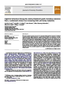
Figure 2. Means and standard errors of end-tidal PCO2 (left) and of band-limited tidal volume oscillation amplitude (right) for panic disorder patients, social phobia patients, and healthy controls at the 3-minute baseline, the 3-minute hyperventilation period (fast breathing with target PCO2 of 20 mm Hg), and the end of the 10-minute recovery period. NOTE: PD = panic disorder; SP = social phobia; CON = healthy controls; BASE = 3-minute baseline; FB = fast breathing; REC = end of 10-minute recovery period.
ery, PD patients reported being more aware of their breathing and shorter of breath, and their PCO2 levels had recovered much more slowly to normal levels than those in the other groups; F(2, 58) = 8.25, p < .001 (see Figure 2). These results indicate that PD patients differ from SP patients and controls in having slower symptomatic and physiological recovery. In spite of abundant laboratory evidence implicating hypocapnia in PD, recent ambulatory results have questioned its significance. In a series of ambulatory studies that assessed transcutaneous PCO2, only some panic attacks were accompanied by hypocapnia (3 out of 5, Hibbert & Pilsbury, 1988; 7 out of 15, Hibbert & Pilsbury, 1989; 1 out of 24, Garssen, Buikhuisen, & van Dyck, 1996). Thus, ambulatorymonitoring results seem to refute the hypothesis that hypocapnia accompanies (or even causes) naturally occurring panic attacks or contributes to their symptoms. However, this evidence cannot be viewed as conclusive. Ambulatory transcutaneous PCO2 monitors have limitations in detecting arterial PCO2 changes: Their typical response time of about 2 minutes results in attenuation and temporal smoothing of PCO2 changes so that small or short hyperventilation
Wilhelm et al. / RESPIRATORY DYSREGULATION
525
Figure 3. End-tidal PCO2 of a patient with panic disorder (Patient No. 10, 50-year-old woman), over the course of 24 hours. Data from individual breaths were averaged minute by minute. Recording started at 10:00 hours. The sleep period started at about 23:15 and ends at about 6:00. The steep drops in PCO2, to about 20 mm Hg, represent five scheduled 3-minute periods of voluntary hyperventilation (at about 10:00, 12:30, 16:30, 20:30, and 10:00 the next day).
episodes may be missed. Furthermore, the large variation in the fraction of panic attacks accompanied by hypocapnia across the studies suggests that the composition of the PD samples differed in their proportions of respiratory subtype patients. To put these ambulatory findings to the test, we recently started recording expired PCO2 waveforms in PD patients over the course of 24 hours. In addition, we recorded respiratory patterns using inductive plethysmography. Preliminary results (Figure 3 represents a typical case) indicate substantial variation in PCO2 in panic patients during day and night, even when they do not report panic attacks. The small number of patients and healthy controls tested does not yet afford meaningful statistical comparisons. Respiratory Instability and Sighing
Respiration is rarely completely regular, except in deep non-REM sleep and under anesthesia. Moderate instability even during quiet sitting reflects a mechanism that was termed dynamic homeostasis, a balancing act of respiratory regulation mechanisms to compensate for continuously changing subtle disturbances (e.g., from transient metabolic changes due to emotional and mental activation) (Bruce & Daubenspeck, 1995). On the other hand, increased levels of baseline respiratory instability are often associated with pathophysiology either at the level of the sensors of the regulation system (such as
526
BEHAVIOR MODIFICATION / September 2001
hypersensitivity or hyposensitivity of peripheral and central CO2 sensors or proprioceptive afferents) or at the effectors (such as stiffness in the diaphragm or intercostal muscles or inhibited nerve transmission to them). In addition, there is a large number of cortical and subcortical projections to the brain stem (the respiratory center) that can potentially influence moment-to-moment breathing. Thus, even the fleeting thought of a stressful event may influence the ongoing breathing pattern profoundly. As a consequence, respiratory instability is not necessarily a sign of physiological damage at the level of chemoreceptors or brain stem but could reflect a thinking style with prevalent, intensely stressful cognitions. One kind of respiratory instability is sighing. Sighing is a fundamental vertebrate behavior. Recently, sigh-like discharges were even isolated in vertebrate brainstem networks, together with sinusoidal and gasping-like signals (Lieske, Thoby-Brisson, Telgkamp, & Ramirez, 2000). Either of two prominent theories of PD, the suffocation false alarm (Klein, 1992) or hyperventilation (Ley, 1985) theories, might have predicted more deep breaths or sighs in PD patients: An individual with an overly sensitive suffocation alarm might be inclined to take periodic deeper breaths to lower the PCO2 safely below the threshold of the CO2 chemoreceptors, whose firing is one source for a feeling of suffocation. In contrast, hyperventilation theories ascribe panic to the lowered PCO2 itself, which deeper sigh breaths could cause. Sigh breaths are often defined as being breaths with a tidal volume exceeding twice the tidal volume of surrounding breaths. Several of the studies below did not measure sighs as distinct events but rather reported a highly correlated measure, the withinsubject variability of tidal volumes, using statistics such as standard deviation, coefficient of variation, or the mean square successive difference (von Neumann statistic). A number of studies evaluated sighing in clinically anxious groups. Finesinger (1943) demonstrated an increased sigh frequency in spirometric measurements among “psychoneurotic” patients. Tobin and colleagues (1983) showed that the sigh frequency measured with inductive plethysmography distinguished chronically anxious patients from patients with asthma, chronic obstructive pulmonary disease, restrictive lung disease, and primary pulmonary hyperten-
Wilhelm et al. / RESPIRATORY DYSREGULATION
527
sion. A panic provocation by lactate infusion increased sighing in PD patients (Schwartz, Goetz, Klein, Endicott, & Gorman, 1996). CO2 inhalation (5%), another panicogen, also increased respiratory instability in PD patients (quantified as length and number of breathing pauses and respiratory rate variability; unfortunately, sighing was not assessed) (Bystritsky, Craske, Maidenberg, Vapnik, & Shapiro, 2000). On the other hand, a recent study by Abelson, Weg, Nesse, and Curtis (2001) showed that the tendency to sigh was highly persistent in PD patients undergoing a panic provocation using the respiratory stimulant doxapram. Patients had large elevations in sigh frequency compared to controls in all phases of the experiment: baseline, doxapram challenge, and recovery. Even a cognitive intervention (essentially telling patients that everyone experiences bodily symptoms during the procedure and that they do not need to worry about them) successfully reduced the panic response but had no effect on this respiratory irregularity. One study looked at the breathing of PD patients during sleep and found increased tidal volume variability and a higher frequency of 5- to 10-second breathing pauses during REM phases (Stein, Millar, Larsen, & Kryger, 1995). Consistent with these results from studies of adults with PD, tidal volume variability differentiated children and adolescents with anxiety disorders from psychiatrically healthy children during a baseline before CO2 inhalation (Pine et al., 1998). Interestingly, anxious individuals who sigh frequently have been observed to have normal total lung capacity but lower vital capacity and higher residual capacity (Aljadeff et al., 1993), which indicates that they were breathing on top of hyperinflated lungs. In recent analyses in our laboratory, we used complex demodulation to quantify tidal volume variability (band-limited tidal volume oscillation amplitude). This method is more precise for quantifying oscillatory variability within distinct frequency bands than the measures used in the cited studies. We found higher sigh frequency (see Figure 4) and tidal volume oscillation amplitude within the spectral band of 0.004 to 0.14 Hz, corresponding to period lengths of 6.6 to 240 seconds in PD patients compared to patients with generalized anxiety disorder or controls during baseline (Wilhelm et al., 2001b). This variability was not entirely due to sighs, because after deletion
528
BEHAVIOR MODIFICATION / September 2001
Sigh frequency (1/min)
0.8
0.6
0.4
0.2
0.0 PD
GAD
CON
Figure 4. Sigh frequency per minute during 30 minutes of rest for patients with panic disorder, generalized anxiety disorder, and controls (left) and ensemble averaged PCO2 levels surrounding sighs for these groups (right). For comparison, PCO2 levels surrounding non-sigh breaths are shown. SOURCE: Reprinted by permission of Elsevier Science from “Characteristics of Sighing in Panic Disorder,” by Frank H. Wilhelm, Werner Trabert, and Walton T. Roth; published in Biological Psychiatry, Vol. 49, Pages 606-614, Copyright 2001 by the Society of Biological Psychiatry. NOTE = PD = panic disorder; GAD = generalized anxiety disorder; CON = control.
and replacement of sigh breaths with interpolated values, PD patients still had higher tidal volume oscillations than controls. Thus, it appears that sighs occur at the background of already unstable respiration. A motivation for this study was to look for unprovoked fluctuation (mini panic attacks) in several biological features known to be associated with full-blown attacks. Cardiovascular, respiratory, and electrodermal variables sensitive to anxiety seemed like good candidates, but surprisingly, only respiratory variables showed group differences. Fluctuations in cardiac output and other hemodynamic variables, as one might expect with intermittent sympathetic discharge, did not distinguish the groups. Thus, respiration disturbance appears to be a prominent feature of PD. In the study described above, we also observed increased sigh frequency and tidal volume oscillation amplitude in PD patients during recovery from hyperventilation (Wilhelm et al., in press) (see Figure 2). Because low PetCO2 in PD patients was not accompanied by increased minute volume, the mechanism for impeded recovery from
Wilhelm et al. / RESPIRATORY DYSREGULATION
529
hypocapnia induced by voluntary hyperventilation may have been sighing breathing interfering with homeostatic dynamics during recovery. This was partly confirmed in a detailed analysis of the respiratory instability study described above (Wilhelm, Trabert, & Roth, 2001a) to examine the role of sigh breaths for respiratory regulation. Surprisingly, sigh frequency was more predictive of individual PetCO2 levels than was minute volume. Ensemble averaging of respiratory variables for sequences of breaths surrounding sighs showed that before sighs, PetCO2 was reduced and tidal volume increased in all groups. Sigh breaths were larger in PD patients than controls. After a sigh, PetCO2 and tidal volume did not return to baseline levels as quickly in PD patients as in controls (see Figure 4). The data indicate that in none of the groups was sighing a homeostatic response (compensating for a rise in PCO2). In summary, hypocapnia in PD patients is related to three processes: increased frequency of sighing, increased depth of sighing, and slower recovery of PCO2 after sighing. As a cautionary note, unfortunately, the current data does not allow us to distinguish whether sighing is a cause or effect of hypocapnia. The slow postsigh PCO2 recovery in PD patients may be the result of a reduced sensitivity of the peripheral CO2 sensor (Khoo & Marmarelis, 1989). However, slow recovery is more likely due to different factors, because other methods of assessment (e.g., exposure to increasing PCO2 concentrations) have often demonstrated increased CO2 sensitivity in this group (Lousberg, Griez, & van den Hout, 1988; Pain, Biddle, & Tiller, 1988). One such factor might be respiratory after-discharge (Folgering, 1999a). Short-term potentiation or after-discharge refers to a persistence in altered breathing beyond when the stimulus for the alteration has ceased, presumably originating in neural networks close to basic respiratory centers. This phenomenon may underlie our results both here and in the slow PCO2 recovery after several minutes of voluntary hyperventilation in PD patients described above (Wilhelm et al., in press). So far, we have discussed biological factors that play a role in sighing behavior, but we suspect that psychological factors also contribute to the pathologically increased sigh frequency seen in anxious patients. Sporadic sighing is an adaptive physiological mechanism that prevents collapsing of alveoli in certain regions of the lung
530
BEHAVIOR MODIFICATION / September 2001
(atelectasis). On the other hand, exaggerated sighing may be conceptualized as a conditioned behavior. Often, sighs are completely unconscious, but sometimes, patients report breathlessness and tightness or pressure in the chest that they try to relieve by expanding their lungs. What follows is a short period of relaxation, which may result from stretching of tight intercostal muscles, a phasic decrease of heart rate (and maybe increased vagal tone), and reduced respiratory drive resulting in attenuated ventilation. This relaxation period may last for periods of seconds to minutes until arterial PCO2 edges its way up to presigh levels and again begins to trigger breathlessness and increased ventilation. Thus, sighing behavior is reliably followed by the positive reinforcement of phasic increases in positive mood and symptom reduction. Consequently, increased sighing behavior occurring on the background of anxiety activation is likely to be maintained or even worsened by instrumental conditioning mechanisms. In the long run, however, frequent sighing would lead to hypocapnia and worsening of mood and symptoms and an increased tendency to sigh. This cycle is similar to ones occurring in well understood habits like nail biting (Wilhelm & Margraf, 1993) or cigarette smoking, where people seek phasic mood enhancement but obtain long-term dependency and negative health consequences. An interesting speculation, maybe the positively reinforcing properties of sighing are the reason why therapists tend to give anxious people the advice to take a deep breath, a practice that should clearly be discouraged. State or Trait?
Respiratory disturbances play an important role in anxiety, but do they represent a state anxiety effect induced by the experimental context or a persisting trait characteristic of anxiety patients? Several of the cited studies (Hegel & Ferguson, 1997; Holt & Andrews, 1998; Munjack et al., 1993; Rapee, Brown, Antony, & Barlow, 1992; van den Hout et al., 1992; Wilhelm et al., 2001b, in press) have evaluated if respiratory abnormalities are specific to one of two equally anxious patient groups (e.g., PD versus generalized anxiety disorder). The results overall (except van den Hout et al., 1992) support the view that respiratory dysregulation is a biological marker for PD. However,
Wilhelm et al. / RESPIRATORY DYSREGULATION
531
results also indicate that other anxiety disorders may show similar abnormalities to a smaller degree. But even these laboratory studies with clinical comparison groups are limited in their ability to document trait characteristics that persist in the absence of anxiety because laboratory baselines are seldom anxiety free. Patients with anxiety disorders may be made especially anxious in the laboratory, in particular under the confining conditions of certain respiratory-testing procedures. Ambulatory monitoring across a variety of settings can help disentangle state versus trait contributions. However, this type of study is seldom attempted. A remarkable exception is a 24-hour ambulatory study from the Columbia University group (Martinez et al., 1996) using a portable respiratory monitor. Tidal volume averaged for 2-minute periods showed larger variability in PD patients than in matched controls. Thus, PD patients appear to exhibit changes in tidal volume over periods of minutes and hours in addition to the breath-by-breath dysregulation observed in the laboratory. Unfortunately, the monitor did not allow recording and analysis of breath-by-breath values, which precluded inspection for certain kinds of artifacts (Wilhelm & Roth, 1998). FCD
Another disorder where respiratory anomalies may mark or at least partially mediate symptoms is FCD. FCD has been defined as chest pain or cardiac discomfort without evidence for cardiac disease. Symptoms usually include chest pain, arrhythmias, shortness of breath, palpitations, dizziness, tremors, sweating, and numbness. It was estimated that 50% to 90% of FCD cases are associated with hyperventilation syndrome (Wheatley, 1975). Others have noted an overlap between PD and FCD (Bass, Chambers, Kiff, Cooper, & Gardner, 1988; Maddock, Carter, Tavano-Hall, & Amsterdam, 1998). In fact, PD is 30 to 50 times more common in FCD patients than in the overall population (Carter et al., 1994). The symptomatology of these disorders is very similar (6 of the 13 diagnostic symptoms of a panic attack are also cardinal features of cardiac disease). In addition, the association between PD and diagnosed coronary heart disease appears to be strongest in patients with atypical chest pain or symptoms that
532
BEHAVIOR MODIFICATION / September 2001
cannot be fully explained by coronary status (Fleet, Lavoie, & Beitman, 2000). Based on this overlap, FCD and PD may share elements of the same pathophysiology. Abnormalities in respiratory control are good candidates. Hypothesized mechanisms underlying the FCD syndrome are coronary artery constriction and spasm, dampened parasympathetic tone, sinus tachycardia, reduced cerebral blood flow, hypophosphatemia, and chronic chest muscle tension. Several of these physiological abnormalities can be caused or worsened by hyperventilation (Fujii et al., 1988; George et al., 1989; Gilbert, 1999; Kennealy et al., 1980). In fact, in an experimental study (Bass, Chambers, & Gardner, 1991), 17 (39%) of 44 patients with noncardiac chest pain had their usual chest pain reproduced during or after 3 minutes of voluntary hyperventilation. These patients with positive hyperventilation tests had not only significantly more hyperventilation-related symptoms and respiratory complaints during the test but also lower baseline PetCO2 and higher respiratory rates than those with negative tests or normal controls. We (DeGuire, Gevirtz, Kawahara, & Maguire, 1992) also found low baseline PetCO2 levels and high respiration rates in FCD patients prior to treatment. Breathing-related abnormalities, especially low PCO2 levels, were found in 65% of chest pain patients without significant coronary disease and in only 13% of those with significant coronary disease (Bass et al., 1983). Among patients with unexplained chest pain, hypocapnia was present at rest in 16 (14%) patients, and a hypocapnic response to treadmill exercise testing was present in 46 (50%) patients, much more frequently than in healthy controls (Chambers, Kiff, Gardner, Jackson, & Bass, 1988). However, in another study comparing noncardiac chest pain patients with healthy controls (Roll, Perski, & Theorell, 1988), no significant differences emerged between the groups regarding PetCO2, neither during a relaxation condition nor during mental stress. In summary, there is some initial evidence that abnormalities in respiratory control may contribute to the experience of noncardiac chest pain, both chronically (at baseline) and acutely (in response to stress). In addition, potential mediating mechanisms have been identified. More experimental research similar to the study by Bass and colleagues (1991) is needed
Wilhelm et al. / RESPIRATORY DYSREGULATION
533
to establish a clear causal link between hyperventilation and FCD and to determine which physiological pathways are relevant. CHRONIC PAIN
Acute pain results in shortness of breath and an increase in ventilation (Nishino, Shimoyama, Ide, & Isono, 1999). A commonly used pain provocation in the laboratory is immersion of a limb into almost freezing water (cold pressor test), which is reliably followed by reductions of PetCO2 among healthy people. (On the other hand, partial or full immersion of the face in cold water causes a modest reduction in ventilation, a component of the diving response). Patients who experience intense chronic pain show these respiratory-related changes over extended periods. For example, migraine headache patients were found to have significantly lowered PetCO2 levels during an attack compared to controls and to migraine-free periods (Hannerz & Jogestrand, 1995), and there were even respiratory abnormalities immediately before an attack (Zhao, Sand, & Sjaastad, 1992). Glynn, Lloyd, & Folkhard (1981) examined arterial pH and PCO2 in 52 chronic pain patients (e.g., back pain, cancer-related pain). PCO2 was markedly lowered in these patients, and nerve blockade of pain resulted in a significant rise in PCO2. Interestingly, blood pH was normal, indicating a long-term blood chemistry compensation for chronic hyperventilation. In a sleep study of fibromyalgia patients, a high incidence of respiratory abnormalities such as periodic breathing were found, and arterial PCO2 was lowered in a subgroup of patients (Sergi et al., 1999). Many clinicians, including one of the present authors (Gevirtz), have had the opportunity to measure PetCO2 levels in hundreds of chronic muscle pain patients, and the clinical impression is that these levels are almost universally low (c.f., Timmons & Ley, 1994). Of course, pain may also play a role in the increased ventilation found in the FCD patients discussed above, especially during acute episodes of chest pain. The increased ventilation during acute pain is likely a component of the fight-flight response, preparing the individual for immediate action and sometimes for being attacked or maybe injured. Interestingly, recent evidence from animal studies indicates that acute hyper-
534
BEHAVIOR MODIFICATION / September 2001
ventilation has anesthetic effects via the adrenergic and endogenous opiate system (Ide et al., 1994a, 1994b). Thus, the increased ventilation that first served to activate an individual for a fight may have the beneficial side effect of relieving pain if the fight is lost. So far, no study we know of has examined if the chronic hyperventilation exhibited by pain patients is of any benefit to their pain experience (and thus a coping strategy), is only a side effect of the intense pain, or makes their pain worse. One would expect that chronic hyperventilation is not healthy in these patients, as it is in other clinical groups, because it interferes with blood homeostatic mechanisms and can lead to a variety of physical symptoms. It has been suggested that by numbing pain, hyperventilation may become a short-term adaptive process with long-term negative consequences (Conway, 1994). Interesting in this context is that opioids are frequently prescribed to chronic pain patients to suppress their pain, and they typically also suppress ventilation via central nervous pathways, sometimes to a lethal extent. In summary, there is some initial evidence that hyperventilation plays a role in chronic pain, and some mediating mechanisms have been identified. However, most of the pain-hypocapnia relationship in chronic pain syndromes is not well understood.
IMPLICATIONS FOR TREATMENT EFFICACY OF BREATHING TRAINING
Based on the above evidence, respiratory feedback and training would seem to be a logical treatment for several disorders. Breathing training is a part of several cognitive-behavioral treatment packages, of most meditative approaches (e.g., yoga, mindfulness meditation), and of other somatic therapies. Paradoxically, despite widespread clinical usage, only limited systematic data exist on the treatment efficacy of breathing training or PetCO2 feedback (Bass, 1994). When breathing therapies have been shown to be effective for PD, critics have claimed that any success was probably the result of cognitive rather than physiological change (de Ruiter, Ryken, Garssen, & Kraaimaat, 1989; Salkovskis, Jones, & Clark, 1986). Bass (1994) pre-
Wilhelm et al. / RESPIRATORY DYSREGULATION
535
sented a brief overview of the controversy regarding the role of breathing training in treatment. In fact, it is probably impossible to teach breathing to patient populations without changing cognitive interpretations (Ley, 1991). On the other hand, based on the abundant evidence for respiratory abnormalities in this group, one could argue that pure cognitive therapies for PD may be successful only because patients unknowingly change their breathing as a consequence of cognitive restructuring. Unfortunately, at the current time, cognitivebehavioral therapists almost always neglect the measurement of physiological treatment outcome measures. Creager and Gevirtz (2000) recently completed a study comparing a standard cognitive protocol for PD treatment with a capnometer feedback–assisted breathing training. Both groups showed dramatic improvement and did not differ from each other. Another study, by Wilhelm, Meuret, and Roth (2000), is examining the efficacy of an intensive capnometer feedback–assisted breathing training performed by PD patients at home. The study assesses, pretherapy and posttherapy, the respiratory characteristics that have shown abnormalities in previous studies—such as baseline PetCO2, speed of PetCO2 recovery after voluntary hyperventilation, and tidal volume variability—in the laboratory and during 24-hour recording. An initial analysis of single cases suggests that the treatment is effective for normalizing PetCO2 (Meuret et al., 2001 [this issue]). Grossman, de Swart, and Defares (1985) assigned 47 patients with symptoms of anxiety and hyperventilation syndrome to one of two groups: breathing training focusing on biofeedback-supported slowpaced breathing or a much simpler breathing-training (placebo) comparison group. The treatment group showed more reduction in symptoms than the comparison group. Furthermore, symptom reduction was related to changes in respiratory parameters. In the work of DeGuire and colleagues (1992), several levels of feedback (capnometer, strain gauge, therapist without equipment) and a control group were compared in their ability to reduce FCD symptoms. All the breathing groups were superior to the control group and not different from each other. Gains were maintained for at least 3 years (DeGuire, Gevirtz, Hawkinson, & Dixon, 1996). Symptom reduction was strongly related to respiration rate reduction (r = .59, p < .001) and PetCO2
536
BEHAVIOR MODIFICATION / September 2001
increase (r = .38, p < .02), indicating that the change in FCD symptomatology was accompanied and potentially mediated by normalization of breathing parameters. Breathing training may also be useful for improving peak performance. Bessel and Gevirtz (1997) assigned female gymnasts experiencing performance anxiety to one of three groups: a control group, a cognitive intervention concentrating on changing self-talk during preperformance, or a capnometer-assisted breathing-training group. The breathing group showed the most dramatic improvement in scores of judges (7.9 pre to 8.8 post) followed by the cognitive group. The controls got slightly worse. Participants rated the breathing skills as very helpful in coping with preperformance anxiety. CLINICAL CONSIDERATIONS
In spite of the paucity of systematic evidence for using respiratory feedback in addition to breathing training alone or as part of cognitive and/or behavioral programs, many clinicians working in this area find the use of capnographic feedback to be invaluable. Detailed descriptions of the specific procedures are given by Fried (1987). In this section, we try to summarize some of the principles experienced clinicians have found useful in the treatment of anxiety disorders, FCD, or chronic pain. There is evidence, at least for PD, that a certain fraction of patients do not show respiratory abnormalities. For these, another treatment procedure may be indicated. Anxiety Disorders
Many clinicians believe that symptoms of PD and, to some degree, those of other anxiety disorders are related to respiratory dysregulation. A variety of clinical protocols, including standard cognitive-behavioral treatment programs (e.g., Barlow, Craske, Cerny, & Klosko, 1989), contain a breathing-training component. Some programs even put their main focus on breathing training. A complete breathing-training program ideally includes the following steps: 1. an analysis of breathing at baseline, during stress, and at recovery using capnometry, strain gauge patterns, or visual observation;
Wilhelm et al. / RESPIRATORY DYSREGULATION
537
2. teaching of background information about the relationship of anxiety and physiological changes in general (fight-flight response patterns) and breathing-relevant physiological changes in specific (e.g., respiratory rate, PCO2, Bohr-effect); 3. instruction in slow diaphragmatic breathing with one hand on the chest and one on the abdomen; 4. weekly sessions of feedback of respiration rate, PetCO2, and respiratory patterns; and 5. homework and generalization training to help patients apply the learned skills in real life situations.
Patients learn how their symptoms can be produced by an interaction of psychological factors (anxiety and the fight-flight response) and specific physiological processes (sympathetic nervous system activation, reduction of arterial PCO2, etc.). This understanding helps patients reduce their fears of these symptoms and reduces concerns that they may be suffering from an untreatable mental or somatic illness. The therapy convinces patients that they have a psychophysiological disorder, which greatly reduces the stigma accompanying their condition. Already, during the first sessions (Step 1 and 2), patients are relieved that their disorder is “real and not imagined,” because their experienced symptomatology can be measured objectively. In addition, using respiratory feedback, patients are able to verify that the home practice is working by monitoring their progress in normalizing PetCO2 levels week by week. FCD
For FCD, the protocol is similar to the one described above. However, the emphasis is on the effect of breathing on coronary arteries and the rest of the cardiovascular system. Again, getting the patient to shift the attribution from undetected heart disease to a model whereby psychological factors can influence physiological systems by means of respiratory changes is the key. We could show that patients who made this shift in attribution improved the most and continued to improve over the next few years (DeGuire et al., 1992, 1996). Capnometry may not be essential, but it appears to promote this process. Wheatley (1975) could show that 15 of 95 patients examined for chest pain showed signs of hyperventilation, and they were success-
538
BEHAVIOR MODIFICATION / September 2001
fully treated (at a 2- to 4-year follow-up assessment) by educating them about the role of breathing in their symptoms. Chronic Pain
Slow abdominal breathing is often taught as a relaxation technique in preparation for acute pain, such as surgery or childbirth, and it also helps patients counteract their tendency to hyperventilate during such events. As described above, the chronic hyperventilation that can accompany long-lasting pain may be especially problematic because it may have long-term negative organismic effects. It is therefore logical that breathing training could be a valuable asset in the overall treatment of chronic pain disorders. However, no data are currently available on the role of breathing training as a systematic intervention in these disorders. It is one author’s (Gevirtz) clinical experience that breathing training is in fact a powerful tool in a comprehensive pain management protocol. This is also a common assumption of most bodywork therapies of pain (c.f., Clifton-Smith, 1998). Here again, the capnometry readings are used to illustrate the physiological basis of the symptomatology. Muscular pain can result from chronically tense muscles. Hubbard, Gevirtz, and their colleagues recently showed that a sympathetically mediated pathway to muscle spindles (trigger points), rather than pathways to muscle fibers, plays an important role in the maintenance of chronic muscular pain (Gerwin, Shannon, Hong, Hubbard, & Gevirtz, 1997; Hubbard & Berkoff, 1993; McNulty, Gevirtz, Hubbard, & Berkoff, 1994). Psychological stress increased the activity of these spindles, which suggests that stress reduction could alleviate chronic muscle pain. Thus, relaxation induced by slow diaphragmatic breathing may have a beneficial effect on the activation of these spindles and reduce general muscle tension.
SUMMARY AND FINAL CONSIDERATIONS
We have presented some basic background information on respiratory physiology and psychophysiology, arguing that respiration is a
Wilhelm et al. / RESPIRATORY DYSREGULATION
539
complex, unique physiological system that is an excellent candidate as a mediator between mind and body for a variety of disorders thought to be psychophysiological in nature. Evidence from three areas—PD, FCD, and chronic pain—is consistent with respiratory abnormalities as either etiological factors (involved in instigating the disorder), mediators (involved in producing symptoms during an acute episode of the disorder), or at the least, markers for these disorders. Treatment procedures specifically targeting these abnormalities are available. Recent technical advances that permit measurement of PetCO2 in naturalistic settings with small portable devices have opened up many avenues for treatment and research. Although there is some supporting evidence for respiratory factors in the diseases outlined, this area is still in its infancy. A strict model of mediation would have to prove that the mediator co-varies with the disorder using a longitudinal study design (Kraemer et al., 1997). With such a design, we might be able to distinguish between respiratory anomalies being risk or etiological factors for the development of a disorder rather than mediators or markers. The cited studies and clinical observations suggest that respiration-focused therapy is effective in treating a disorder. However, this does not prove that the mechanism for improvement is respiratory without validation by physiological measurements. Furthermore, unless a respiratory training is demonstrated to be superior to an equally plausible comparison treatment, even the strongest treatment effect could be entirely nonspecific, a placebo effect. If such nonspecific effects were as large and sustained as that of the current standard therapies, they would be of great theoretical interest. Such effects would also be of considerable practical utility if the treatment that produces them proves to be easier to disseminate, more cost effective, and leading to fewer side effects than competing therapies. Respiration training may fit that bill considering that psychopharmacological treatment requires costly continued drug administration, often resulting in undesirable side effects (although new drugs such as selective serotonin reuptake inhibitors fare somewhat better), and that complex cognitive-behavioral treatment programs are difficult to disseminate, causing a shortage of adequately trained practitioners (a situation that has somewhat improved over the
540
BEHAVIOR MODIFICATION / September 2001
past years). In any case, an increased focus on respiration in the disorders discussed appears warranted. We think that at the current time the understanding of the complex relationships between psychological and somatic factors is not yet sufficient to optimally help a variety of patient groups. With improved mediational models—and possibly by identifying diagnostic subtypes with respiratory anomalies—will come improved treatment procedures. Applied psychophysiology should be especially useful in making these treatments theoretically and practically acceptable in medical settings, perhaps overcoming the stigma that often accompanies mental and psychosomatic disorders.
REFERENCES Abelson, J. L., Weg, J. G., Nesse, R. M., & Curtis, G. C. (2001). Persistent respiratory irregularity in patients with panic disorder. Biological Psychiatry, 49(7), 588-595. Aljadeff, G., Molho, M., Katz, I., Benzaray, S., Yemini, Z., & Shiner, R. J. (1993). Pattern of lung volumes in patients with sighing breathing. Thorax, 48, 809-811. Anderson, D., Dhokalia, A., Parsons, D., & Bagrov, A. (1996). High end tidal CO2 association with blood pressure response to sodium loading in older adults. Journal of Hypertension, 14(9), 1073-1079. Aronson, T. A., & Logue, C. M. (1988). Phenomenology of panic attacks: A descriptive study of panic disorder patients’ self-reports. Journal of Clinical Psychiatry, 49(1), 8-13. Barlow, D. H., Craske, M. G., Cerny, J. A., & Klosko, J. S. (1989). Behavioral treatment of panic disorder. Behavior Therapy, 20, 261-282. Bass, C. (1994). Management of patients with hyperventilation-related disorders. In B. H. Timmons & R. Ley (Eds.), Behavioral and psychological approaches to breathing disorders (pp. 149156). New York: Plenum. Bass, C., Cawley, R., Wade, C., Ryan, K., Gardner, W., Hutchison, D., & Jackson, G. (1983). Unexplained breathlessness and psychiatric morbidity in patients with normal and abnormal coronary arteries. Lancet, 1(8325), 605-609. Bass, C., Chambers, J., & Gardner, W. (1991). Hyperventilation provocation in patients with chest pain and a negative treadmill exercise test. Journal of Psychosomatic Research, 35(1), 83-89. Bass, C., Chambers, J., Kiff, P., Cooper, D., & Gardner, W. (1988). Panic anxiety and hyperventilation in patients with chest pain: A controlled study. Quarterly Journal of Medicine, 69(260), 949-959. Bessel, J., & Gevirtz, R. (1997). Effects of breathing retraining versus cognitive techniques on cognitive and somatic components of state anxiety and on performance of female gymnasts. Paper presented at the International Society for the Advancement of Respiratory Psychophysiology, Cape Cod, MA. Biber, B., & Alkin, T. (1999). Panic disorder subtypes: Differential responses to CO2 challenge. American Journal of Psychiatry, 156(5), 739-744.
Wilhelm et al. / RESPIRATORY DYSREGULATION
541
Briggs, A. C., Stretch, D. D., & Brandon, S. (1993). Subtyping of panic disorder by symptom profile. British Journal of Psychiatry, 163, 201-209. Bruce, E. N., & Daubenspeck, J. A. (1995). Mechanisms and analysis of ventilatory stability. In J. A. Dempsey & A. I. Pack (Eds.), Regulation of breathing (pp. 285-313). New York: Marcel Dekker. Bystritsky, A., Craske, M., Maidenberg, E., Vapnik, T., & Shapiro, D. (2000). Autonomic reactivity of panic patients during a CO2 inhalation procedure. Depression and Anxiety, 11(1), 15-26. Carter, C., Maddock, R., Zoglio, M., Lutrin, C., Jella, S., & Amsterdam, E. (1994). Panic disorder and chest pain: A study of cardiac stress scintigraphy patients. American Journal of Cardiology, 74(3), 296-298. Chambers, J., Kiff, P., Gardner, W., Jackson, G., & Bass, C. (1988). Value of measuring end tidal partial pressure of carbon dioxide as an adjunct to treadmill exercise testing. British Medical Journal (Clinical Reserach Edition), 296(6632), 1281-1285. Clifton-Smith, T. (1998). Breathing works. Auckland, New Zealand: Penguin. Conway, A. V. (1994). Breathing and feeling. In B. H. Timmons & R. Ley (Eds.), Behavioral and psychological approaches to breathing disorders (pp. 243-252). New York: Plenum. Creager, B., & Gevirtz, R. (2000). The treatment of panic disorder: A comparative study between breathing retraining and cognitive behavioral therapy. Talk presented at the 7th Annual Meeting of the International Society for the Advancement of Respiratory Psychophysiology, San Diego, CA. DeGuire, S., Gevirtz, R., Hawkinson, D., & Dixon, K. (1996). Breathing retraining: A three-year follow-up study of treatment for hyperventilation syndrome and associated functional cardiac symptoms. Biofeedback and Self-Regulation, 21(2), 191-198. DeGuire, S., Gevirtz, R., Kawahara, Y., & Maguire, W. (1992). Hyperventilation syndrome and the assessment of treatment for functional cardiac symptoms. American Journal of Cardiology, 70(6), 673-677. Dempsey, J. A., & Pack, A. I. (1995). Regulation of breathing. New York: Marcel Dekker. de Ruiter, C., Ryken, H., Garssen, B., & Kraaimaat, F. (1989). Breathing retraining, exposure and a combination of both, in the treatment of panic disorder with agoraphobia. Behaviour Research and Therapy, 27(6), 647-655. Finesinger, J. E. (1943). The spirogram in certain psychiatric disorders. American Journal of Psychiatry, 100, 159-169. Fleet, R., Lavoie, K., & Beitman, B. D. (2000). Is panic disorder associated with coronary artery disease? A critical review of the literature. Journal of Psychosomatic Research, 48(4-5), 347-356. Folgering, H. (1999a). The hyperventilation syndrome. In M. D. Altose & Y. Kawakami (Eds.), Control of breathing in health and disease (pp. 633-660). New York: Marcel Dekker. Folgering, H. (1999b). The pathophysiology of hyperventilation syndrome. Monaldi Archives for Chest Disease, 54(4), 365-372. Forster, H., & Pan, L. (1988). Breathing during exercise: Demands, regulation, limitations. Advances in Experimental Medicine and Biology, 227, 257-276. Fried, R. (1987). The hyperventilation syndrome: Research and clinical treatment. Baltimore: Johns Hopkins University Press. Fujii, H., Yasue, H., Okumura, K., Matsuyama, K., Morikami, Y., Miyagi, H., & Ogawa, H. (1988). Hyperventilation-induced simultaneous multivessel coronary spasm in patients with variant angina: An echocardiographic and arteriographic study. Journal of the American College of Cardiology, 12(5), 1184-1192.
542
BEHAVIOR MODIFICATION / September 2001
Gardner, W. N. (1994). Diagnosis and organic causes of symptomatic hyperventilation. In B. H. Timmons & R. Ley (Eds.), Behavioral and psychological approaches to breathing disorders (pp. 99-112). New York: Plenum. Garssen, B., Buikhuisen, M., Hornsveld, H., Klaver, C., & van Doornen, L. (1994). Ambulatory measurement of transcutaneous PCO2. Journal of Psychophysiology, 8, 231-240. Garssen, B., Buikhuisen, M., & van Dyck, R. (1996). Hyperventilation and panic attacks. American Journal of Psychiatry, 153(4), 513-518. George, D. T., Nutt, D. J., Walker, W. V., Porges, S. W., Adinoff, B., & Linnoila, M. (1989). Lactate and hyperventilation substantially attenuate vagal tone in normal volunteers. A possible mechanism of panic provocation? Archives of General Psychiatry, 46(2), 153-156. Gerwin, R. D., Shannon, S., Hong, C. Z., Hubbard, D., & Gevirtz, R. (1997). Interrater reliability in myofascial trigger point examination. Pain, 69(1-2), 65-73. Gilbert, C. (1999). Hyperventilation and the body. Accident and Emergency Nursing, 7(3), 130140. Glynn, C., Lloyd, J., & Folkhard, S. (1981). Ventilatory response to intractable pain. Pain, 11(2), 201-211. Gorman, J. M., Askanazi, J., Liebowitz, M. R., Fyer, A., Stein, J., Kinney, J. M., & Klein, D. F. (1984). Response to hyperventilation in a group of patients with panic disorder. American Journal of Psychiatry, 41, 857-861. Gorman, J. M., Fyer, M. R., Goetz, R., Askanazi, J., Liebowitz, M. R., & Fyer, A. J. (1988). Ventilatory physiology of patients with panic disorder. Archives of General Psychiatry, 45, 31-39. Gorman, J. M., Goetz, R. R., Uy, J., Ross, D., Martinez, J., Fyer, A. J., Liebowitz, M. R., & Klein, D. F. (1988). Hyperventilation occurs during lactate-induced panic. Journal of Anxiety Disorders, 2, 193-202. Grossman, P., de Swart, J. C., & Defares, P. B. (1985). A controlled study of a breathing therapy for treatment of hyperventilation syndrome. Journal of Psychosomatic Research, 29(1), 4958. Guyton, A. (1995). Textbook of medical physiology (9th ed.). Philadelphia: W. B. Saunders. Hannerz, J., & Jogestrand, T. (1995). Provocation of unilateral pain in cluster headache patients by breathing CO2. Headache, 35(1), 38-43. Hegel, M. T., & Ferguson, R. J. (1997). Psychophysiological assessment of respiratory function in panic disorder: Evidence for a hyperventilation subtype. Psychomatic Medicine, 59(3), 224-230. Hibbert, G., & Pilsbury, D. (1989). Hyperventilation—Is it a cause of panic attacks. British Journal of Psychiatry, 155, 805-809. Hibbert, G. A., & Pilsbury, D. (1988). Hyperventilation in panic attacks. Ambulant monitoring of transcutaneous carbon dioxide. British Journal of Psychiatry, 153, 76-80. Hoffmann, U., Essfeld, D., & Stegemann, J. (1990). Comparison of arterial, end-tidal and transcutaneous PCO2 during moderate exercise and external CO2 loading in humans. European Journal of Applied Physiology, 61(1-2), 1-4. Holt, P. D., & Andrews, G. (1998). Hyperventilation and anxiety in panic disorder, social phobia, GAD, and normal controls. Behaviour Research and Therapy, 27, 453-460. Hubbard, D. R., & Berkoff, G. M. (1993). Myofascial trigger points show spontaneous needle EMG activity. Spine, 18(13), 1803-1807. Ide, Y., Hanaoka, K., Tagami, M., Nagase, M., Numata, K., & Yamamura, H. (1994a). The effects of hyperventilation upon the spinal pain modulating system (second report). Masui, 43(3), 294-298.
Wilhelm et al. / RESPIRATORY DYSREGULATION
543
Ide, Y., Hanaoka, K., Tagami, M., Nagase, M., Numata, K., & Yamamura, H. (1994b). Effects of hyperventilation upon the spinal pain modulating system (third report). Masui, 43(10), 1461-1466. Kennealy, J. A., McLennan, J. E., Loudon, R. G., & McLaurin, R. L. (1980). Hyperventilationinduced cerebral hypoxia. American Review of Respiratory Disease, 122(3), 407-412. Kerr, W. J., Dalton, J. W., & Gliebe, P. A. (1937). Some physical phenomena associated with the anxiety states and their relation to hyperventilation. Annals of Internal Medicine, 11, 961992. Khoo, M. C., & Marmarelis, V. Z. (1989). Estimation of peripheral chemoreflex gain from spontaneous sigh responses. Annals of Biomedical Engineering, 17(6), 557-570. Klein, D. F. (1992). False suffocation alarms, spontaneous panics, and related conditions. An integrative hypothesis. Archives of General Psychiatry, 50, 306-317. Kraemer, H., Kazdin, A., Offord, D., Kessler, R., Jensen, P., & Kupfer, D. (1997). Coming to terms with the terms of risk. Archives of General Psychiatry, 54(4), 337-343. Kroeze, S., & van den Hout, M. (1998). No superior perception of hyperventilatory sensations in panic disorder. Behaviour Research and Therapy, 36(3), 285-295. Ley, R. (1985). Blood, breath, and fears: A hyperventilation theory of panic attacks and agoraphobia. Clinical Psychology Review, 5, 271-285. Ley, R. (1991). The efficacy of breathing retraining and the centrality of hyperventilation in panic disorder: A reinterpretation of experimental findings. Behaviour Research and Therapy, 29(3), 301-304. Ley, R., & Yelich, G. (1998). Fractional end-tidal CO2 as an index of the effects of stress on math performance and verbal memory of test-anxious adolescents. Biological Psychology, 49(12), 83-94. Lieske, S. P., Thoby-Brisson, M., Telgkamp, P., & Ramirez, J. M. (2000). Reconfiguration of the neural network controlling multiple breathing patterns: Eupnea, sighs and gasps. Nature Neuroscience, 3(6), 600-607. Lousberg, H., Griez, E., & van den Hout, M. A. (1988). Carbon dioxide chemosensitivity in panic disorder. Acta Psychiatrica Scandinavica, 77, 214-218. Lum, L. C. (1987). Hyperventilation syndromes in medicine and psychiatry: A review. Journal of the Royal Society of Medicine, 80(4), 229-231. Maddock, R., Carter, C., Tavano-Hall, L., & Amsterdam, E. (1998). Hypocapnia associated with cardiac stress scintigraphy in chest pain patients with panic disorder. Psychomatic Medicine, 60(1), 52-55. Maddock, R. J., & Carter, C. S. (1991). Hyperventilation-induced panic attacks in panic disorder with agoraphobia. Biological Psychiatry, 29, 843-854. Margraf, J., & Ehlers, A. (1990). Biological models of panic disorder and agoraphobia—Theory and evidence. Neurobiology of Anxiety, 3, 79-139. Martinez, J. M., Papp, L. A., Coplan, J. D., Anderson, D. E., Mueller, C. M., Klein, D. F., & Gorman, J. M. (1996). Ambulatory monitoring of respiration in anxiety. Anxiety, 2(6), 296302. McNally, R. J., Hornig, C. D., & Donnell, C. D. (1995). Clinical vs. nonclinical panic: A test of the suffocation false alarm theory. Behavior Therapy and Research, 33(2), 127-131. McNulty, W. H., Gevirtz, R. N., Hubbard, D. R., & Berkoff, G. M. (1994). Needle electromyographic evaluation of trigger point response to a psychological stressor. Psychophysiology, 31(3), 313-316. Meuret, A. E., Wilhelm, F. H., & Roth, W. T. (2001). Respiratory biofeedback assisted therapy for panic disorder. Behavior Modification, 25, 584-605.
544
BEHAVIOR MODIFICATION / September 2001
Munjack, D. J., Brown, R. A., & McDowell, D. E. (1993). Existence of hyperventilation in panic disorder with and without agoraphobia, GAD, and normals: Implications for a cognitive theory of panic. Journal of Anxiety Disorders, 7, 37-48. Nishino, T., Shimoyama, N., Ide, T., & Isono, S. (1999). Experimental pain augments experimental dyspnea, but not vice versa in human volunteers. Anesthesiology, 91(6), 1633-1638. Pain, M.C.F., Biddle, N., & Tiller, J.W.G. (1988). Panic disorder, the ventilatory response to carbon dioxide and respiratory variables. Psychosomatic Medicine, 50, 541-548. Papp, L. A., Martinez, J. M., Klein, D. F., Coplan, J. D., Norman, R. G., Cole, R., de Jesus, M. J., Ross, D., Goetz, R., & Gorman, J. M. (1997). Respiratory psychophysiology of panic disorder: Three respiratory challenges in 98 subjects. American Journal of Psychiatry, 154(11), 1557-1565. Perez, W., & Tobin, M. J. (1985). Separation of factors responsible for change in breathing pattern induced by instrumentation. Journal of Applied Physiology, 59(5), 1515-1520. Pine, D. S., Coplan, J. D., Papp, L. A., Klein, R. G., Martinez, J. M., Kovalenko, P., Tancer, N., Moreau, D., Dummit, E. S., Shaffer, D., Klein, D. F., & Gorman, J. M. (1998). Ventilatory physiology of children and adolescents with anxiety disorders. Archives of General Psychiatry, 55, 123-129. Rapee, R. (1986). Differential response to hyperventilation in panic disorder and generalized anxiety disorder. Journal of Abnormal Psychology, 95, 24-28. Rapee, R. M., Brown, T. A., Antony, M. M., & Barlow, D. H. (1992). Response to hyperventilation and inhalation of 5.5% carbon dioxide–enriched air across the DSM-III-R anxiety disorders. Journal of Abnormal Psychology, 101(3), 538-552. Roll, M., Perski, A., & Theorell, T. (1988). Acute chest pain without obvious organic cause before the age of 40. Respiratory and circulatory response to mental stress. Acta Medica Scandinavica, 224(3), 237-243. Salkovskis, P. M., Jones, D. R., & Clark, D. M. (1986). Respiratory control in the treatment of panic attacks: Replication and extension with concurrent measurement of behaviour and PCO2. British Journal of Psychiatry, 148, 526-532. Schleifer, L., & Ley, R. (1994). End-tidal pCO2 as an index of psychophysiological activity during VDT data-entry work and relaxation. Ergonomics, 37, 245-254. Schwartz, G. E., Goetz, R. R., Klein, D. F., Endicott, J., & Gorman, J. M. (1996). Tidal volume of respiration and “sighing” as indicators of breathing irregularities in panic disorder patients. Anxiety, 2, 145-148. Sergi, M., Rizzi, M., Braghiroli, A., Puttini, P., Greco, M., Cazzola, M., & Andreoli, A. (1999). Periodic breathing during sleep in patients affected by fibromyalgia syndrome. European Respiratory Journal, 14(1), 203-208. Shioiri, T., Someya, T., Murashita, J., & Takahashi, S. (1996). The symptom structure of panic disorder: A trial using factor and cluster analysis. Acta Psychiatrica Scandinavica, 93(2), 80-86. Stein, M. B., Millar, T. W., Larsen, D. K., & Kryger, M. H. (1995). Irregular breathing patterns during sleep in patients with panic disorder. American Journal of Psychiatry, 152, 11681173. Timmons, B. H., & Ley, R. (Eds.). (1994). Behavioral and psychological approaches to breathing disorders. New York: Plenum. Tobin, M. J., Chadha, T. S., Jenouri, G., Birch, J. S., Gazeroglu, H. B., & Sackner, M. A. (1983). Breathing patterns. 2. Diseased subjects. Chest, 84, 286-294. van den Hout, M. A., Hoekstra, R., Arntz, A., Christiaanse, M., Ranschaert, W., & Schouten, E. (1992). Hyperventilation is not diagnostically specific to panic patients. Psychosomatic Medicine, 53, 182-191.
Wilhelm et al. / RESPIRATORY DYSREGULATION
545
Wheatley, C. E. (1975). Hyperventilation syndrome: A frequent cause of chest pain. Chest, 68(2), 195-199. Wilhelm, F. H., Alpers, G. W., Meuret, A. E., & Roth, W. T. (2001). Respiratory pathophysiology of clinical anxiety outside the laboratory: Assessment of end-tidal pCO2, respiratory pattern variability, and transfer function RSA. In J. Fahrenberg (Ed.), Progress in ambulatory assessment (pp. 313-343). Göttingen, Germany: Hogrefe & Huber. Wilhelm, F. H., Gerlach, A. L., & Roth, W. T. (in press). Slow recovery from voluntary hyperventilation in panic disorder. Psychosomatic Medicine. Wilhelm, F., & Margraf, J. (1993). Nail-biting: Description, etiological models, and treatment. Verhaltenstherapie, 3, 176-196. Wilhelm, F. H., Meuret, A. E., & Roth, W. T. (2000). A new treatment for panic disorder: PCO2 and respiratory rate feedback assisted breathing training at home. Talk presented at the 7th Annual Meeting of the International Society for the Advancement of Respiratory Psychophysiology, San Diego, CA. Wilhelm, F. H., & Roth, W. T. (1998). Trusting computerized data reduction too much: A critique of Anderson’s ambulatory respiratory monitor. Biological Psychology, 49(1-2), 215-222. Wilhelm, F. H., Trabert, W., & Roth, W. T. (2001a). Characteristics of sighing in panic disorder. Biological Psychiatry, 49(7), 606-614. Wilhelm, F. H., Trabert, W., & Roth, W. T. (2001b). Physiological instability in panic disorder and generalized anxiety disorder. Biological Psychiatry, 49(7), 596-605. Woods, S. W., Charney, D. S., Loke, J., Goodman, W. K., Redmond, D. E., & Heninger, G. R. (1986). Carbon dioxide sensitivity in panic anxiety. Ventilatory and anxiogenic response to carbon dioxide in healthy subjects and patients with panic anxiety before and after alprazolam treatment. Archives of General Psychiatry, 43, 900-909. Zhao, J., Sand, T., & Sjaastad, O. (1992). Cluster headache: Oxygen saturation and end-tidal CO2 during and without attack. Headache, 32(3), 126-131.
Frank H. Wilhelm, Ph.D., is a senior research scholar at the Department of Psychiatry and Behavioral Sciences at Stanford University School of Medicine and an associate director at the Laboratory of Clinical Psychopharmacology and Psychophysiology at Stanford University. In addition, he has ongoing collaborations with the Stanford Psychophysiology Lab and the Stanford Mood and Anxiety Disorders Lab. Wilhelm is an international expert on ambulatory psychophysiological monitoring, analysis of respiration, and the noninvasive assessment of autonomic nervous system function. In a series of National Institutes of Health–funded projects, he is studying psychophysiological mechanisms in anxiety disorders. Richard Gevirtz, Ph.D., is a professor of health psychology at California School of Professional Psychology and Alliant University in San Diego. He has written extensively on topics in applied psychophysiology and biofeedback, especially in reference to respiratory psychophysiology and mechanisms of chronic muscle pain. Walton T. Roth, M.D., is a professor of psychiatry and behavioral sciences at the Stanford University School of Medicine and the chief of the Psychiatric Consultation Service at the Veterans Administration Palo Alto Health Care System. He has published over 100 scientific articles on clinical psychophysiology.