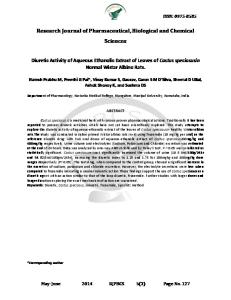
ISSN: 0975-8585
Research Journal of Pharmaceutical, Biological and Chemical Sciences
Detection and eradication of major microbial contaminants during callus culture of Sugarcane (Saccharum offcinarum L.) genotype Co 86032. Virdhaval M Nalavade1, 2*, Mokshadaa R Naidu1, Ravindra R Kale3, Avinash S Thorat1 and K Harinath Babu1, 2 1Molecular
Biology and Genetic Engineering Section, Vasantdada Sugar Institute, Manjari (Bk) Pune, Maharashtra, India. of Biotechnology, Shivaji University, Kolhapur, Maharashtra, India. 3Institute of Biotechnology, PJTSAU, Hyderabad, T.S, India. 2Department
ABSTRACT The present investigation focuses on development of economical and reliable protocol in effective control of microbial contamination during callus culture of sugarcane genotype Co 86032. Major microbial contamination was observed after first week of callus initiation and subsequent sub culturing. Maximum microbial contamination was occurred due to different bacterial contaminants compared to fungal contaminants, fungi observed were Aspergillus Spp. and Rhizopus Spp.. The contaminants were isolated and identified on basis of their morphological and biochemical characteristics. These contaminant microorganisms were identified as Saccharomyces Spp., Herbaspirillium Spp., Burkholderia Spp., Acetobacter Spp., Azorcus Spp., and Serretia Spp. Out of the six isolates four of them Azorcus Spp., Burkholderia Spp., Herbaspirillium Spp. and Acetobacter Spp. are nitrogen fixing endophytes associated with sugarcane. In order to control this kind of microbial contamination problem in tissue culture process, attempt was made to see the effect of different antibiotics (Cefotaxime, Gentamycin, Kanamycin and Tetracycline) to control these contaminants. Among four antibiotics used to check growth of contaminants, Cefotaxime (250 mg/L) was found to be effective in controlling the growth of various microbial contaminants effectively. Effective antibiotic (Cefotaxime 250 mg/L) was incorporated in CIM and sugarcane leaf roll disc were inoculated for callus induction. It was observed that microbial contamination percentage was reduced from 35.11 ± 0.47% to 7.85 ± 0.93% due to use of Cefotaxime in CIM. This protocol can be used to generate healthy and contamination free Co 86032 callus for further use. Keywords: Sugarcane, callus, callus induction media (CIM), antibiotic, contamination.
*Corresponding author:
January – February
2017
RJPBCS
8(1)
Page No. 1153
ISSN: 0975-8585 INTRODUCTION Sugarcane (Saccharum offcinarum L.) is an important cash crop grown in different parts of the world from the tropics to subtropics. The importance of sugarcane has increased with use as industrial raw material for sugar and allied industries producing alcohol, acetic acid, butanol, paper, plywood, industrial enzymes, biomass production and animal feed. Traditional plant breeding technique, together with chemical, biotechnological approaches, have been extensively used to increase crop yields by selecting improved varieties which are more productive and resistant to diseases and pathogens. However, conventional sugarcane breeding requires 10-15 year identifying and releasing a new clone into a variety. Sugarcane [1], would greatly benefit from biotechnological improvements due to its complex polyploid–aneuploid genome, narrow genetic base, poor fertility, susceptibility to various diseases and pests. For genetic transformation establishment of tissue culture is important. The early-established robust tissue culture regeneration system of sugarcane became a foundation for efficient genetic transformation of this crop. The explant most frequently used for transformation experiments in sugarcane is embryogenic callus. This callus is produced from the culture of immature leaf whorls above the apical meristem. Plant-tissue culture is an important method in study of plant metabolism, plant genetics, plant morphogenesis and plant physiology, in genetic transformation of plants, in elimination of plant pathogens, in preservation of important plant species in limited space and in multiplication of plants in vitro. Microbial contamination is one of the major challenges facing plant in in-vitro propagation during different stages of culture processes such as culture initiation and sub-culturing. Tissue culture media acts as source of nutrient for growth of various microbial contaminations in addition to plant growth. These microbes compete with plant tissue for nutrients and some of them produce phytotoxins which result in culture mortality, tissue necrosis, and reduced shoot proliferation and rooting. The list of organisms described as contaminants in plant-tissue cultures includes viruses, bacteria, yeasts, fungi, mites and thrips . Pathogenic fastidious bacteria, viruses, and viroids are thought only to be introduced with the initial explant and have to our knowledge not been reported to cause significant plant losses in vitro. They can, however, be latent contaminants in plant tissue cultures and cause disease when plants are transferred back to soil [2]. Mites and thrips found in tissue cultures do not usually harm the plants directly but introduce other contaminants such as fungi, yeast and bacteria into sterile plant cultures. Visually detecting contaminants has proven to be very ineffective as most bacteria show only a very weak growth on standard tissue culture media only after long cultivation periods . In contrast when the explants have been wounded seriously , visual detection of bacterial contamination in or on substrates which are prone to microbial growth does give satisfactory results. Contamination by endophytic bacteria is one of the most serious problems in tissue culture [3]. Persistent endophytic microorganisms may be transferred unknowingly at each subculture and may become apparent weeks or months after the material was initiated in vitro. Many endophytes have been reported earlier in sugarcane which play important role in sugarcane ecology and physiology [4]. The detection and differentiation of microbial contamination is important to establish a effective tissue culture system. Ascertaining the Gram reaction, purity of the isolate, colony growth, and a few defining biochemical characteristics along with screening for responses to selected antibiotics should prove helpful in predicting whether treatment to eliminate a contaminant will succeed [5]. Earlier studied were made to eliminate microbial contamination in sugarcane during apical meristem and axillay bud culture using plant preservative mixture™ (PPM), sodium hypochlorite (NaOCl), and chitosan by [6, 7]. The present study aimed to characterize and identify microbial contaminants from in-vitro sugarcane callus culture and management and control of the microbial contaminants. MATERIAL AND METHOD Establishment of callus culture and isolation of microbial contaminants: Leaf whorls of 6 to 12 months old cane stalk of healthy sugarcane plants was used as explant for callus initiation. The experiment were conducted on high yielding commercial sugarcane (Saccharum officinarum L) variety Co 86032 collected from experimental fields of Vasantdada Sugar Institute, Pune, India.
January – February
2017
RJPBCS
8(1)
Page No. 1154
ISSN: 0975-8585 Stem tops were collected from field. after removing expanded leaves were washed under running tap water. The outer mature leaves of shoot tops were removed, additional outer leaves were removed to isolate immature leaf rolls (about 1 to 1.5 cm diameter) formed by the innermost 5–6 tightly furled spindle leaves. An 8–10 cm long basal portion of the leaf roll, starting from the leaf base just above the apical meristem was excised. Then the leaf rolls were pre-treated with solution containing 0.2% ascorbic acid, 0.4% citric acid, 0.1% bavistin and 0.1% streptomycin for 30 min followed by 0.1% mercuric chloride treatment for 15 min. Then under laminar flow cabinet leaf roll were aseptically rinsed with sterile distilled water for 2-3 times. The outer 2 to 3 layers of leaf sheath were removed aseptically and made into thin slices of explants (1 to 2 mm) and cultured on callus induction medium. Callus induction medium (CIM) consisting of Murashige and Skoog (MS) salts and vitamins [8], 500 mg/L casein hydrolysate, 100 mL/L young coconut water, 25g/L sucrose, 3 mg/L 2,4D, 20 mg/L inositol, 1 mg/L Thiamine HCl, 100 mg/L MES buffer, 100 mg/L PVP, 8 g/L Agar and pH 5.7. Subculturing of callus was done at 2 weeks interval to get embryogenic callus. Different kinds of microbial contaminants were observed after one week of callus culture initiation. These contaminants were marked and then isolated on callus induction medium at 28°C. Further the isolated pure colonies were maintained on LB medium for further study. Identification of microbial contaminants: Morphological analysis, colony characteristics and motility: Morphological characterization of isolated contaminants was done on the basis of simple staining and gram staining methods [9]. Colonies of isolates were screened for characters like size, shape, colour, margin, elevation, opacity, consistency. To study the motility 48 hours old culture was taken for observing motility by hanging drop method under oil immersion objective. The contaminating fungi were identified by colony characteristics (appearance, colour and pigmentation), morphology of vegetative hyphae and macroconidia produced by comparing with standards enlisted by [10]. Biochemical analysis: Biochemical characterization of isolates was done by using a series of biochemical tests, like Nitrate reduction, Catalase, Starch hydrolysis, Casein hydrolysis, Gelatin hydrolysis, Hydrogen Sulphide gas (H 2S) production, sugar fermentation ability with acid and gas production for various isolates [9]. The characterized bacterial strains were compared with standard in Bergey’s Manual of Bacteriology [11, 12, 13]. Identification of endophytic bacteria on selective media: Isolated microbial contaminants were streaked on selective medium for endophytic bacteria in sugarcane like, LGI medium for Acetobacter spp. [14], LMG medium for Azorcus spp. [15], JMV medium for Burkholderia spp. [16] and LGIP medium for Herbaspirillium spp. [17]. Antibiotic sensitivity test of isolated contaminants: Various antibiotics like Cefotaxime, Gentamycin, Kanamycin and Tetracycline were used to study the inhibition of microbial growth. Stocks of 50 mg/mL were prepared and stored at -20°C. LB medium with different antibiotic at following concentrations 50, 100, 150, 200 and 250 mg/L were prepared. Isolated microbial contaminants were streaked over LB medium with above mentioned antibiotic concentrations to check their antibiotic sensitivity. The effective antibiotic concentration was used in callus induction medium and percent contamination was recorded. Use of effective antibiotic in CIM: Cefotaxime at 250 mg/L was added to callus induction medium. About 10 leaf roll disk of Co 86032 were inoculated on CIM petri plate. Experiments were performed in triplicate and readings were taken after 20 days. Microbial contamination percentage was recorded for CIM with and without Cefotaxime.
January – February
2017
RJPBCS
8(1)
Page No. 1155
ISSN: 0975-8585 RESULTS Callus induction and isolation of microbial contaminants: The callus induction of sugarcane variety Co 86032 was carried out in callus induction medium supplemented with nutrients and hormones augmenting proliferation of callus and tissue morphogenesis. Callus generally became visible on the cut surfaces within 5-7 days of initiation on CIM. Few inoculated leaf roll disc turned brownish black or reddish brown colored and failed to proliferate during incubation as a result of tissue necrosis or secretion of poly-phenolic compounds. Adverse effects on callus growth were minimized by shortening the interval between subcultures in proportion to the intensity of browning, which generally subsided after several subcultures. Along with prominent growth of calli, contamination was a major hurdle in early as well as late stages of incubation due to growth of bacteria and fungi on tissue as well as in the medium. Fungal contamination percentage was low due to treatment of explant with Bavistin, but major contamination was due to bacterial populations, fungi observed were Aspergillus Spp. and Rhizopus Spp.. Hence the morphologically distinct bacteria were isolated initially on CIM and then maintained on LB medium. In total 8 isolates based on phenotypic character were isolated on callus induction medium and maintained on LB medium. Out of 8 isolates, Isolate 2 & 8 and 4 & 6 showed similar characteristics with each other (Fig. 1 & 2).
Fig. 1:- Microbial contamination during callus culture a & b- Bacterial contamination and c & d- Fungal contamination
Fig 2:- Isolated different microbial contamination from callus induction medium
January – February
2017
RJPBCS
8(1)
Page No. 1156
ISSN: 0975-8585 Identification of microbial contaminants : The isolated six microbial contaminants were analysed for their morphology, colony characteristic, motility and various biochemical characters (Table 1 & 2) according to BERGY’S manual [11, 12, 13]. All the six microbial contaminants were streaked on specific medium for endophytic bacteria in sugarcane. These contaminant microorganisms were identified as Yeast, Herbaspirillium Spp., Burkholderia Spp., Acetobacter Spp., Azorcus Spp., and Serretia Spp. (Table 3). Out of six contaminants four were identified as sugarcane endophytes Azorcus Spp., Burkholderia Spp., Herbaspirillium Spp. and Acetobacter Spp. (Table 4). Table 1: Morphology, colony characteristic and motility of isolated microbial contaminants
Particulars
Isolate 1
Size Shape Colour Margin Elevation Opacity Consistency
1.0 mm Circular Cream Entire Convex Opaque Moist Negarive (pink rods) Motile
Gram staining Motility
Isolated Microbial contaminants Isolate 2 Isolate 3 Isolate 4 Isolate 5 Colony Characteristics 1.0 mm 1.0 mm 0.1 mm 1-2 mm Circular Circular Circular Circular Light cream Cream Yellow Cream Entire Entire Entire Entire Convex Flat Convex Low Convex Opaque Opaque Opaque Translucent Smooth Smooth Moist Moist Negarive Negarive Negarive Negarive (pink rods) (pink rods) (pink rods) (pink rods) Motile Motile Motile Motile
Isolate 6 0.2 mm Circular Pink Entire Convex Opaque Moist Negarive (pink rods) Motile
Table 2: Various biochemical characters of isolated microbial contaminants
Particulars Glucose Sucrose Fructose Lactose Mannose Dextrose Trehalose Xylose Sorbitol Arabinose Starch Catalase Gelatin Nitrate Reduction Production of H2S Casein
Isolated Microbial contaminants Isolate 1 Isolate 2 Isolate 3 Isolate 4 Isolate 5 Sugar Fermentation Capability + + + + + + + + + + + + + + + + + + + + + + + Other biochemical characters + + + + + + + -
Isolate 6 + + + + + + + + + + + + -
Table 3: Identified microbial contaminants ISOLATES Isolate 1 Isolate 2 Isolate 3 Isolate 4 Isolate 5 Isolate 6
January – February
2017
Genus Saccharomyces Spp. Herbaspirillum Spp. Burkholderia Spp. Acetobacter Spp. Azoarcus Spp. Serretia Spp.
RJPBCS
8(1)
Page No. 1157
ISSN: 0975-8585 Table 4 :- Morphological characteristics of the different identified endophytes. Sr. NO.
Specific Media
Colony Characteristics
1
LGI media
Round redissh orange colony
2
Herbaspirillum media
Whitish colony
3
Azoarcus media
Creamy white/Whitish brown colony
4
Burkholderia media
Pinpoint brown colony
Morphological Characteristics
Special Characters
Isolate identified to Genus
Gram negative short rods,0.1 to 1.4 mm, circular Gram negative vibroid shape, 1 to 2 mm, circular Gram negative rods, 1 to 1.4 mm, Circular/Irregular Gram negative rods, 1 to 1.4 mm, Circular/Irregular
Grows on PDA with brownish colonies
Acetobacter
Chalky white colonies
Herbaspirillum
Brown pigmentation
Azoarcus
Tiny brownish colony
Burkholderia
Antibiotic sensitivity test: Various antibiotics like Cefotaxime, Gentamycin, Kanamycin and Tetracycline at following concentrations 50, 100, 150, 200 and 250 mg/L in LB medium were used to study the growth inhibition of isolated six microbial contaminants microbial growth. It was observed that addition of Cefotaxime at concentration of 250 mg/ L was satisfactory to control the growth of all six microbial contaminants (Table 5). Table 5:- Growth of isolated microbial contaminants on various antibiotic concentrations Antibiotic and their concentration(mg/L)
Cefotaxime
Gentamycin
Kanamycin
Tetracycline
50 100 150 200 250 50 100 150 200 250 50 100 150 200 250 50 100 150 200 250
Microbial contaminants Isolate 1 +++ +++ +++ ++ + +++ +++ +++ ++ + +++ +++ +++ ++ ++ +++ +++ +++ ++ ++
Isolate 2 +++ +++ +++ ++ +++ +++ +++ ++ + +++ +++ +++ ++ +++ +++ +++ ++ -
Isolate 3 +++ +++ ++ + +++ +++ +++ ++ +++ +++ +++ ++ + +++ +++ +++ ++ +
Isolate 4 +++ ++ ++ +++ +++ +++ ++ ++ +++ +++ +++ ++ +++ +++ +++ ++ +
Isolate 5 ++ ++ + + +++ +++ +++ ++ +++ +++ +++ ++ + +++ +++ +++ ++ ++
Isolate 6 +++ +++ ++ + +++ +++ +++ ++ + +++ +++ +++ ++ +++ +++ +++ ++ -
Note: +++: abundant, ++: good , +: slow growth, - : no growth
Use of effective antibiotic in CIM: The antibacterial property of cefotaxime has been harnessed to enhance the frequency of plant regeneration in finger millet [18], wheat [19], pearl millet [20], sorghum [21] and rice [22]. In sugarcane, the influence of cefotaxime (250-500 mg/L) on shoot multiplication and elongation has been studied and found to be effective in promoting growth in sugarcane tissue culture by [23]. Effective antibiotic (Cefotaxime 250
January – February
2017
RJPBCS
8(1)
Page No. 1158
ISSN: 0975-8585 mg/L) was incorporated in CIM and sugarcane leaf roll disc were inoculated for callus induction. Our results indicate that microbial contamination percentage was reduced from 35.11 ± 0.47% to 7.85 ± 0.93% due use of cefotaxime in CIM (Table 6). Table 6:- Effect of Cefotaxime on microbial contamination percentage in CIM Antibiotic treatment
Cefotaxime Without Cefotaxime
Number of Petri plates inoculated
Number of contaminated petri plates
Contamination (%)
27 31 31 29 24 16
2 3 2 10 8 6
7.41 9.68 6.44 34.49 33.34 37.5
Average (%)
7.85 ± 0.93
35.11 ± 0.47
CONCLUSION During in-vitro culture of sugarcane leaf different type of bacterial contamination was observed. Six bacterial contaminants were isolated and identified by using morphological and biochemical characterisitcs. Major microbial contamination was observed during callus initiation and subsequent sub culturing. In order to control this kind of microbial contamination problem in tissue culture process, attempt was made to see the effect of antibiotics (Cefotaxime, Gentamycin, Kanamycin and Tetracycline) to control these contaminants. Maximum microbial contamination was caused due to different bacterial contaminants compared to fungal contaminants, fungi observed belonged to Aspergillus Spp. and Rhizopus Spp.. Contaminant microorganisms were identified as Saccharomyces Spp., Herbaspirillium Spp., Burkholderia Spp., Acetobacter Spp., Azorcus Spp., and Serretia Spp. Out of the six isolates four of them Azorcus Spp., Burkholderia Spp., Herbaspirillium Spp. and Acetobacter Spp. are nitrogen fixing endophytes associated with sugarcane. Among four antibiotics used to check growth of contaminants, Cefotaxime (250 mg/L) was found to be effective in controlling the growth of various microbial contaminants effectively. Effective antibiotic (Cefotaxime 250 mg/L) was incorporated in CIM to check efficiency for controlling the contamination percentage during callus induction. It was observed that microbial contamination percentage was reduced from 35.11 ± 0.47% to 7.85 ± 0.93% due use of Cefotaxime in CIM. ACKNOWLEDGEMENTS The authors are thankful to Shri Shivajirao Deshmukh Director General VSI, Pune, for providing necessary facilities to carry out the research work. REFERENCES [1] [2] [3] [4] [5] [6] [7] [8] [9] [10]
Bakker H. Sugar cane cultivation and management. New York: Kluwer Academic/Plenum Publishers 1999. Leifert C, Morris CE, Waites WM. Critical Reviews in Plant Sciences 1994; 13(2):139-183. Kneifel W, Leonhardt W. Plant Cell, Tissue and Organ Culture 1992; 29(2):139-144. Boddey RM, Urquiaga S, Alves BJR, Reis V. Plant and Soil 2003; 252: 139-149. Buckley PM, Dewilde TN, Reed BM. In Vitro Cell. Dev. Biol. 1995; 31(1):58-64. Sawant RA, Tawar PN. Sugar Tech 2011; 13(1): 27-35. Thorat AS, Muley AB, Shingote PR, Nalavade VM, Babu KH, Research J Pharmaceutical Biological and Chemical Sciences 2016; 7(2): 1122-1135. Murashige T, Skoog F. Physiologia Plantarum 1962; 15(3): 473–497. Sherman N, Cappuccino JG. Microbiology: a laboratory manual. 8 edition; Pearson Publishing Company, 2008. Barnett HL, Hunter BB. Illustrated genera of imperfect fungi. Burgress, Minneapolis MN, USA 241, 1972.
January – February
2017
RJPBCS
8(1)
Page No. 1159
ISSN: 0975-8585 [11] [12] [13] [14] [15] [16]
[17] [18] [19] [20] [21] [22] [23]
Bergey DH, Holt JG, Krieg NR, Sneath PHA. Bergey's Manual of Determinative Bacteriology, 9th ed., (Breed RS, Murray EGD and Smith NR, eds.) Williams and Wilkims, Baltimore, 1994. Buchanan RE, Gibbons NE. Bergey’s Manual of Determinative Bacteriology. Science Publisher, 8th ed., Beijing, 1984. Holt JG, Krieg NR, Senath, PHA Staley JT, Williams ST. Bergey’s Manual of Determinative Bacteriology 9th Ed. Baltimore Md Williams and Wilkins1994. Fuentes-Ramirez LE, Caballero-Mellado J, Sepulveda J, Martinez- Romero E. FEMS Microbiology Ecology 1999; 29(2): 117-128. Ronald M. Atlas Hand book of media for environment microbiology 2 nd edition CRC Press, 2005. Reis VM, Estrada-delos Santos P,Tenori-Salgado S, Vogel J, Stoffels M,Guyon S, Mavingui P, Baldani VLD, Schmid M, Baldani JI, Balandreau J,Hartmann A, Caballero-Mellado J. Int. J. Syst. Evol. Microbial. 2004; 54: 2155-2162. Njoloma JP, Moriya O, Kazuhiko T, Yuichi S, Shoichiro A. African J of Biotechnology 2006; 5(10): 836841. Eapen S, George L. Plant Cell Tissue Org Cult 1990; 22: 87-93. Borrelli GM, di Fonzo N, Lupotto E. J Plant Physiol 1992; 140(3): 372-374. Pius J, George L, Eapen S, Rao PS. Plant Cell Tissue Org Cult 1993; 32(1): 91-96. Rao AM, Sree KP, Kishor PBK. Plant Cell Rep 1995; 15(1): 72-75. Grewal D, Gill R, Gosal SS. Biotech J 2006; 1(10): 1158 - 1162. Kaur A, Gill MS, Ruma D, Gosal SS. Sugar Tech 2008; 10(1): 60-64.
January – February
2017
RJPBCS
8(1)
Page No. 1160