Renal Functions Lecture-4
The Kidneys Dr. Khalid Al-Ani Department of Clinical Pharmacy Faculty of Pharmacy
Renal Functions Endocrine Functions - renin – erythropoietin – Calcitriol ( activation of vitamin D)
Excretion of waste -Production of urine -elimination of metabolic end products (Urea, Creatinine, uric acid …etc) -elimination of foreign materials (Drugs) Control of volume & composition of ECF -Water and electrolyte balance -Acid/Base status Dr. Khalid Al-Ani
Why Test Renal Function? u To
identify renal dysfunction. u To diagnose renal disease. u To monitor disease progress. u To monitor response to treatment. u To assess changes in function that may impact on therapy (e.g.Digoxin, chemotherapy).
Dr. Khalid Al-Ani
1
Renal Anatomy and Physiology
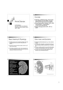
Renal Anatomy and Physiology u Consists
of renal cortex and renal medulla.
u pair
of fist-sized organs located on either side of the spinal column just behind the lower abdomen (L 1-3)
u The
Cortex
functional unit of the kidney is the nephron;
Pelvis
u 106
nephrons /Kidney.
Capsule Medulla
To the bladder
Afferent arteriole Glomerulus
Proximal tubule Distal tubule
Bowman’s capsule Collecting duct Renal artery Henle’s Loop
Dr. Khalid Al-Ani
2
Blood is separated from the lumen of the nephron by three layers,
-Capillary endothelial -basement membrane -epithelial cell The glomerular filtrate is an ultra filtrate of plasma, that has similar composition of plasma, except for proteins Dr. Khalid Al-Ani
What gets filtered in the glomerulus? u Freely
endothelium is impermeable to blood cells as well as large proteins. Proteins with MWt lower than that of albumin (68KDa) are filterable.
urine
filtered
– H2O – Na+, K+, Cl-, HCO3-, Ca++, Mg+, PO4, etc. – Glucose – Urea – Creatinine – Insulin
u None
filtered – Proteins >68KDa – Immunoglobulins – Ferritin – Blood Cells
Dr. Khalid Al-Ani
3
Kidneys receive ∼ 2,000 L/ day (25% of cardiac output)
•The filtration is passive process. •The filtration rate of the kidneys depend on the difference between blood pressure in the glomerular capillary and the hydrostatic pressure in the lumen of the nephron
200 Liters Of plasma ultra filtrate formed per day
•GFR= 110 ml/min
2 liters
Reabsorption from glomerular filtrate % Reabsorbed Water Sodium Potassium Chloride Bicarbonate Glucose Albumin Urea Creatinine
99.2 99.6 92.9 99.5 99.9 100 95-99 50-60 0 (or negative)
Reabsorption can be active or passive, and occurs in virtually all segments of the nephron
4
Nephrone performed three functions. 1.Glomerular filtration 2. Tubular secretion 3. Tubular reabsorption
Biochemical Tests of Renal Function Diseases affecting kidneys can be selectively damage glomerular or tubular function
Biochemical Tests of Renal Function
u Test
of glomerular function Measurement of GFR –Clearance tests –Plasma creatinine –Blood urea
u Tubular u
function tests
Urinalysis Dr. Khalid Al-Ani
urine
Measurement of Glomerular Filtration Rate (GFR)
Measurement of Glomerular Filtration Rate (GFR) u Measurement
reflects no of functional nephrones
5
is based on concept of clearance: “Measuring urinary excretion of a substance (X) that is completely filtered from the blood by the glomeruli.
Measurement of Glomerular Filtration Rate (GFR)-conti
Determination of Clearance u If
clearance = GFR then substance x properties: – freely filtered by glomeruli – Not secreted or reabsorbed or metabolized by tubular cells – Non-toxic and easily measurable
u Clearance
= (U x V)/P Where U is the urinary concentration of substance x V is the rate of urine formation (mL/min) P is the plasma concentration of substance x
Dr. Khalid Al-Ani
Inulin Clearance Standard u Plant polysaccharide u measurement of inulin clearence requires the infusion of inulin into blood u clinically is not suitable
Creatinine Clearance
u Gold
6
u Creatine
is a nitrogen containing compound u formed from glycine, arginine, methionine in the liver
Creatinine Clearance conti
Creatinine Clearance conti.
stored in muscle as creatine phosphate
u 1-2%
of muscle creatine converted to creatinine (Cr) each day
Dr. Khalid Al-Ani
u Amount
of Cr produced relates to muscle mass
u
Freely filtered at the glomerulus
u Creatinine u Corrected
clearence =110ml/min
to standard body surface area of 1.73m2
7
u Some u This
GFR
active tubular excretion(10%).
is of little significant for normal
u When
GFR< 10 ml/min, GFR is over estimated
Creatinine Clearance: advantage and disadvantage Timed urine collection for creatinine measurement (usually 24h) Problems: u Practical problems of accurate urine collection and volume measurement. u Time consuming, inconvenient and potentially unreliable u Carried out for transplanted kidneys & degree of renal impairment u
Plasma Creatinine Concentration conti.
Problems u Plasma
Cr can increase by 30% 7 hrs after meal.
Plasma Creatinine Concentration u Most
reliable simple biochemical test of GFR
u
plasma Cr level remains fairly constant through adult life
Plasma Creatinine Concentration conti
u Cr
level can be changed independently to renal disease decreased in -starvation -wasting disease -pregnancy -immediately after surgery -steroid therapy Dr. Khalid Al-Ani
8
Plasma Creatinine Concentration conti
Plasma Creatinine Concentration conti.
u Plasma u Cr
Normal reference value 60-120 μmole/l Or 0.7-1.4 mg/dl
u Concentration
inversely related to GFR.
Cr level can be misleading
u GFR
can decrease by 50% before plasma Cr rise beyond normal range
Plasma Creatinine Concentration conti.
u Normal
Cr level does not imply normal renal function
Blood Urea u Urea
is nitrogen containing compound formed in the liver as the end product of protein metabolism and digestion.
u eliminate
in urine as a major nitrogen waste product (85%)
9
UREA conti.
Blood Urea
u freely
filtered but about 50% reabsorbed by through passive diffusion
u
tubular reabsorption increases at low rate of urine flow
u
often used an index of renal function along with plasma Cr
u Blood
Urea level can be changed independently to renal disease high protein intake GIT hemorrhage hypovolumia, burns dehydration congestive heart failure Catabolic state
Dr. Khalid Al-Ani
Blood Urea
u blood
Urea level reduced in starvation Low protein diet Sever liver disease
The causes can be subdivided to Prerenal
u Thus,
Renal intrinsic renal disease
u The
Postrenal obstruction to urine outflow
BU needs to be compared to cr to determine true renal dysfunction levels of urea and Cr almost always are paralleled to each other
10
High plasma Urea (Uremia or Azotemia) (azotemia = elevated BU)
Other Methods for Assessing GFR-conti.
Other Methods for Assessing GFR u 51Cr-EDTA, 99Tc-DTPA
–Exogenous ∴ need to be administered –Not readily available –Radioactive
β2-Microglobulin (BMG) u Small
protein (MW=11.8K) u not affected by muscle mass or diet u BMG is filtered in the glomerulus, but is reabsorbed in the renal tubules. – Urinary BMG levels are a sensitive measure of renal tubular function u Increased in renal failure
Cystatin-C protease inhibitor (MW13 kDa) u freely filtered at glomerulus u Reabsorbed and degraded by proximal tubule u Plasma concentration reflects GFR u Constant production rate by all nucleated cells u No known extra-renal excretion routes u Not influenced by muscle mass, diet or subjects sex u
Tests of Tubular Function performed less frequently u Proximal Tubular Function – Aminoaciduria – Glycosuria with normal blood glucose u Distal
Tubular Function
Dr. Khalid Al-Ani
11
Urinalysis (UA)-conti
Urinalysis (UA) General urine examination (GUE) u it
is a general test for evaluation of renal function u Fresh sample = Valid sample u Physical, u chemical
and u macroscopic examination
Physical examination includes u Appearance
Colour, turbidity
u
pH
u Specific
gravity and osmolality
Urinalysis (UA)
Appearance – clear u Microscopic
examination includes Sediments RBCS WBC Crystals
12
q Turbidity:
(infection, nephrotic syndrome, proteinuria)
q Colour:
amber light Coloured-haemoglobin, myoglobin, Jaundice, drugs, beet
Urine pH acidic Normal Acidic Alkaline
Urine Osmolality u Normal
Average: 400-900 mOsm/kg H2O Max 1200 mOsm/kg
u Normally
4.5-8 4.5-5.5 6.5-8
u pH
that is >8 or less 300mg/L u
14
overflow (raised plasma Low MW Proteins, Bence Jones, myoglobin) Renal diseases
Urine glucose Normally –ve u +ve urine glucose –Increased blood glucose –Low renal threshold or other tubular disorders u
u Ketone
bodies –Ve u bilirubin –ve u Nitrite –Ve , +ve during UTI by gram +ve bacteria
u False
+ve –Ascorbic acid Dr. Khalid Al-Ani
Microscopic examination Urine sediment Freshly passed urine. looking for
Cells
q Cells,
q RBCS
q Casts
q
15
Microscopic examination Urine sediment-cont
(Tamm-Horsfall protein)
Crystals
q WBC
q
epithelial
WBC 0-1 HPF
•The presence of more than 5 WBC's / hpf may suggest -infection, pyelonephritis or inflammation of the genitourinary tract
Epithelial 0-2 HPF increased in bladder inflammation
RBCS 0-1 HPF
•Large no. of RBC's in the urine may be associated with (i) renal disease, (ii) lower urinary tract disease, (iii) external disease, (iv) physiologic causes including exercise.
Microscopic examination Urine sediment-cont
Casts (Tamm-Horsfall protein)
Dr. Khalid Al-Ani
16
Granular cast
Red Cell casts
White cell cast + polymorphs +Bacteruria = pyelonephritis hematuria - glomerular disease
White blood Cell casts
17
Hylan cast
Crystals
Triple phosphate and amorphous phosphate of normal urine amorphous Triple phosphate
crystals such as phosphates, urates, and oxalates occur in normal urine sediment, and are of limited clinical significance
Calicum oxalate crystal
Calcium oxalates appear at any pH . They are octahedrons that resemble envelopes
18
Triple phosphate crystals are seen only in alkaline urine. They have a characteristic crystal shape, often referred to as "coffin lids ."
Urate crystals
Renal Disorders-conti
Renal Disorders Failure of renal function may occurs rapidly or over a period of time u Acute
u
renal failure (ARF)
u Acute
renal failure (ARF) potentially reversible occurs during sever illness
u Chronic
renal failure (CRF) not reversible leading to end stage renal failure
Chronic renal failure (CRF)
Dr. Khalid Al-Ani
ARF
Signs and Symptoms of Renal Failure u Symptoms
of Uraemia (nausea, vomiting, lethargy) u Disorders of Urine volume (polyuria, oliguria, anuria) u Alterations in urine composition (haematuria, proteinuria, calculi) u Pain u Oedema
19
Divided in to three categories failure due to decreased blood supply u Pre-renal
u Renal-
intrarenal due to intinsic damage to kidney
u Post
renal u due to urinary tract obstruction
Causes of pre-renal failure Kidney hypo-perfusion (circulatory insufficiency) -sever haemorrhage -burns -dehydration -cardiac failure -hypotension
Causes of pre-renal failure-cont.
consequence GFR and Increased RAS
u reduced u urine
osmolality high low in Na ( 600 mmol/L
(as response to hypovolumia)
Dr. Khalid Al-Ani
21
Pre-Renal ARF? Pre = functioning tubules Test
Chronic Renal Failure: Causes u Glomerulonephritis
Result Pre-renal
Renal
Urine Na+ (mmol/L
40
Ratio urine/plasma osmolality Ratio urine urea/plasma urea concentration
>1.5
10