Journal of Neurology, Neurosurgery, and Psychiatry, 1978, 41, 784-790
Refractory period of human motor nerve fibres J. KIMURA, T. YAMADA, AND R. L. RODNITZKY From the Division of Clinical Electrophysiology, Department of Neurology, University of Iowa, Iowa City, USA S Y Using a collision technique, the ulnar nerve was made refractory for a shorter distance normally covered in 0.5 ms or a longer distance covered in 1.5 ms. Studying the shorter refractory segment with paired shocks of maximal intensity, the test response first appeared (more than 5% of unconditioned response) at an interstimulus interval of 1.16±0.18 ms (mean ±SD in 20 ulnar nerves). The conduction velocity of the test impulse then was 55.3%+± 19.2% of normal. Recovery in amplitude of the test response was nearly complete (more than 95%) at 2.11±0.50 ms, when it was conducting at a speed of 81.2±17.4% of normal. The conduction velocity recovered to a level above 95% of normal at 2.65±+0.65 ms. Whereas recovery in amplitude of the test response was unrelated to the length of the refractory segment, change in latency was greater with the longer refractory segment, although not in proportion to the distance. U M M A R
After passage of an impulse, an axon becomes totally inexcitable for a fraction of a millisecond, followed by progressive recovery to prestimulus level within the ensuing few milliseconds. These two phases of decreased excitability, the absolute and relative refractory periods, have been studied in experimental animals by measuring the nerve action potentials elicited by paired stimuli (Bishop and Heinbecker, 1930; Graham, 1935; Tasaki, 1953; Hodgkin, 1967; Bergmans, 1973). In 1963, Gilliatt and Willison reported the first comprehensive study on the refractory periods of human peripheral nerves. This and subsequent human studies, however, have been limited to sensory and mixed fibres (Buchthal and Rosenfalck, 1966; Lowitzsch et al., 1973; Hopf et al., 1974; Tackmann and Lehmann, 1974; Betts et al., 1976; Hopf et al., 1976). Muscle action potentials elicited by pairs of stimuli applied to motor fibres could not be measured accurately at short interstimulus intervals because of the overlap between conditioning and test responses (Wagman and Flick, 1951). To study motor fibres using the paired shock technique, therefore, it is necessary to block the effect of the first stimulus of the pair without affecting the second. This can be achieved by "collision" if a third stimulus is delivered to the
nerve at a point distal to the paired stimuli. Using the collision technique, we showed previously that serial changes in amplitude of the test response at increasing interstimulus intervals indicated the range of the absolute refractory periods of different human motor fibres (Kimura, 1976). We have now determined changes in latency of the test response elicited at various intervals after a conditioning impulse. The course of the relative refractory period of motor fibres was measured by changes in conduction velocity.
Methods
The ulnar nerve was stimulated by paired shocks at the axilla. A single shock was given at the wrist to block the first axillary stimulus of the pair. The compound muscle action potential was recorded by surface electrodes placed on the hypothenar eminance. A stimulator with an output impedance of 250 ohms was coupled with a transformer. The stimulus was a slightly distorted square pulse of 0.05 ms duration. In preliminary experiments, the intensity of the second axillary shock was altered systematically to determine the voltage change necessary to excite the lowest threshold fibres at different times after the first shock. At near threshold, however, the results obtained were very Address for reprint requests: Jun Kimura, MD, Chief, Division of inconsistent from one trial to the next in the same Clinical Electrophysiology, University Hospitals, Iowa City, Iowa subject. Consequently, in this and previous studies 52242, USA. (Kimura, 1976) we elected to use the same axillary Accepted 18 April 1978 784
Refractory period of human motor nerve fibres
785
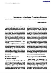
shock intensity throughout the experiment. The ditioning stimulus before it was extinguished by first (conditioning) and second (test) shocks were the antidromic impulse. To compare short versus identical, and of just maximal intensity. The maxi- long refractory segments, two different values, 0.5 mal stimulus, determined by delivering single and 1.5 ms were chosen arbitrarily as the time of shocks in increasing intensity, varied between collision after delivery of the conditioning individuals from 200 to 350 V. A single shock stimulus. If motor conduction velocity is assumed delivered at the wrist was always 50% supra- to be 60 m/s, therefore, the lengths of the short maximal to ensure a complete block of the first of and long segments made refractory by the conthe paired axillary stimuli. ditioning impulse travelling to the point of When paired shocks at the axilla were combined collision would be 3 and 9 cm respectively. The with a single shock at the wrist (Fig. 1), the anti- interval between a pair of axillary stimuli was dromic impulse from the wrist was eliminated by varied from 0.1 ms to 4.5 ms, in increments of collision with the orthodromic impulse of the 0.1 ms up to 2.2 ms, then in increments of 0.2 ms conditioning axillary stimulus. Because the motor up to 3.4 ms, followed by 4.0 and 4.5 ms. In some axons were now cleared of antidromic activity, the subjects, interstimulus intervals of 5.0 and 5.5 ms impulse of the test axillary stimulus was trans- were also tested. mitted distally, but only if the axons were excitable Although three stimuli were delivered, only two after the passage of the conditioning stimulus. muscle potentials were recorded, because the conWith this technique, it was possible to adjust the ditioning shock given at the axilla was blocked. point of collision and consequently the length The two responses recorded were designated of the nerve segment made refractory by the con- M(A2), elicited by the test axillary stimulus, and M(W), evoked by the shock given at the wrist. These two potentials were clearly separate and Collision Technique Site of their characteristics easily measured (Fig. 2). When Stimulation the F wave (recurrent discharge of anterior horn cells after antidromic invasion) was elicited by the Axi la J test stimulus, it appeared at the tail end of M (A2), z I-i7_ S(A) thus not altering its latency and affecting its amplitude very little (Kimura, 1974). The appearCollision ance of the F wave in the absence of M(A2) was more confusing but these two potentials were Wrist and 5L easily distinguished on the basis of their latency. Axilla r ( If a recurrent discharge was evoked by the conS(A) S(W) ditioning stimulus, it could not reach the muscle because of collision with the antidromic impulse Collision of the test stimulus. M(W)V Wrist and 5s2S The peak-to-peak amplitude of M(A2) was Mto. M (I Paired Axilla i presumably proportional to the number of axons no longer refractory when the test stimulus was S(A) + S(A2) applied. The muscle response M(A), evoked by a Lmv single axillary shock alone, represented the total S(W)S(Ai)S(A2) S(AO)S(A2) 5ims number of axons available in the nerve. Hence, Fig. 1 Compound muscle action potentials recorded the amplitude ratio, M(A2)/M(A), gave the proby surface electrodes placed over the abductor digiti portion of nerve fibres already conducting in minimi after percutaneous stimulation of the ulnar response to the stimulus; that is, the degree of nerve. Figures on left are schematic diagrams showing orthodromic (solid arrows) and antidromic (dotted nerve recovery from the refractoriness induced arrows) impulses. Axillary stimulation, S(A), was by the conditioning shock (Fig. 3). The initial given 6.0 ms after the stimulus at the wrist, S(W), amplitude recovery of the test response was which triggered sweeps on the oscilloscope. With defined as a return of M(A2) to more than 5% single stimulation at the wrist and the axilla (the of M(A), which was generally easier to determine middle tracing), the orthodromic impulse from the axilla was extinguished by collision with the antidromic than the very first, often equivocal, potential we used previously (Kimura, 1976). Full recovery impulse from the wrist. When paired shocks were was defined as the return of M(A2) to 95 % of delivered at the axilla (bottom tracing), M(A2) M(A) amplitude (Table). appeared because the first axillary stimulus, S(A,), cleared the path for the orthodromic impulse of the The latency of M(A2) was measured from the second stimulus, S(A 2). stimulus artefact of the test shock to the initial (A,)(SW)
S(W)
J. Kimura, T. Yamada, and R. L. Rodnitzky
786
100 n
E -
12
80
~0a
-6
13
'a
E
a
1.4
60a 0
Change
C -
15
16
0
in
0
5ms
00
0ms
a
meanI
o
amplitude of test
response
during
40
point
to the
of
-to
the point of SE in 20 nervi
collision collision
ves
20
1.8 E
1 3 4 2 /Interstmulus interval between pared shocks at the oxilla (ms)
2-0 22
c,
/
Fig. 3 The time
course
of
recovery
5
in amplitude of
M(A2) after the passage of a preceding impulse in
24
/____
E
2-6
/
28
___/
3-0
10 healthy subjects (20 responses considering right and left sides together). The response to the second shock of the pair, M(A,), was converted into a percentage of the response to a single stimulus, M(A), at each interstimulus interval of paired axillary stimuli. The return of M(A) was practically identical whether the shorter segment
-M(A2)
longer
segment
normally covered
in 0.5
normally covered in 1.5
ms or
ms was
the
made
refractory. The gradual increase of M(A2) indicates LrnV
01 mV V.
5ms 0 2ms Fig. 2 Paired axillary shocks of just max-imal intensity were combined with a single shoc Ik at the wrist (cf bottom tracing in Fig. 1). The fir,st axillary stimulation, S(A,), was given 6.0 ms after the shock at the wrist, S(W), so that the impulses of these two stimuli always collided 1.5 ms after the deilivery of S(A ). The second axillary shock S(A,) wass given at intervals ranging from 1.2 to 3.0 ms after,S(A,). For amplitude mea:urements (left half of the Ifigure), slow sweep was triggered by S(W). To det latencies (right half), a fast sweep was trigg ered by S(A,) and the response displayed after a prredetermined delay of 11.0 ims. a
that the absolute refractory periods of the different motor
fibres
vary
considerably, full
being
recovery
achieved when the least excitable fibres
are no
longer
refractory.
deflection of the evoked response, using
triggered after
sweep
was a
by
the
shock
a
a
fast
predetermined delay
compared to the latency single axillary shock. Since
(Fig. 4). This value of M(A) evoked by M(A2) followed
a
muscle potential, M(W), elicited
given
at
the
wrist,
change
in
muscular excitability after M(W) might have affected the latency of M(A2). When paired stimuli were delivered at the wrist to allow a second appear with the same time delay as
M(W) to
Table Interstimulus intervals of the paired shocks and conduction velocity of the test response (mean±SD) Length of refractory segment
Full recovery in
unconditioned response)
Full recovery in amplitude (test response greater than 95 % of unconditioned response)
Interstimulus interval Conduction velocity between pairedshocks oftest impulse (ms) (Y% of normal)
Interstimulus interval Conduction velocity between pairedshocks oftest impulse (% of normal) (ms)
Interstimulus interval between paired shocks
Initial recovery in amplitude (test response greater than 5 % of
conduction velocity (test response conducts at speed greater than 95 % of
normal)
Distance normally covered in 0.5 ms Distance normally covered in 1.5 ms
(ms)
1.16±0.18
55.3±19.2
2.11 ±0.50
81.2±17.4
2.65±0.65
1.18±0.16
70.3±13.5
2.16±0.52
87.3±14.2
2.36±0.45
787
Refractory period of human motor nerve fibres 0-6 a
0-5 t 04 SQ
'l
0-4
l
Charxge in latency of test response durng 15 ms to the point of collision 5.06ms to the point of collision -,Last lOims of 1-5 ms segment j mean * SE in 20 nerves
_
-
032 a02 1
.C
0.1
1
2
3
4
5
Interstirmuus Interval between poired shocks
at the axillo (ins)
Fig. 4 The time course of recovery in latency of M(A2) in the same subjects as shown in Fig. 3. The latency of M(A), the response to a single axillary shock, was subtracted from that of M(A,), the response to the second axillary shock of the pair. The recovery was significantly slower when the longer segment normally covered in 1.5 ms was made refractory as compared to the shorter segment normally covered in 0.5 ms. The bottom curve (triangles) was obtained by plotting the difference between the delay that occurred during the time period of 1.5 ms and that during 0.5 ms at each interstimulus interval. The values so calculated represented the delay attributable to the last two-thirds of the longer segments, that is the distance covered during the last 1.0 ms of the 1.5 ms.
M(A2), however, the first and second M(W) were nearly identical in latency, indicating that there was no significant alteration in neuromuscular excitability (Kimura, 1976). Because of rapid recovery of nerve excitability, changes induced by the antidromic impulse from the wrist must be small by the time the test impulse reached the point of collision. If the effect of antidromic activity on the conduction of the test impulse was considered negligible then the latency difference between M(A2) and M(A) represented a delay that occurred in the segment proximal to the point of collision. Since the time, T, normally required to cover the short and long refractory segment was 0.5 and 1.5 ms respectively, conduction velocity of the test impulse could be expressed as a percentage of normal as follows, even though the exact length of the refractory segments, D, was unknown: Velocity of test impulse X 100 Normal conduction velocity T D/(T+delay) - D/T XO -T+delayXl
where T=0.5 and 1.5 ms for the short and long refractory segments respectively. Results Twenty ulnar nerves were studied in 10 healthy subjects (five males) with a mean age of 30 years. In each nerve tested, the amplitude change of M(A2) obtained with shocks of maximal intensity followed a predictable time course. The amplitude recovery curves of M(A2) were practically identical whether the shorter or longer segment was made refractory (Fig. 3, Table). The impulse was conducted at a slower speed than normal, if transmitted at all, during the relative refractory period (Figs. 4, 5). Slowing of conduction was greatest near the absolute refractory period, followed by progressive recovery to normal as the interstimulus interval between conditioning and test stimuli increased. When the amplitude of M(A2) recovered to 95% of that of M(A), conduction velocities were still slower than normal. With further increase in the interstimulus interval, therefore, recovery of the conduction velocity of M(A2) continued. The difference between the interstimulus intervals required to achieve 95% recovery in amplitude and 100
r
C 0
D 90 C 0
a 80 c0 u ._
70
.-
a#' 70
j
Change in velocity of test response
0 u aJ
0
during 60
SE it
F
'e-
1-i5ms to the point of collision 05ms to the point of collision Last 10 ms of 15 ms segment mean ± SE in 20 nerves
S
50
1
2
3
4
Interstimulus interval between paired shocks at the axil la ( ms )
5
Fig. 5 The time course of recovery in conduction velocity of M(A,) in the same subjects as shown in Figs. 3 and 4. The conduction velocities were calculated assuming that the delay of M(A2) occurred primarily in the segment proximal to the point of collision, a distance normally covered in 0.5 ms or 1.5 ms. In contrast to recovery in latency (cf Fig. 4) the recovery in conduction velocity was significantly faster when the refractory segment was longer. The top curve (triangles) was obtained using the calculated delay of M(A2) over the segment of nerve covered during the last 1.0 ms of the 1.5 ms.
788 those necessary for 95% recovery in conduction velocity, while present for both short and long refractory segments, was statistically significant (P