Research Report
Real-time ultrasound feedback and abdominal hollowing exercises for people with low back pain Sonya G Anderson Worth, PGDipHSc (Manip Physio), BPhEd, B Phty Clinical Instructor Department of Rehabilitation and Movement Science, University of Vermont Sharon M. Henry, PhD, PT, ATC1 Associate Professor Department of Rehabilitation and Movement Science, University of Vermont Janice Y. Bunn, PhD Research Assistant Professor Department of Biometry and Medical Biostatistics, University of Vermont
ABSTRACT: Purpose: This study examined whether or not supplementing typical clinical instruction with visual feedback from real-time ultrasound images of the anterolateral abdominal wall facilitates performance of the abdominal hollowing exercise for people with low back pain. Methods: Nineteen patients with low back pain were randomly divided into two feedback groups. Group 1 received typical clinical instruction whilst attempting the abdominal hollowing exercise, whereas Group 2 additionally received visual feedback from the ultrasound image. A retention test was performed on each patient within four days of the initial testing session. Three consecutive correct repetitions out of ten attempts within each 20-minute session were defined as successful learning. Results: During the initial testing session, significantly more patients in Group 2 reached the criteria for consistency of performance compared to Group 1 (p = 0.01), and Group 2 had fewer trials to performance criteria compared to Group 1 (p=0.0002). During retention testing there was no significant difference (p=0.09) in the number of patients who reached the criteria for consistency of performance; however Group 2 reached performance criteria in fewer number of trials (p = 0.05). Conclusion: Visual feedback provided by ultrasound imaging can enhance the learning of the abdominal hollowing exercise in patients with low back pain, making it a useful clinical teaching tool for physiotherapists. Anderson Worth SG, Henry SM, Bunn JY (2007): Real-time ultrasound feedback and abdominal hollowing exercises for people with low back pain. New Zealand Journal of Physiotherapy 35(1): 4-11. Keywords: trunk stabilization, trunk exercises, motor learning, real-time ultrasound
INTRODUCTION Physiotherapy treatment of patients with recurrent episodic low back pain (LBP) has evolved to often include the abdominal hollowing exercise (AHE) (Richardson et al., 2004). A correctly performed AHE is believed to involve preferential recruitment of the transversus abdominis (TA) and internal oblique (IO) muscles over the external oblique (EO) and rectus abdominis (RA) muscles (Richardson et al., 2004). TA and IO muscles are hypothesized to play a role in increasing lumbar stability (O'Sullivan et al., 1997), therefore when their function is compromised, and there is increased tissue stress, LBP may result. The AHE is used as part of treatment for patients with LBP because these patients are reported to have poor control of the deep anterolateral abdominal muscles, in particular, TA and IO (Hodges and Richardson, 1996). Whether or not the poor control of these muscles is a cause of or a consequence of LBP remains to be elucidated. 4
There is conflicting evidence regarding the benefit of the AHE in the rehabilitation of patients with LBP. O’Sullivan et al (1997) reported reduced pain and improved function in 21 patients with LBP (and a radiographic diagnosis of spondylolisthesis or spondylolysis) who received the AHE protocol. In contrast, Koumantakis et al (2005) reported that patients with chronic LBP who received the AHE in addition to general exercises did not have a superior outcome compared to those patients who received general exercise alone. If the TA and IO muscles are indeed a useful contributor to lumbar spine control, then measurement of the function of the TA and IO muscles is important prior to and following physiotherapy intervention to quantify a patient’s response to treatment. In-dwelling fine wire electromyography (EMG) of the TA muscle is a useful measure of muscle activity but is not feasible clinically. Surface EMG biofeedback tools can be used, but the overlapping orientation of the deep NZ Journal of Physiotherapy – March 2007, Vol. 35 (1)
abdominal muscles can create cross talk in the EMG signals making the signals difficult to interpret. An alternative is real time ultrasound (RTUS) imaging which is a non invasive method of quantifying muscle function as seen by thickening of the muscle layers on the ultrasound screen (Bunce et al., 2004; Teyhen et al., 2005). The thickening of the TA muscle in the ultrasound image has also been correlated with increased signal in fine wire EMG (Hodges et al., 2003; McMeeken et al., 2004). A number of issues make the AHE difficult to teach clinically. The TA muscle is deep and not directly palpable, therefore, the TA and IO muscles are palpated together just medial to the anterior superior iliac spine. Only a low load isometric contraction, which is difficult to detect through palpation, is needed to perform the AHE correctly. Confirmation of a correct AHE by palpation through the subcutaneous tissue can be difficult for the physiotherapist, or the patient who is learning the exercise and is using tactile information from the fingertips via palpation for feedback. It is also difficult to monitor the magnitude of contraction and to differentiate EO and/or RA muscle contractions from TA/IO muscle contractions while palpating through the subcutaneous tissue. Many patients activate TA, IO and EO muscles due to their inexperience at interpreting the sensory information from their fingers and at producing a selective isolated low level isometric contraction of just the IO and TA muscles. Few patients with episodic LBP are consciously able to isolate a contraction of their deep anterolateral abdominal muscles without some form of training. Some patients still have difficulty recruiting their deep anterolateral abdominal muscles after a typical clinical training session involving education about the anatomy of the abdominal musculature, palpation, verbal cueing and demonstration by a clinician. Therefore, additional forms of feedback are needed to assist these patients to successfully learn this challenging skill. To help teach the challenging skill, an AHE, researchers and clinicians have begun using RTUS to provide visual biofeedback about the state of the anterolateral abdominal muscle layers (Henry and Westervelt, 2005; Teyhen et al., 2005). RTUS gives immediate visual feedback of AHE performance to both the patient and clinician. The muscle layers can be seen thickening and moving in response to the patient’s attempts at muscle recruitment. It has been previously found that augmenting typical clinical instruction with visual feedback of the anterolateral abdominal wall using RTUS reduced the number of trials needed for subjects without LBP to learn the AHE (Henry and Westervelt, 2005).
Similar to Henry and Westervelt (2005), the purpose of this pilot study was to test the hypothesis that supplementing typical clinical instruction with visual feedback from RTUS imaging would also assist patients with LBP to learn the AHE in fewer trials.
METHODS Subjects We aimed to recruit a cross sectional sample of 20 subjects with LBP from a local physiotherapy clinic. Volunteers were excluded if they had: 1) spinal surgery; 2) spinal deformities; 3) known neuromuscular or joint disease; 4) a history of cancer, or, 5) if they were pregnant. Any volunteers who had any prior experience with the AHE were also excluded from the study. Each volunteer gave his/her informed consent by signing a lay summary and consent form approved by the University of Vermont’s Institutional Review Board and the rights of the volunteers were protected. Operational definition of a correct AHE The operational definition of a correct AHE used in this study included the following: 1) an observable thickening and lateral movement of the TA muscle and thickening of the IO muscle, which was verified by imaging the anterolateral abdominal wall with RTUS; 2) no contraction of the EO muscle, which was verified by an absence of muscle thickening on the RTUS image and by palpation of this muscle by the physiotherapist; 3) minimal to no movement of the pelvis in the posterior direction, which was verified by visual inspection and palpation of the pelvis; 4) no increased weight bearing through the subjects’ heels, which was verified by visual inspection; and (5) no deep inspiration followed by breath holding as determined by visual inspection and palpation of the anterior thorax. To reach the criteria for consistency of performance, the subject needed to perform three consecutive correct AHEs out of ten attempts. Three consecutive correct AHEs were chosen as the performance criteria in keeping with other published methodology (Henry and Westervelt, 2005).
“Real-time ultrasound gives
immediate visual feedback of abdominal hollowing exercise performance to both the patient and clinician”
NZ Journal of Physiotherapy – March 2007, Vol. 35 (1)
Examiner’s ability to detect correct AHEs and common substitution patterns Prior to recruitment of participants, it was important to establish that the examiner (SW), a physiotherapist, could identify accurately and repeatedly a correct versus an incorrect AHE and the four common substitution patterns. To establish the examiner’s proficiency in this task, we trained a physiotherapist familiar with the segmental stabilization program to simulate a 5
subject performing a correct and incorrect AHE and the four substitution patterns (described below). A skilled simulator has been used before in a similar study examining the AHE (Henry and Westervelt 2005) and the use of a simulator can be a valid and reliable method to test clinical competence (Vu et al., 1992). To ascertain the correct performance of an AHE, the examiner observed the thickening and lateral movement of the TA muscle and thickening of the IO muscle using a Genesis II US machine (Biosound Inc., Indianapolis, Indiana, USA) with a 7.5 MHz mechanical transducer. A second physiotherapist (SH), who was skilled in the use of RTUS and the AHE technique, simultaneously assessed the simulator's performance of the AHE and the RTUS images. Having not conferred with each other during the simulated trials, the examiner and the second physiotherapist then compared findings after each set of ten trials. The examiner then repeated this process comparing her findings with those of a third physiotherapist skilled in the use of RTUS and the AHE. We monitored four common substitution patterns indicating incorrect performance of the AHE: 1) excessive EO muscle activity (Richardson et al., 2004); 2) posterior pelvic tilting (Glass et al., 1999; Richardson et al., 2004; Richardson et al., 1992); 3) increased weight bearing through the heels (Richardson et al., 1992); and 4) breath holding (Glass et al., 1999; Hagins et al., 1999; Richardson et al., 1992). Contraction of the EO muscle was assessed by palpation and by using RTUS to ascertain whether or not the muscle thickened. The presence of EO muscle contraction was considered an incorrect AHE. Posterior tilting of the pelvis was assessed using visual inspection and palpation of the pelvis. Increased weight bearing through the heels was assessed by visual inspection and palpation with a hand on the heel to assess how the foot moved before and during a contraction. Inspiration followed by breath holding was assessed by visual inspection and palpation of the anterior thorax and compared to readings from a tape measure placed around the simulator’s thorax at the level of the xyphoid process. An independent examiner monitored and recorded the changes in circumference of the thorax in centimetres. An incorrect AHE was recorded if there was no decrease in rib cage circumference following a deep inspiration and subsequent exhale. When all the simulated trials were complete, the examiner and the second and third physiotherapist then compared assessments.
randomly assigned (through the use of a random number generator to assign group) to one of two groups, Typical Clinical Instruction (TCI) or Real Time Ultrasound (TCI + US) and subsequently participated in an approximately 20-30 minute AHE teaching session. The examiner taught the AHE and tested with the subject in a supine position with hips flexed. The knee and hip joint angles of each subject were measured with a goniometer and recorded. During initial testing, the left anterolateral abdominal wall was palpated approximately 2.5 cm medial to the anterior superior iliac spine, in an attempt by the examiner to feel the tensioning in the TA and IO muscles as they contracted. Both groups received the TCI in how to perform an AHE including a basic anatomical description of the abdominal muscles, how to palpate the TA and IO muscles, and their function. Typical clinical instructions included feedback consisting of verbal descriptive feedback of any observed substitution patterns, verbal corrective feedback, and cutaneous feedback from palpation by the examiner and by the subject of his/her anterolateral abdominal wall. Feedback was given after every trial. If the subject appeared to be having difficulty performing the AHE, then the verbal corrective feedback also included a rewording of the instructions to promote understanding. An AHE was classified as correct if it was performed as per the operational definition and held for ten seconds while the subject breathed normally. A ten second hold is the duration of time often used clinically during the initial testing sessions of the AHE (O'Sullivan et al., 1997). Group 2, the TCI+US group, received the typical clinical feedback provided to the TCI group as well as visual feedback from the real time US image generated from an US head placed on the left anterolateral abdominal wall approximately 2.5 cm inferior to the inferior angle of the ribs and approximately 15 cm lateral to the umbilicus. The RTUS image was shown on the ultrasound machine LCD screen placed in a position that was easily visible for the subject from the supine position. Before starting the AHE, subjects were instructed to cough so they could see movement of their abdominal muscles on the monitor. These images were used to educate subjects regarding the muscle layers visible on the RTUS image. Subjects in the TCI+US group received feedback regarding their performance as reflected by RTUS image after every trial. To examine the number of trials until the criteria for consistency of performance were achieved, all subjects participated in a teaching session for 20-30 minutes during which time they completed 10 trials. A 20-30 minute time limit represents the length of time often available in a single physiotherapy treatment session and is short enough to minimize
“We monitored four common
substitution patterns indicating incorrect performance of the abdominal hollowing exercise…”
Procedure for initial testing All subjects admitted to the study completed a demographic data sheet. Subjects were then were 6
NZ Journal of Physiotherapy – March 2007, Vol. 35 (1)
the adverse influence that fatigue may introduce. Procedure for retention testing To assess learning, all subjects were invited back for a retention test within four days. Each subject was given two warm up trials of the AHE, followed by ten trials of the AHE that were assessed as correct or incorrect by the examiner. Ten trials were assessed in the initial testing session, and for comparison purposes, ten trials were completed in the retention test. The instructions were repeated before every trial, but no feedback was given to any of the subjects after each trial (Magill 1997; Magill and Wood, 1986). The subjects’ ability to perform an AHE was assessed by the examiner with the same methods used during the initial testing session (described previously): RTUS imaging, palpation, and inspection of the subject’s abdomen, pelvis and feet. Subjects who successfully performed three consecutive correct AHEs on the retention test were considered to have retained the ability to perform a correct AHE.
DATA ANALYSIS Descriptive statistics The means and standard deviations were calculated for the various subject characteristics (age, height, weight, duration of symptoms, hip and knee joint angles during crook-lying, McGill Pain Questionnaire, Oswestry Disability Scale, and Numeric Pain Index). Examiner agreement reliability We calculated percent agreement values for the comparison among the examiner’s visual and palpatory assessment of an AHE, the simulator's list of correct and incorrect AHEs performed, and the two skilled physiotherapists’ visual and palpatory assessment of an AHE. Comparative statistics To examine group differences at initial and retention testing with regard to the number of subjects who reached the criteria for consistency of performance we used Fisher’s exact test. To examine group differences in the number of trials needed to meet the performance criteria, we used a Time to Response Analysis, also referred to as a Survival Analysis (Steinberg, 1998). We used Time to Response Analysis because it accounts for those subjects who did not succeed at demonstrating three consecutive correct AHE trials on initial testing by including their results, but marking them as unsuccessful at the end of the set of ten attempts. The Kaplan-Meier estimator (Steinberg, 1998) was used to determine the probability of successfully meeting the performance criteria after each attempt for each group, with the log-rank test used to determine statistically significant differences between the groups at both initial and retention testing. To determine whether there was a group difference in the number of correct trials out of ten attempts we NZ Journal of Physiotherapy – March 2007, Vol. 35 (1)
Table 1. Subject characteristics (mean ± standard deviation). Gender Male Female Age (years) Height (m) Weight (kg) Duration of symptoms (months) Hip flexion angle (degrees) Knee angle (degrees) McGill Pain Questionnaire “# of words circled” Oswestry Disability Scale (%) NPI
TCI
TCI+US
6 (60.0%) 4 (40.0%) 37.0 ± 11.5 1.74 ± 0.14 79.0 ± 9.08 105.5
4 (44.4%) 5 (55.6%) 33.1 ± 13.5 1.73 ± 0.12 73.2 ± 14.89 76.4
51.4 ± 6.3 105.7 ± 13.4 9.33 ± 4.69
52.8 ± 5.1 110.0 ± 12.2 7.33 ± 4.06
17.5 ± 7.2
20.5 ± 9.6
3.67 ± 2.60
3.28 ± 2.31
NPI= Numeric Pain Index TCI = Typical Clinical Instruction Group TCI + US = Typical Clinical Instruction augmented with Real Time Ultrasound Group
used Fisher’s exact test. The requirement of three consecutive correct AHEs was selected to represent maintenance of the performance level demonstrated during the initial testing session. All statistics were calculated using SAS statistical software (SAS Institute Inc., Cary, North Carolina, USA) and statistical significance was set at the 0.05 level.
RESULTS Subject characteristics Nineteen patients with recurrent, episodic low back pain were recruited and completed this study. Characteristics of the participants are reported in Table 1. Author's ability to detect correct AHEs and common substitution patterns Over four different testing sessions, there was 90% agreement between the examiner and the second therapist, and 100% agreement between the examiner and the simulator for identifying the four different substitution patterns tested individually. There was also 90% agreement between the examiner and the second therapist for identifying a correct AHE versus an incorrect AHE on the fifth testing session when the simulator could choose any individual or combined substitution pattern(s). In addition, the examiner had 100% agreement with the third therapist, who previously had demonstrated reliable detection of a correct and incorrect AHE (Henry and Westervelt, 2005) (Table 2). Performance on initial testing session Significantly more subjects, nine out of nine (100%), in the TCI+US group reached the criteria for consistency of performance (3 consecutive correct AHEs) whereas only 4 out of the 10 subjects (40%) in the TCI group were successful (p = 0.01) (Table 3). There was a difference between groups in the number of trials until criteria for consistency of 7
Table 2: Ability to detect correct AHEs and common substitution patterns
Session #
Rater
Patient Simulator
Substitution Pattern Tested for:
% Agreement of Examiner with:
1
Examiner / PT 3
AW
Push through feet
100 w/AW; 100 w/PT3
2
Examiner / PT 3 / PT 2
AW
EO muscle activity
100 w/AW; 100 w/PT 3; 90 w/PT 2
3
Examiner / PT 3
AW
Posterior pelvic tilt
100 w/AW; 100 w/PT 3
4
Examiner / PT 2
AW
Upper chest breathing
100 w/AW; 90 w/PT 2
5
Examiner / PT 3
AW
Any pattern (individual or combined)
100 w/AW; 100 w/PT 3
PT = Physiotherapist EO = external oblique muscle Table 3. Initial and Retention testing: Reaching criterion
Table 4. Initial and Retention testing: Number of correct AHE trials
Number of subjects who reached criterion TCI
TCI+US
n (%) 4 (40.0) 6 (60.0)
n (%) 9 (100.0)
Reached Criterion
6 (60.0)
9 (100.0)
Did not reach criterion
4 (40.0)
0 (0.0)
Initial testing Reached Criterion Did not reach criterion
Number of Correct AHE Trials pvalue** Initial Testing 0-2 3-4 5-6 7-8 9-10 Retention Testing 0-2 3-4 5-6 7-8 9-10
0.01
0 (0.0)
Retention testing
**
0.09
Fisher’s exact test
**
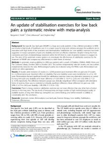
Figure 1. Initial Testing. The number of trials needed to reach the consistency of performance criteria is indicated on the X-axis and the proportion of subjects who failed to meet the performance criteria are indicated on the Y-axis. TCI + US = typical clinical instruction + real-time ultrasound imaging feedback; TCI = typical clinical instruction.
performance were reached as demonstrated by the results of the log-rank test (p = 0.0002) (Figure 1). 8
Number of subjects from each group TCI
TCI+US
pvalue**
3 3 2 2 0
0 0 4 3 2
0.20
3 0 1 4 2
0 0 0 4 5
0.59
Fisher’s exact test
The median number of trials out of 10 until the performance criteria in the TCI+US group was 5.0 (interquartile range=2.0); since fewer than half of the TCI group reached the performance criteria, the median number of trials cannot be calculated. Interestingly, the overall number of trials during which the AHE was performed correctly (but not necessarily three trials sequentially) was not statistically different between the groups (Fisher’s exact test, p = 0.20) (Table 4). Retention testing Six out of 10 TCI group subjects (60%) reached the criteria for consistency of performance whereas nine out of the nine (100%) subjects in the TCI+US group were successful (Table 3), however this difference in number of subjects who reached performance criteria at the retention testing session did not reach statistical significance (p = 0.09). There was a statistical difference between groups in the number of trials until criteria for consistency of performance were reached as demonstrated by the log-rank test (p = 0.05) (Figure 2). The median number of trials until performance criteria for the TCI group was 5.50 (interquartile range cannot NZ Journal of Physiotherapy – March 2007, Vol. 35 (1)
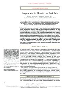
Figure 2. Retention Testing. The number of trials needed to reach the consistency of performance criteria is indicated on the X-axis and the proportion of subjects who failed to meet the performance criteria are indicated on the Y-axis. TCI + US = typical clinical instruction + real-time ultrasound imaging feedback; TCI = typical clinical instruction.
be calculated), and the TCI+US group was 3.0 (interquartile range=2.0). As with the initial testing, the overall number of trials during which the AHE was performed correctly (but not necessarily three trials sequentially) was not statistically different between the groups (Fisher’s exact test, p = 0.59) (Table 4).
DISCUSSION Initial testing session This pilot study demonstrated significant differences in the performance of the AHE between the two feedback groups. In the 20-30 minutes initial testing session, significantly more of the subjects assigned to the TCI+US group reached the criteria for consistency of performance in significantly fewer number of trials than those subjects assigned to the TCI group. As there were no initial differences between the two groups, these data suggest that the type of feedback can account for the improved performance of the TCI+US group. The precise visual image produced by the real time ultrasound may have been a particularly beneficial form of feedback, given that both visual feedback (Kim and Kramer, 1997) and precise feedback (Wright et al., 1997) have been shown to accelerate skill acquisition. The recommendation for the use of real time ultrasound feedback to teach the AHE by others (Henry and Westervelt, 2005; Hides et al., 1998; Hides et al., 1995) is supported by the results of this study. The benefit of RTUS in learning the correct AHE was evidenced in the fact that the two groups had the same feedback schedule (after every other trial), yet the TCI+US group still had a significantly greater proportion of subjects who achieved the criteria for consistency of performance in significantly fewer trials compared to the NZ Journal of Physiotherapy – March 2007, Vol. 35 (1)
TCI group. Given the constraints on number of treatment sessions allowed in managed health care systems, the use of RTUS to facilitate faster learning of the AHE skill, therefore allowing the progression onto integration of the AHE into patients’ specific functional impairments, seems justifiable. In contrast to our results, a recent study by Teyhen et al (2005) does not support the use of RTUS imaging as a beneficial tool when teaching the AHE to patients who have LBP. Teyhen et al (2005) found no benefit to adding RTUS biofeedback to the typical clinical instruction when teaching the AHE to patients with LBP. Teyhen et al (2005) used RTUS to measure the thickness of TA, IO and EO muscles prior to training, immediately after training, and four days after training in patients who had been seeking treatment for their LBP for three months or less. After only minimal instruction (“Take a deep breath in and, as you exhale, pull your belly button up and in towards your spine”), all patients were able to increase the thickness of TA with only minimal change in thickness of IO and EO muscles. After continued training with or without the use of RTUS imaging, no further changes in TA muscle thickness were seen. The LBP population in Teyhen’s (2005) study were patients who had sought treatment for their LBP within the previous approximately three months, whereas in the current study, the mean duration of patients’ symptoms was 76 – 105 months; interestingly, the Oswestry Disability Scores were similar for both cohorts. The patients in our study may have suffered with LBP long enough to have developed alternate muscle recruitment patterns, thus making it more difficult to learn how to perform a low load isometric contraction of selective muscles in their anterolateral abdominal wall. Thus, the more chronic patients in our study benefited from the additional visual feedback provided by the US image, whereas those in the study by Teyhen (2005) did not. Additionally, our patients performed 10 attempts of the AHE without any pre-training whereas those in the study by Teyhen (2005) received some pre-training and then a total of 15 attempts. The additional training trials in the study by Teyhan (2005) may have allowed more subjects to master the AHE during the initial testing session, thereby diminishing any potential benefit of feedback from the US image. Retention testing The results from the retention testing are inconclusive. Two of the three statistical tests used showed no significant difference between the two groups on the retention test. For one of these tests, which examined the difference in the number of subjects in each group that achieved consistency of performance criteria, there was a trend toward significance at retention testing (p = .09). The third statistical test did demonstrate that the TCI + US group did achieve the consistency to performance criteria in statistically fewer trials than the TCI group. 9
The retention testing results of our study and those of Henry and Westervelt (2005) do need to be interpreted with care due to the small sample numbers, which may not be sufficient to detect a true difference. These results suggest that RTUS feedback may be most valuable during the initial learning of the AHE skill. A recent study by Henry and Westervelt (2005) also found that RTUS imaging was beneficial during the learning of the AHE but not for the retention of the skill four days later. The results of this study show that 2 more patients in the TCI group reached performance criteria during the retention test compared to the initial testing. This suggests that TCI is effective in helping patients learn the AHE but that TCI+US may be a more efficient approach for teaching a skill as complex as the AHE. With the visual feedback for the patient and the physiotherapist, the skill can be learned more efficiently, and accurately, allowing progression to the integration of the AHE into more specific impairment based movement reeducation to achieve the patient’s functional goals in a timely fashion. Our results did indicate that the TCI + US group achieved consistency to performance criteria in fewer numbers of trials on the retention test suggesting that the addition of the RTUS during the initial testing session influenced retention of the AHE skill. More research is needed to address the optimum number of practice trials per session as well as the optimal feedback schedule. Perhaps those patients who achieved the criteria for consistency of performance at retention testing, but not at initial testing, just needed a greater number of training trials than our protocol allowed. One limitation with the method used to establish the examiner's ability to assess a correct or incorrect AHE was that in all but one of the simulation sessions, the examiner practiced identifying each of the four substitution patterns individually. This differs from the clinical setting because patients learning the AHE may substitute with any combination or variation of patterns while trying to master the correct manoeuvre. In an attempt to mimic the clinical setting, one simulation session involved the simulator using a random selection of the four common substitution patterns or any combination of the substitution patterns. However, a patient who is learning the AHE probably will not perform in such a deliberate fashion. It also must be acknowledged that it is not yet known whether the methodology of using a trained simulator is a valid way to train examiners on the detection of a correct or incorrect AHE. Other limitations of the study include that the person taking the initial and retention test measures was the same person teaching the patients the AHE, leading to possible bias on the examiner’s part in terms of teaching and of measuring performance of the AHE. Inadequate masking of group allocation was also a limitation of the study. In summary, quicker mastery of the basic AHE may allow clinicians to progress their patients 10
quickly into a patient specific rehabilitation program integrating the AHE into the retraining of the patient’s performance impairments and functional goals. The segmental stabilization protocol has been shown to be an effective rehabilitation protocol for patients with chronic recurrent LBP secondary to spondylolysis or spondylolisthesis (O'Sullivan et al., 1997). The evidence for whether or not a similar protocol will be effective for patients with chronic, recurrent idiopathic LBP is conflicting (Koumantakis et al., 2005; Goldby et al., 2006; Hicks et al. 2005) and is the focus of an ongoing study in our clinics.
CONCLUSIONS In this pilot study in patients with recurrent episodic LBP, the use of real RTUS imaging to visualize the anterolateral abdominal wall enhanced the learning of the AHE. The retention testing results were inconclusive, however, with no group differences found in performance of the AHE in two of the three tests used to detect group differences, perhaps because of insufficient statistical power in our small sample size. Key Points • Visual feedback provided by RTUS imaging of the anterolateral abdominal wall whilst learning the AHE decreased the number of trials needed to achieve the consistency of performance criteria (defined as 3 consecutive correct AHE) in people with chronic recurrent LBP. • The effect of RTUS imaging on the retention of performance of the AHE is unclear based on these results.
ACKNOWLEDGEMENTS We would like to thank Long Trail Physical Therapy and Timberlane Physical Therapy for the use of their clinical facilities for the purposes of this clinical research. Grant support was received from the Dean’s Research Incentive Fund, College of Nursing and Health Sciences, University of Vermont. This research was previously presented in poster format at the American Physical Therapy Association Annual Exposition, Chicago, IL June 2004. Ethical approval was granted by the Institutional Review Board, 201 Rowell Bldg., University of Vermont, Burlington, VT 05405. Written informed consent was obtained from each study participant and the rights of the participants were protected.
REFERENCES
Bunce SM, Hough AD and Moore AP (2004): Measurement of abdominal muscle thickness using M-mode ultrasound imaging during functional activities. Manual Therapy 9: 41-44. Glass K, Allison GT and Edmondston S (1999): Physiotherapists' ability to differentiate anterolateral abdominal control strategies. Doctorate of Philosophy dissertation, Curtin University of Technology, Perth, WA, Australia.
NZ Journal of Physiotherapy – March 2007, Vol. 35 (1)
Golby LJ, Moore AP, Doust J, and Trew ME (2006): A randomized controlled trial investigating the efficacy of musculoskeletal physiotherapy on chronic low back disorder. Spine 31(10): 1083-1093. Hagins M, Adler K, Cash M, Daugherty J and Mitrani G (1999): Effects of practice on the ability to perform lumbar stabilization exercises. J Orthop Sports Phys Ther 29: 546555. Henry SM and Westervelt KC (2005): The use of real-time ultrasound feedback in teaching abdominal hollowing exercises to healthy subjects. J Orthop Sports Phys Ther 35: 338-345. Hicks GE, Fritz JM, Delitto A, McGill SM (2005): Preliminary development of a clinical prediction rule for determining which patients with low back pain will respond to a stabilization exercise program. Archives of Physical Medicine and Rehabilitation 86 (9):1753-62. Hides JA, Richardson CA and Jull GA (1998): Masterclass: Use of real-time ultrasound imaging for feedback in rehabilitation. Man Ther 3: 125-131. Hides JA, Richardson CA, Jull GA and Davies S (1995): Ultrasound imaging in rehabilitation. Australian Journal of Physiotherapy 41(3): 187-193. Hodges PW, Pengel LHM, Herbert RD and Gandevia SC (2003): Measurement of muscle contraction with ultrasound imaging. Muscle and Nerve 27: 682-692. Hodges PW and Richardson CA (1996): Inefficient muscular stabilization of the lumbar spine associated with low back pain: A motor control evaluation of transversus abdominis. Spine 21(22): 2640-2650. Kim H and Kramer J (1997): Effectiveness of visual feedback during isokinetic exercise. J Orthop Sports Phys Ther 26: 318-323. Koumantakis G, Watson P and Oldham J (2005): Trunk muscle stabilization training plus general exercise versus general exercise only: randomized controlled trial of patients with recurrent low back pain. Phys Ther 85: 209-225. Magill R (1997): Motor Learning: Concepts and Applications. Boston: WCB McGraw-Hill. Magill R and Wood C (1986): Knowledge of results precision as a learning variable in motor skill acquisition. Res Q Exerc Sport 57: 170-173. McMeeken JM, Beith ID, Newham DJ, Milligan P and Critchley DJ (2004): The relationship between EMG and change in thickness of transversus abdominis. Clinical Biomechanics 19: 337-342. O'Sullivan PB, Twomey LT and Allison GT (1997): Evaluation of specific stabilizing exercises in the treatment of chronic low back pain with radiologic diagnosis of spondylolysis or spondylolisthesis. Spine 22 (24): 2959-2967. Richardson C, Hodges P and Hides JA (2004): Therapeutic exercise for spinal segmental stabilization in low back pain: Scientific basis and clinical approach. (2nd ed.) Edinburgh: Churchill Livingstone. Richardson C, Jull G, Toppenberg R and Comerford M (1992): Techniques for active lumbar stabilization for spinal protection: a pilot study. Australian Physiotherapy 38 (2): 105-112. Steinberg M (1998): Kaplan-Meier Survival Analysis Examples. SPSS Statisitical Software Manual. Chicago: SPSS Inc.: 271282. Teyhen DS, Miltenberger CE, Deiters HM, Del Toro YM, Pulliam JN, Childs JD, Boyles RE and Flynn TW (2005): The use of ultrasound imaging of the abdominal drawing-in maneuver in subjects with low back pain. J Orthop Sports Phys Ther 36: 346-355. Vu NV, Barrows HS, Marcy ML, Verhulst SJ, Colliver JA and Travis T (1992): Six years of comprehensive, clinical, performance-based assessment using standardized patients at the Southern Illinois University School of Medicine. Acad Med 67: 42-50. Wright DL, Smith-Munyon VL and Sidaway B (1997): How close is too close for precise knowledge of results? Res Q Exerc Sport 68: 172-176.
ADDRESS FOR CORRESPONDENCE
Sharon Henry, Department of Rehabilitation and Movement Science, 305 Rowell Building, University of Vermont, Burlington, Vermont 05405-0068, USA. Phone number: (802) 656 - 8146. Fax number: (802) 656 - 6586. E-mail:
[email protected]
NZ Journal of Physiotherapy – March 2007, Vol. 35 (1)
11