NATIONAL CENTER FOR CASE STUDY TEACHING IN SCIENCE
Putting the Pieces Together: The Discovery of DNA Structure and Replication by Kevin M. Bonney Liberal Studies, Faculty of Arts and Sciences New York University, New York, NY
Part I – Setting the Stage In the autumn of 1952, while the weather in London, England, was cooling down, the race to discover the structure of DNA was heating up inside two rival laboratories: the Cavendish Lab at Cambridge University and the laboratory of J.T. Randall at nearby King’s College. DNA had recently been identified as the hereditary material that organisms use to transmit genetic traits from one generation to another. This finding is largely attributed to the work of two groups of American scientists, Oswald Avery, Colin MacLeod, and Macyln McCarty of the Rockefeller Institute, and Alfred Hershey and Martha Chase of Cold Spring Harbor Laboratory, though many important experiments conducted by other scientists were crucial to developing this understanding. The next great challenge was to identify the structure of DNA so that we could understand how the information contained in DNA is copied and passed from cell to cell, and how that information is translated into the instructions for building the proteins necessary for life. The groundbreaking research of the Cavendish and Randall laboratories describing the structure of DNA was published in a series of papers in the journal Nature on April 25, 1953 [Wilkins et al., 1953; Franklin et al., 1953]. These would become known as some of the most important scientific papers ever published, as well as a symbol of great controversy involving allegations of misogyny and ethics violations among scientists who would go on to win a Nobel Prize.
Preparing for Your Mission It’s now your job to figure out the next piece of the puzzle: How do cells make new copies of their DNA? Before you can figure out how DNA is replicated, you must understand the structure of DNA. To prepare for your task, answer the following questions to assess your understanding of DNA structure on your own, without using any outside sources. You will then be asked to view a video.
Questions 1. The structure of DNA is a: A. alpha helix B. single helix C. double helix D. triple helix 2. DNA is made of repeating units called: A. amino acids B. monosaccharides C. nucleotides D. peptides
“Putting the Pieces Together” by Kevin M. Bonney
Page 1
NATIONAL CENTER FOR CASE STUDY TEACHING IN SCIENCE 3. The “D” in DNA stands for: A. Darwin B. Deoxyribose C. Dimethyl D. Dinucleic acid 4. The bonds that hold the backbone of each strand of DNA together are called: A. hydrogen bonds B. ionic bonds C. peptide bonds D. phosphodiester bonds 5. The information contained in DNA is encoded by the: A. hydrogen bonds on the inside of the DNA molecule B. nitrogenous bases on the inside of the DNA molecule C. phosphate groups on the outside of the DNA molecule D. sugars on the outside of the DNA molecule 6. In which direction is DNA read? A. 0' to 1' B. 1' to 3' C. 3' to 5' D. 5' to 3' 7. The letters in the genetic code of DNA are A, C, G, and T. The A stands for: A. amino B. adenine C. alanine D. aspartame 8. If one strand of DNA is AGCTA, the complementary strand would be: A. AGCTA B. ATCGA C. CTAGC D. TCGAT Whether you are a DNA novice or a pro, watching the following video should help you put the pieces together. After watching the video, answer the questions again to demonstrate what you know now. You may wish to watch the video more than once before moving on to the questions. The Chemical Structure of DNA http://www.hhmi.org/biointeractive/chemical-structure-dna
References Franklin, R.E. and Gosling, R.G. 1953. Molecular configuration in sodium thymonucleate. Nature 171(4356):740–1. Howard Hughes Medical Institute. The Chemical Structure of DNA. http://www.hhmi.org/biointeractive/chemicalstructure-dna. Wilkins, M.H., et al. 1953. Molecular structure of deoxypentose nucleic acids. Nature 171(4356):738–40.
“Putting the Pieces Together” by Kevin M. Bonney
Page 2
NATIONAL CENTER FOR CASE STUDY TEACHING IN SCIENCE
Part II – Discovering the Structure of DNA The story of how DNA structure was discovered is not only one of the most important tales in the history of science, it is also the root of a great controversy that will live on for many years. It is your job to research the collaborations and controversies that made the discovery of DNA structure possible, and determine once and for all who deserves the credit for this groundbreaking feat of science. The main characters in the story are named Francis Crick, Rosalind Franklin, James Watson, and Maurice Wilkins. To understand this story, read Watson and Crick’s original scientific paper: Watson, J.D. and Crick, F.H. (1953) Molecular structure of nucleic acids; a structure for deoxyribose nucleic acid. Nature. 1953 Apr 25;171(4356):737-8. http://www.nature.com/nature/journal/v171/n4356/ pdf/171737a0.pdf. After you have read Watson and Crick’s paper, watch this short video to strengthen your understanding of the implications of that work: The Secret of Life—Discovery of DNA Structure. Produced by Virginia Commonwealth University. Running time: 8:36 min. Date: 2014. http://youtu.be/JjO6n85wq8A. You may also wish to access the following resource for more information: The structure of DNA: Cooperation and competition. Understanding Science Team. The University of California, Berkeley website. http://undsci.berkeley.edu/article/dna_01. With the help of your peers, analyze, evaluate, and debate some of the important ethical implications of the work conducted by Crick, Franklin, Watson, and Wilkins.
Questions 1. Why is understanding the structure of DNA and how it is replicated important? List some technologies and practical applications that could not have been developed without this understanding. 2. How does the structure of DNA identified by Watson and Crick differ from the model previously proposed by Linus Pauling and others? 3. What discovery by Erwin Chargaff helped Watson and Crick build their model of DNA structure? How was this piece of information helpful to them? 4. What is the main scientific weakness you notice in Watson and Crick’s paper? 5. The conclusion of the paper published by Watson and Crick includes the now rather famous statement: “It has not escaped our notice that the specific pairing we have postulated immediately suggests a possible copying mechanism for the genetic material.” Describe what this statement means and why it is significant. 6. Describe the specific contribution of each of the following researchers to the discovery of DNA structure: • Francis Crick • Rosalind Franklin • James Watson • Maurice Wilkins 7. In their paper, how do Watson and Crick acknowledge Rosalind Franklin for her contribution to their work ? 8. Is Watson and Crick’s acknowledgement of Franklin satisfactory, or did she deserve more credit? Your instructor will divide you into two groups to debate this question. • If you are in the Watson and Crick group, write a one-paragraph argument from their perspective. Defend the way you have treated Rosalind Franklin and provide an argument that you have given her all of the credit she deserves. Support your claims using information from the paper and video you just watched. Prepare to debate your position with members of the other group.
“Putting the Pieces Together” by Kevin M. Bonney
Page 3
NATIONAL CENTER FOR CASE STUDY TEACHING IN SCIENCE • If you are in the Franklin group, write a one-paragraph argument from your perspective. Defend yourself against the treatment you received from Watson and Crick, and argue why you deserve more credit for the discovery of DNA structure than you have been given. Support your claims using information from the paper and video you just watched. Prepare to debate your position with members of the other group.
“Putting the Pieces Together” by Kevin M. Bonney
Page 4
NATIONAL CENTER FOR CASE STUDY TEACHING IN SCIENCE
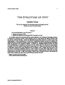
Part III – Building a Model of DNA Replication Now that you can illustrate the structure of DNA and describe the experiments and ethical issues related to its discovery, you are prepared for your mission: To determine how cells make new copies of their DNA. Remember, it is still the 1950s. At this time, three hypothetical models have been proposed to explain how DNA is replicated. These models, known as the conservative model, the dispersive model, and the semi-conservative model, are illustrated in Figure 1.
Figure 1. Hypothetical models of DNA replication.
In the conservative model, both original template DNA strands form one double helix after replication, while the two newly synthesized strands form a second double helix. In the dispersive model, the template DNA is broken into double-stranded segments that act as templates for the synthesis of new double helix molecules; the two resulting double helix molecules each contain half of the original template DNA and half of the newly synthesized DNA. In the semi-conservative model, the original DNA strands are separated, and each acts as a template for the synthesis of a new strand of DNA to complete the double helix. To figure out which model of DNA replication correctly illustrates how living organisms make new copies of their DNA, you must first find a way to distinguish the newly synthesized DNA from the original template DNA. Next, you want to figure out what would happen in each model and compare that to the actual experimental data. Step 1. To start, make a double-stranded DNA molecule to represent the original template DNA. Make each strand of the molecule six nucleotides long, using gumdrops to represent the nucleotides of the original template DNA. Use toothpicks to form bonds that connect the gumdrops in each strand and hold the two strands together. Step 2. Choose one model of replication (conservative, dispersive, or semi-conservative) as the mechanism for copying your DNA molecule. From now on use marshmallows to represent newly synthesized DNA and
“Putting the Pieces Together” by Kevin M. Bonney
Page 5
NATIONAL CENTER FOR CASE STUDY TEACHING IN SCIENCE toothpicks to form bonds. Make a copy of the original template DNA molecule by following the principles of the model of replication that you chose. Step 3. Repeat Steps 1 and 2 for the other two models of DNA replication. Step 4. Record the percentage of gumdrops and marshmallows in each DNA molecule using the table below. (Divide the number of each by the total number of both and multiply by 100 to calculate the percent.) After one round of replication: Model
DNA molecule A
DNA molecule B
________% gumdrops
________% gumdrops
________ % marshmallows
________ % marshmallows
________% gumdrops
________% gumdrops
________ % marshmallows
________ % marshmallows
________% gumdrops
________% gumdrops
________ % marshmallows
________ % marshmallows
Conservative
Dispersive
Semi-Conservative
Step 5. Using only marshmallows and toothpicks, make a copy of each DNA molecule you have from Step 3 following the principles of the three models of replication. Step 6. Record the percentage of gumdrops and marshmallows in each DNA molecule in the table below. (Divide the number of each by the total number of both and multiply by 100 to calculate the percent.) After two rounds of replication: Model
DNA molecule A
DNA molecule B
DNA molecule C
DNA molecule D
______% gumdrops
______% gumdrops
______% gumdrops
______% gumdrops
Conservative
______ % marshmallows ______ % marshmallows ______ % marshmallows ______ % marshmallows ______% gumdrops
______% gumdrops
______% gumdrops
______% gumdrops
Dispersive
______ % marshmallows ______ % marshmallows ______ % marshmallows ______ % marshmallows SemiConservative
______% gumdrops
______% gumdrops
______% gumdrops
______% gumdrops
______ % marshmallows ______ % marshmallows ______ % marshmallows ______ % marshmallows
Questions 1. Describe what occurs in each of the following models of DNA replication: conservative, dispersive, and semiconservative. 2. Describe how each model of DNA replication differs in terms of the observations you made using gumdrops and marshmallows. How can you tell each model apart based on the relative amount of gumdrops and marshmallows in each DNA molecule? “Putting the Pieces Together” by Kevin M. Bonney
Page 6
NATIONAL CENTER FOR CASE STUDY TEACHING IN SCIENCE
Optional Exercise A short video entitled Building a Model of DNA Replication demonstrating the activity above can be viewed at: https://youtu.be/SAXHwkKe6Bc After watching the video, answer the following questions: 1. Which of the following statements about the use of gumdrops and marshmallows in this activity is true? A. Both are used to represent nucleotides. B. Gumdrops represent nitrogen and marshmallows represent phosphorus. C. Gumdrops represent DNA and marshmallows represent RNA. D. Gumdrops represent nucleotides and marshmallows represent phosphodiester bonds. 2. In this activity, DNA molecules containing only original template DNA are represented using: A. gumdrops and toothpicks only. B. marshmallows and toothpicks only. C. gumdrops and marshmallows only. D. toothpicks only. 3. After one round of replication, what has been produced by the conservative model of replication? A. One molecule that contains 0% gumdrops and 100% marshmallows, and one molecule that contains 100% gumdrops and 0% marshmallows. B. Two molecules that contain 100% marshmallows and 0% gumdrops. C. Two molecules that both contain 100% gumdrops and 0% marshmallows. D. Two molecules that both contain 50% gumdrops and 50% marshmallows. 4. After two rounds of replication, which of the following is true of the four DNA molecules produced by the semi-conservative model? A. All four contain 25% gumdrops and 75% marshmallows. B. All four contain 50% gumdrops and 50% marshmallows. C. Two contain 50% gumdrops and 50% marshmallows, and two contain 0% gumdrops and 100% marshmallows. D. Two contain 100% gumdrops and 0% marshmallows, and two contain 0% gumdrops and 100% marshmallows. 5. Which model(s) of DNA replication never produce(s) a DNA molecule that contains both gumdrops and marshmallows? A. The conservative model only. B. The dispersive model only. C. Both the conservative model and the dispersive model. D. Both the dispersive and the semi-conservative model.
“Putting the Pieces Together” by Kevin M. Bonney
Page 7
NATIONAL CENTER FOR CASE STUDY TEACHING IN SCIENCE
Part IV—Testing the Model of DNA Replication Now that you can describe what occurs in each of the three hypothetical models of DNA replication and identify which type of replication has occurred by labeling the nucleotides of the original and newly synthesized DNA, you are ready to complete your mission and determine which model of replication correctly explains how cells make new copies of their DNA. However, because real DNA does not incorporate gumdrops or marshmallows, and is actually too small to see with your eyes, you will want to use data collected by two other famous scientists, Matthew Meselson and Franklin Stahl, to help you complete your mission. To distinguish between newly replicated DNA and the original template DNA from which it was copied, Meselson and Stahl used isotopes of nitrogen called 14N and 15N to label the nitrogenous bases of DNA. Isotopes of nitrogen differ in the number of neutrons they contain; 15N, having one more neutron than 14N is denser than 14N, since neutrons comprise much of the mass of atoms. Thus, DNA containing 15N can be distinguished from DNA containing 14N by comparing the density of each DNA molecule. Think of 14N as the marshmallows you used when making your models, and 15N as the denser gumdrops. Meselson and Stahl used Escherichia coli bacteria as a source of cells to study DNA replication. The Escherichia coli were first grown in nutrient broth containing 15N as the only source of nitrogen until all of the nitrogen in the bacteria’s DNA was 15N. Next, the bacteria were placed in media containing only 14N and allowed to divide, so the newly synthesized DNA would use and incorporate the 14N into its structure. During each division, the amount of DNA was doubled as one newly synthesized DNA molecule was made from each template DNA molecule. Meselson and Stahl knew that DNA containing 15N could be separated from DNA containing 14N by centrifuging the DNA molecules in a density gradient, which causes molecules of higher density to move toward the bottom of the gradient and molecules of lower density to move toward the top. Unlike the models you just made, Meselson and Stahl could not see real DNA molecules with their eyes or even using a microscope. However, they knew that DNA absorbs ultraviolet (UV) light, and that by shining UV light on the density gradients they made they could determine where in the gradient the DNA was located because a shadow would be cast in the location where DNA was absorbing the light. The data that Meselson and Stahl collected in this way is represented in Figure 2. Remember that the bacteria started out with 15N comprising 100% of the nitrogen in their DNA, then they were supplied only 14N so that any newly synthesized DNA would contain 14N. Use the knowledge you gained from building the three hypothetical models of DNA replication with gumdrops and marshmallows to complete your mission by answering the following questions.
Questions 1. Before analyzing the results of this experiment, Meselson and Stahl needed to know where DNA containing 100% 14 N or 15N would be in the density gradient. Predict what Meselson and Stahl did to figure this out. “Putting the Pieces Together” by Kevin M. Bonney
Figure 2. Representation of data collected by Meselson and Stahl.
Page 8
NATIONAL CENTER FOR CASE STUDY TEACHING IN SCIENCE 2. How can you interpret the data collected after one round of division? In other words, what do the number and location of the band(s) present signify? 3. Which model(s) of DNA replication are consistent with the data collected after the first round of replication? Which model(s) can be ruled out? Why? 4. For any models of DNA replication that were ruled out after the first round of replication, what would the data have looked like if the DNA had, in fact, been replicated according to that model? 5. Which model of DNA replication is consistent with the data collected after the second round of replication? Which model can be ruled out? Why? 6. What would the data have looked like if the DNA had, in fact, been replicated according to the model just ruled out? 7. According to the model of DNA replication identified as correct, what would the data look like after a third round of a replication? How about after a fourth round? 8. Why are bacteria like Escherichia coli good for studying DNA replication (as opposed to using other types of cells)?
• Case copyright held by the National Center for Case Study Teaching in Science, University at Buffalo, State University of New York. Originally published July 28, 2015. Please see our usage guidelines, which outline our policy concerning permissible reproduction of this work. Licensed photograph of scientist with DNA in title block ©Sergey Nivens | Fotolia, ID#59039425. “Putting the Pieces Together” by Kevin M. Bonney
Page 9