YOUNG INVESTIGATORS WRITING COMPETITION WINNERS
Pulsed Dye Laser and Pulsed Dye Laser–Mediated Photodynamic Therapy in the Treatment of Dermatologic Disorders AUSTIN LIU, MD,* RONALD L. MOY, MD,† EDWARD VICTOR ROSS, MD,‡ ILTEFAT HAMZAVI, MD,* AND DAVID M. OZOG, MD*
BACKGROUND The pulsed dye laser (PDL) is used for treating cutaneous vascular disorders. Recent reports have also shown its effectiveness in conditions of other etiologies, although the precise mechanisms of action are unknown. PDL has also been used in photodynamic therapy (PDT) for many dermatologic conditions. We review the broad array of disorders that can be effectively managed using the PDL. OBJECTIVES AND METHODS A review of the literature on the application of the PDL and PDL-mediated PDT in dermatologic disorders. A literature-based search was performed using PubMed from 1997 to 2010. Search terms included: “pulsed dye laser,” “pulsed dye laser photodynamic therapy,” and “pulsed dye laser indications.” RESULTS The PDL was initially designed for cutaneous vascular disorders. Recent investigations have demonstrated successful results when treating malignant, inflammatory, viral, and collagenous conditions. Side effects, including pain, purpura, edema, and postinflammatory hyperpigmentation, were mild, well tolerated, and transient. CONCLUSIONS PDL is accepted as first-line therapy for vascular disorders including port-wine stains, telangiectasias, and hemangiomas. PDL causes selective photothermolysis of dermal vasculature. This mechanism also allows it to be applicable for disorders of other etiologies. Recent studies suggest that the PDL may induce cytokine expression and collagen formation, further increasing its applicability in dermatology. The authors have indicated no significant interest with commercial supporters.
T
he U.S. Food and Drug Administration (FDA) approved the pulsed dye laser (PDL) in 1986 for the treatment of cutaneous vascular disorders. The PDL emits light within the visible (yellow) spectrum, which oxyhemoglobin selectively absorbs, resulting in selective photothermolysis. Anderson and Parrish introduced the concept of selective photothermolysis in 1983,1 and the PDLs were among the first designed after the acceptance of this discovery. With PDL treatment, vascular tissue is targeted while surrounding tissue remains intact. Currently, its efficacy is well established, and it is widely used in the treatment of numerous
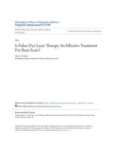
vascular conditions, including port-wine stains (PWS), telangiectasias, hemangiomas, and other vascular growths. It has also been found to be effective for cutaneous erythema of various etiologies (Figure 1). It has also proven to be effective for numerous dermatologic conditions that are not primarily of vascular etiology.2 More recently, the PDL has also gained FDA approval for the treatment of numerous nonvascular conditions, including benign epidermal pigmented lesions, rhytides, and benign cutaneous lesions such as acne, warts, psoriasis, and scars. Although speculation exists regarding its mechanism of action in treating
* Department of Dermatology, Division of Mohs Micrographic Surgery, Henry Ford Hospital, Detroit, Michigan; † David Geffen School of Medicine, University of California at Los Angeles, Los Angeles, California; ‡Division of Dermatology, Scripps Clinic, San Diego, California © 2012 by the American Society for Dermatologic Surgery, Inc. � Published by Wiley Periodicals, Inc. � ISSN: 1076-0512 � Dermatol Surg 2012;38:351–366 � DOI: 10.1111/j.1524-4725.2011.02293.x 351
PDL AND PDL-MEDIATED PHOTODYNAMIC THERAPY
(A)
(B)
Figure 1. Clinical photograph of persistent erythema of the neck associated with high tryptase level (A) before and (B) after treatment with the pulsed dye laser.
nonvascular disorders, no definitive answers have been elucidated. A possible and somewhat partial explanation may be the targeting of underlying vasculature of these various conditions. Originally, the PDL was designed to emit light at a wavelength of 577 nm. It was subsequently modified to 585 and 595 nm, allowing for greater depth of vascular injury—to approximately 1.2 mm. Newer-generation PDLs offer higher fluences, larger spot sizes, longer pulse widths, and cryogen and air cooling. For selective photothermolysis to occur, certain requirements must be met. Specifically, the chromophore target needs to absorb the wavelength that the laser emits, the pulse duration should be equal to or less than the thermal relaxation time of the target, and a sufficient fluence is needed to reach temperatures capable of damaging the target. Possible side effects after PDL treatment include erythema, purpura, edema, pigmentary changes, and rarely, scarring. The frequency and severity of these effects depend on individual patient characteristics and the specific laser settings used.
352
(ALA) or methyl-ALA, with light in the visible wavelength spectrum. After topical incubation of the skin with 5-ALA, rapidly dividing cells take up the 5-ALA and convert it into protoporphyrin IX (PpIX), an endogenous porphyrin. Upon exposure to a light source, reactive oxygen species with cytotoxic effects are generated. Although PpIX has peak absorption in the Soret band (400–410 nm), absorption occurs at the PDL wavelengths, with observable effects. The activation of porphyrins may be less with a pulsed light source than with continuous light exposure.3 Although many different light sources have been used to activate PpIX, some have certain disadvantages. For example, PDT using an incoherent light source such as intense pulsed light (IPL) can cause erythema and crusting and requires longer, more-painful treatment sessions but, PDL-mediated PDT is generally faster, with fewer side effects, making it an ideal light source for treating certain conditions. Herein, we review the treatment of various dermatologic conditions using the PDL and PDL-mediated PDT (Table 1).
Efficacy for some conditions requires purpura as an endpoint, whereas nonpurpuric settings are effective for others.
Vascular Disorders
More recently, PDL has also been used in photodynamic therapy (PDT). This process combines a photosensitizer, usually 5-aminolevulinic acid
A significant amount of research and experience has demonstrated the effectiveness of the PDL in treating primarily vascular-based disorders.4 PWS are
DERMATOLOGIC SURGERY
Vascular Disorders Commonly Treated Using PDL
LIU ET AL
TABLE 1. Current Indications for the Use of Pulsed Dye Lasers (PDL) FDA Approved Indications – Conditions Commonly Treated with PDL ● Benign cutaneous vascular lesions Telangiectasias, port-wine stains, hemangiomas, angiomas, rosacea, poikiloderma of Civatte ● Benign cutaneous lesions Verruca vulgaris, scars, striae, psoriasis, acne vulgaris, sebaceous hyperplasia ● Rhytids FDA approved indications – conditions less commonly treated with PDL ● Benign epidermal pigmented lesions ● Benign cutaneous vascular lesions Angioma serpiginosum, glomus tumor, unilateral nevoid telangiectasia, angiokeratoma of Fordyce, dermatomyositis (poikilodermatous erythema), angiofibromas, keratosis pilaris rubra/keratosis pilaris atrophicans faciei ● Benign cutaneous lesions Atopic dermatitis, granuloma faciale, granuloma annular, dermatomyositis (Gottron papules), molluscum contagiosum Off-label (non-FDA approved) indications ● Actinic keratosis ● Actinic cheilitis ● Bowen’s disease ● Basal cell carcinoma FDA = Food and Drug Administration.
congenital dermal vascular malformations that are responsive to PDL treatment (Figure 2). The PDL has been compared with other treatments, including PDL-mediated PDT5 and IPL.6 In a study of eight patients with PWS, no difference in effectiveness was found between treatment with PDL alone and PDL-mediated PDT.5 Both modalities led to improvement in all patients. The lack of difference in clinical results may be because of the achieve(A)
ment of maximum efficacy with PDL alone. Further possibilities include insufficiencies in the photosensitizer dosage or PDL settings to initiate an adequate photodynamic reaction. IPL has been shown to be less effective than the PDL in treating PWS.6 Specifically, 20 patients with PWS were treated in a single session with a 595-nm PDL or IPL. Significantly better clinical results and higher clearance rates were seen in PDL-treated patients, and as a result, (B)
Figure 2. Clinical photograph of port-wine stain (A) before and (B) after pulsed dye laser treatment. Photograph courtesy of Iltefat H. Hamzavi, MD.
38:3:MARCH 2012
353
PDL AND PDL-MEDIATED PHOTODYNAMIC THERAPY
(A)
(B)
Figure 3. Clinical photograph of telangiectasias at site of surgical flap (A) before and (B) after treatment with the pulsed dye laser. Photograph courtesy of David M. Ozog, MD.
the authors recommended IPL only as a second-line option for patients unresponsive to PDL treatments. Similarly, PDL improves the appearance of hemangiomas and telangiectasias (Figure 3). The treatment sessions are short, and pain is generally well tolerated, further making it an ideal method of intervention especially in children. Uncommon but reported side effects after PDL treatment of hemangiomas include atrophy, pigmentary changes, and scarring. Scarring is rare with cryogen cooling and may be an inevitable result of the resolving hemangioma as opposed to an adverse event of PDL. Hemangiomas in various locations, including those in critical anatomic sites, have been successfully treated. A recent retrospective study treated 22 patients with superficial infantile eyelid hemangiomas using a 595-nm PDL.7 After an average of 5.6 sessions, most patients had near-complete to complete resolution. Adverse effects such as hypopigmentation, atrophy, scarring, ulceration, and infection were not observed. Two patients had residual hyperpigmentation. Overall, the use of PDL in the management of hemangiomas should be limited to superficial hemangiomas. Although systemic medications including corticosteroids and propranolol are effective in treating hemangiomas, they are associated with endocrinologic and hemodynamic effects. Nevertheless, they can be more effective, especially for deeper hemangiomas that do not respond well to PDL treatments because of inadequate depth of penetration. With regard to management of telangiectasia, many light therapy systems, including but not lim-
354
DERMATOLOGIC SURGERY
ited to pulsed dye, diode, neodymium-doped, and alexandrite lasers and IPL are currently available.8 The choice of any particular system depends on various factors, including location, size, and depth of the telangiectasias and patient skin type. A recent randomized controlled split-face study of 40 patients with facial telangiectasias compared a 595-nm PDL with IPL.9 Three treatments were performed at 6-week intervals. Although both treatments were effective, the PDL-treated sides had significantly more reduction in overall telangiectasias and significantly less pain than with IPL. As a result, most patients in the study preferred PDL. Lower extremity spider veins are generally larger and deeper than facial vessels. Venous lesions also have more deoxyhemoglobin, a different chromophore target than the pulsed dye is designed for. Thus, they can be more difficult to eradicate. Lower extremity spider veins have also been documented to improve significantly after three treatments at 6-week intervals using a 595-nm high-energy, long pulse duration PDL.10 Mild to moderate side effects, including pain, edema, erythema, purpura, and hyperpigmentation, were observed. Although some efficacy is seen, this is not considered a treatment of choice for lower limb telangiectasia and venulectasia.
Miscellaneous Vascular Disorders It has also been reported in the literature that numerous less-common conditions of vascular etiology can be effectively treated using PDL through
LIU ET AL
selective photothermolysis of vascular tissue. For example, angioma serpiginosum is an infrequently seen vascular disorder appearing as punctate, red to violaceous macules with serpiginous configurations. The 58511 and 595-nm12 PDLs have been useful in clearing these cutaneous lesions after approximately three to four treatment sessions. A retrospective study involving 12 angioma serpiginosum sites treated using a 595-nm PDL found excellent responses (> 80% improvement) to complete resolution in nine of the 12 sites after an average of 3.75 treatments.12 The 585- and 595-nm PDLs have also been reported to be effective for conditions such as glomus tumors,13 unilateral nevoid telangiectasia,14 angiokeratoma of Fordyce,15 postoperative ecchymoses after facial cosmetic procedures,16 Kaposi’s sarcoma,17 and erythematous lesions secondary to Goltz syndrome.18 The number of treatment sessions was variable and ranged from one to six. Generally, side effects were transient and included mild pigmentary changes, pain, and purpura. In a study involving 12 men with angiokeratoma of Fordyce, after two to six sessions with a 585-nm PDL, seven experienced 75% to 100% clearance, and the remaining five demonstrated 50% to 75% clearance.15 Temporary side effects included pain and purpura, and no permanent adverse effects were noted. The authors recommended PDL as an effective and safe alternative to other therapies such as cryotherapy, surgical excision, and electrocoagulation.
Nonvascular Disorders Although PDL is most commonly used to treat vascular lesions, numerous reports have demonstrated the value of PDL for nonvascular conditions, including dermatosis papulosa nigra (DPN)19 and lentigines.20 Other nonvascular conditions successfully treated with the PDL include inflammatory linear verrucous epidermal nevus,21 nodular amyloidosis,22 eccrine hidrocystoma,23 and porokeratosis.24 Although oxyhemoglobin is known to absorb PDL-emitted energy, melanin possesses this ability
as well. In a randomized split-face controlled study, the 595-nm PDL was evaluated in the treatment of 10 patients of Fitzpatrick skin types III and IV with lentigines.20 Clinically significant improvement was observed by global assessment after three monthly sessions. Although participants were of darker skin types, only transient post-treatment hyperpigmentation was seen in two individuals. Another randomized controlled study of 10 patients with DPN compared the efficacy of treatment with curettage, electrodesiccation, and PDL.19 All three methods proved equally effective in the treatment of DPN. A 585-nm PDL was used with a 7-mm spot size, 10 J/cm2, and a 10-ms pulse duration. Commonly used modalities for DPN, including cryotherapy, electrosurgery, and curettage, are associated with pigmentary changes. In this study, the most common side effect after PDL sessions was transient hyperpigmentation, at a similar rate to that seen after electrodesiccation and curettage. Advantages of the PDL over the standard modalities include shorter treatment sessions and no need for anesthesia, although the PDL has certain limitations. Specifically, the minimum spot size of 3 mm is larger than many lesions, and in darker skinned patients, there is often inadequate contrast between the lesion and surrounding skin for safe removal. The 595-nm PDL has also been compared with a 607-nm study prototype PDL for the treatment of benign epidermal pigmented lesions.25 In 10 patients with solar lentigines and macular seborrheic keratoses, both wavelengths were found to be equally effective, with no persistent side effects. The 607-nm PDL was slightly less painful according to the study participants.
Malignancies Actinic Keratosis, Actinic Cheilitis, and Bowen’s Disease Traditional interventions for precancerous lesions include cryotherapy, electrodesiccation, curettage,
38:3:MARCH 2012
355
PDL AND PDL-MEDIATED PHOTODYNAMIC THERAPY
and field treatment with topical agents and PDT with incoherent light sources. Disadvantages of such modalities include scarring, pigmentary changes, need for adherence, irritation, and pain. PDT with activation by a PDL light source is known to be effective for treating photodamaged skin, actinic keratoses (AKs), actinic cheilitis (AC), and acne vulgaris.26 PDL-mediated PDT has also been compared with PDL alone for management of precancerous conditions. In a prospective study, 40 patients with AKs were treated using a 595-nm PDL alone or PDT using topical 5-ALA with activation by PDL.27 After a single treatment session and follow-up period of 8 months, significant improvement was seen in the PDL-mediated PDT group, whereas there was no clearance at all in the control group (PDL alone). Additional advantages of PDL-mediated PDT over traditional methods are shorter treatment and recovery times and less discomfort. The response of AC to PDL alone and PDL-mediated PDT has also been examined.28 Specifically, 21 patients with AC were treated with one to three sessions using a 595-nm long-pulse PDL alone (control group) or PDT with topical 5-ALA followed by PDL photoactivation (treatment group). After an average follow-up of 4.1 months, the control group demonstrated no clearance, whereas the treatment group showed complete clearance in 68% of patients after an average of only 1.8 treatments. Side effects were mild and included discomfort and erythema. No crusting, purpura, or scarring was observed. PDL-mediated PDT has also been assessed for treating Bowen’s disease. The mechanism of action has been attributed to a combination of photothermal effects on vessels and phototoxic effects by oxygen radicals on tumors cells and vasculature. A study reported in 2005 involved 13 patients with a total of 17 Bowen’s disease lesions treated in a single session of PDT using topical 5-ALA and a 585-nm PDL.29 After 1 year of follow-up, 82% of the lesions were completely cleared. The remaining
356
DERMATOLOGIC SURGERY
lesions were not re-treated because of refusal by the two patients. The authors concluded that PDLmediated PDT is an effective option for Bowen’s disease, although post-treatment side effects can be greater than those from conventional treatments. These included crusting, one episode of cellulitis, and discomfort, although no anesthesia was used in the study. Guture studies designed to investigate the optimal and effective fluences required for treating Bowen’s disease may develop ways to decrease the incidence of these side effects. Basal Cell Carcinoma Superficial basal cell carcinomas (BCCs) are also amenable to therapy with PDL and have obvious cosmetic advantages over modalities such as surgical excision. Although the exact mechanism of action in treating BCCs with the PDL is unknown, it is likely that the destruction of underlying supporting vasculature plays a major role. Twenty patients with superficial BCCs were treated in five monthly sessions using a 595-nm flashlamppumped PDL.30,31 After 1 to 2 years of follow-up, 16 patients (80%) had complete clearance, and three recurrences and one lack of response were observed. No scarring or persistent pigmentary changes occurred. Another study reported in 2009 used a 595-nm PDL (four treatments total, at 2-week intervals) on 20 patients with superficial, nodular, micronodular, and keratinizing BCCs.32 Eleven of 12 BCCs smaller than 1.5 cm in diameter had complete resolution that was confirmed histologically, although only two of the eight BCCs larger than 1.5 cm in diameter were completely cleared, although a smaller tumor burden, with a 71% to 99% decrease in tumor size, was seen. The authors suggested PDL as an alternative option for BCCs smaller than 1.5 cm in diameter. For larger BCCs, PDL may be an effective method of debulking the lesions, allowing for less-extensive and -invasive surgical excision. Division of Mohs Micrographic Surgery, Department of Dermatology, Henry Ford Hospital
LIU ET AL
(Detroit, MI) has been using a 595-nm PDL with PDT for multifocal superficial and small (