Quarterly Journal of Medicine, New Series 62, No. 238, pp. 127-141, February 1987
Psoriatic Arthritis (PSA) - An Analysis of 220 Patients D. D. GLADMAN, R. SHUCKETT, M. L. RUSSELL, J. C. THORNE, and R. K. SCHACHTER From the University of Toronto Rheumatic Disease Unit, Women's College Hospital, and the Psoriasis Education and Research Center, Women's College Hospital, Toronto, Ontario Accepted 11 September 1986
SUMMARY
INTRODUCTION The concept of psoriatic arthritis as an entity distinct from rheumatoid arthritis has evolved over the past three to four decades. Epidemiological studies have shown an increased frequency of psoriasis among arthritis patients, and conversely, an increased prevalence of arthritis among patients with psoriasis [ 1]. Clinically, it has been noted that unlike rheumatoid arthritis, there is no female preponderance in the arthritis associated with psoriasis. The description of rheumatoid factor and its association with rheumatoid arthritis further supported the concept that psoriatic arthritis was a separate entity, since it tended to occur in patients who were seronegative. The emergence of the clinical patterns unique to this form of arthritis resulted from the Address correspondence to Dr Dafna D. Oladman, Women's College Hospital, Burton Hall, 60 Grosvenor Street, Suite 423, Toronto, Ontario, M5S IBS, Canada. Supported In part by the Canadian Arthritis Society, and by Women's College Hospital Research Fund. © Oxford University Press 1987
Downloaded from by guest on November 25, 2014
Since 1978,220 patients with psoriatic arthritis have undergone detailed study at the Women's College Hospital In Toronto, Canada. Clinical, radiological and biochemical data were subjected to computer analysis in order to determine clinical-biochemical correlations within subsets of patients with psoriatic arthritis. Our findings indicate a spectrum of disease patterns and severity. Overall, we found a 40 per cent incidence of deforming, erosive arthropathy, with 17 per cent of patients having five or more deformed joints. ARA stage 3 and 4 radiological joint change occurred in 28 and 14 per cent respectively, and 11 per cent of patients had ARA Class HI or IV functional impairment. The asymmetric ollgoarthritis previously reported to account for the majority of cases of psoriatic arthritis was not a dominant pattern in our own experience, occurring In only 28 per cent of the series. Polyarthritis was the most common joint pattern, present in 61 per cent with symmetric and asymmetric patterns occurring equally. Our experience suggests that polyarthritis, symmetric or asymmetric, is a more common presentation of the disease than is generally acknowledged. Furthermore, the frequency of deforming destructive arthropathy challenges the concept of psoriatic arthritis as a benign arthropathy.
128
D. D. Gladman and others
descriptive studies of Wright [2-4] and Moll and Wright [5,6], and the radiological studies of Avila el al. [7] and Baker [8]. These were recently summarized by Moll [9]. At present, psoriatic arthritis is defined as an inflammatory arthritis, usually rheumatoid factor negative, associated with psoriasis [6,10]. Moll and Wright described five clinical patterns: (i) arthritis of the distal joints; (ii) arthritis mutilans); (iii) symmetric polyarthritis, indistinguishable from rheumatoid arthritis; (iv) asymmetric oligoarthritis; and (v) spondylo-arthropathy. There have been several studies supporting this concept of psoriatic arthritis and its clinical patterns [10-16]. However, in a number of studies groups were 'lumped' together and the true incidence of each pattern became obscure [11,14,15]. Moreover, in some patients more than one pattern was recognized and it is unclear in what group they were placed. The predominant clinical pattern in the current literature appears to be an asymmetric oligoarthritis, although the exact number of affected joints varies [6, 10-16]. In order to characterize psoriatic arthritis further we have initiated a long-term study of a large group of patients fulfilling its current definition, with a study period of up to six years. This report details clinical, biochemical and radiological findings at the time of initial assessment of the first 220 patients. PATIENTS AND METHODS
Downloaded from by guest on November 25, 2014
All patients had an inflammatory arthropathy, associated with psoriasis and most were seronegative. In accordance with recent studies of psoriatic arthritis and the known presence of rheumatoid factor in up to 20 per cent of the general population [15, 17], seropositivity alone was not an exclusion criterion to the diagnosis. However patients with rheumatoid nodules, classical rheumatoid arthritis, crystal-induced arthritis (proven by synovial fluid analysis), grade IV osteoarthritis, Reiter's syndrome and obvious inflammatory bowel disease were excluded. All patients were seen by a rheumatologist at the psoriatic arthritis clinic at Women's College Hospital, a University-based center. The majority of the patients were referred by dermatologists, either directly (47 per cent), or through the Psoriasis Education and Research Center of Women's College Hospital (19 per cent). Other rheumatologists referred 15 per cent, family practitioners 14 per cent, and the remaining 5 per cent were sent to the clinic by other physicians. Patients were evaluated on admission to the study and at six-month intervals thereafter, Assessments included detailed history, physical examination and biochemical and radiological evaluation according to a standard data retrieval protocol, Information was obtained regarding age of onset for both skin and joint disease, pattern of joint disease at onset, relationship between skin and joint manifestations, family history, nail lesions and other extra-articular features including eye disease, cardiac disease and inflammatory bowel symptoms, Inquiry was also made into the presence and duration of morning stiffness, constitutional symptoms, symptoms of inflammatory spinal disease, previous medication, ARA functional level [18] and general medical history. Physical examination consisted of a general medical examination with particular attention to skin, nails, ocular, cardiac and most important, peripheral and axial joints. Both ARA [19] and Lansbury {20] joint counts were used to assess inflammatory activity. The number of deformed joints (ankylosis, subluxation or decreased range of motion attributable to joint damage rather than activity) was recorded. Spinal disease was evaluated by testing for sacro-iliac stress pain, using at least three techniques [21], The Gaenslen's maneuver, performed with the patient supine, with one leg flexed and the other allowed to drop over the edge of the examining table, was considered positive if pain was elicited in the sacroiliac area. The Patrick-FABERE test, performed by applying pressure to the flexed knee, as well as to the opposite anterior superior iliac spine, with the hip in extreme flexion, abduction and external rotation, was considered positive if pain was elicited in the sacro-iliac area.
Psoriatic Arthritis (PSA) - An Analysis of 220 Patients
129
TABLE 1. Clinical features in 200 patients with psoriatic arthritis
Number of females (%) Number of males (%) Mean age at presentation (range) Mean age at onset of skin lesions (range) Mean age at onset of joint disease (range) Duration of psoriasis (range) Duration of psorjatic arthritis Family history of psoriasis/psoriatic arthritis Skin and joints flaring simultaneously Iritis Psoriasis pattern Vulgaris Guttate Nail lesions
116 (53) 104 (47) 46(14-89) 29 (0-75) 37 (10-80) 16.6 (0-60) 9(0-48) 40% 35% 7% 94% 4% 83%
Downloaded from by guest on November 25, 2014
Compression and direct palpation of sacro-iliac joints was also carried out. Spinal mobility was assessed by measurement of 10 cm segments [22] and by the finger-to-floor distance for flexionextension. Finger-fibula distances for lateral flexion, direct measurement of thoracic rotation, and cervical spine range of movement were recorded. Radiological evaluation included radiographs of hands, feet, cervical, thoracic, and lumbar spine and sacro-iliac joints. ARA criteria [18] were used to identify the radiological stage of peripheral joints. Sacro-iliac radiographs were graded according to the New York criteria [23]. The presence of both classic and paramarginal syndesmophytes was recorded. The latter were defined as large, bulky ossifications extending between vertebral bodies, usually asymmetrically distributed. Originally described by Bywaters and Dixon [24], they are considered typical of psoriatic arthritis [25]. All radiographs were reported by at least two rheumatologists, without knowledge of patient identity. As a test of inter-observer variation, 40 radiographs were reported without knowledge of patient identity by a third rheumatologist with complete agreement. Based on the clinical and radiological features, patients were classified into one of the following patterns: (i) distal (distal interphalangeal joints only affected); (ii) oligoarthritis ( s 4 joints); (iii) polyarthritis (2:5 joints); (iv) back (radiological evidence of sacro-iliitis and/or classical syndesmophytes and inflammatory back pain, but without any peripheral joint disease); (v) distal with back; (vi) oligoarthritis with back; (vii) polyarthritis with affected back. Arthritis was divided further into symmetric (identical joints affected on both sides of the body) and asymmetric distribution. Laboratory evaluation included complete blood counts and differential counts and erythrocyte sedimentation rate (ESR) by the Westergren method. Biochemical tests of kidney and liver function, serum uric acid and serum protein electrophoresis were also performed. Serological tests included latex fixation test for rheumatoid factor, fluorescent antibody test for antinuclear antibody (using Hep 2 substrate, titer above 1:40 considered positive), and serum complement levels. All information was entered on an IBM 3600 computer. Statistical analysis was carried out using the SAS (Statistical Analysis System) program, and included Student's t test for continuous variables and the^ 2 test or Fisher's exact test for comparing frequencies between the various patient groups.
130
D. D. Gladman and others
RESULTS General features Among the first 220 patients entered into the psoriatic arthritis clinic there were 116 females and 104 males (female to male ratio of 1.1:1.0). Two hundred and seventeen of these patients were Caucasian and three were black. The demographic data of these patients at presentation are outlined in Table 1. There were no significant differences in any of the variables between males and females. Sixty-eight per cent of the patients developed arthritis an average of 12.8 years after the onset of psoriasis; 15 per cent had a simultaneous onset of skin and joint disease (within one year), while in 17 per .cent the arthritis preceded psoriasis by a mean of 7.4 years (range 1-33 years). Ninety-four per cent of the patients had psoriasis vulgaris at the time of first assessment and 4 per cefft had guttate psoriasis. In 2 per cent of the patients no skin lesions were detected at initial assessment, but they had nail lesions (including nail pits and/or onycholysis) and had had psoriasis vulgaris in the past. Clinical features of psoriatic arthritis
Treatment The majority of patients were treated with non-steroidal anti-inflammatory drugs. However, disease-remittive drugs were used in some patients. Gold was used in 16 per cent of the patients, and it is of note that patients treated with gold were more likely to have evidence of damage, possibly because physicians tended to treat more aggressively those patients with more severe disease. Antimalarials were used in 5 per cent, and azathioprine in 4 per cent of the patients. Weekly methotrexate was used in 24 per cent, and oral corticosteroids by 19 per cent of the patients. Five patients were taking retinoic acid while seven patients were treated with PUVA.
TABLE 2. Clinical features of psoriatic arthritis Morning stiffness ARA functional class III/IV Inflammatory neck pain and stiffness Inflammatory back pain and stiffness Actively inflamed joints Deformities 2:1 2:5
Dactylitis Distal interphalangeal joint disease SacToiliac stress pain
52% 11% 23% 18.6% 97% 43% 16% 33% 54% 10%
Downloaded from by guest on November 25, 2014
On initial visit to the clinic the majority of the patients had evidence of inflammatory joint disease, based on history and physical examination. These features are outlined in Table 2, and in part (a) of Figs. 1—4. Eleven per cent of the patients reported marked restriction of daily activities because of arthritis (ARA functional class III/IV). Indeed, at least one deformity was detected in 43 per cent of the patients, while in 16 per cent, more than five deformed joints were detected, with either significant restriction of movement and/or ankylosis, and/or telescoping of the fingers, the last being classified as arthritis mutilans. Over a quarter of the patients had clinical evidence of either sacro-iliitis or spondylitis.
Psoriatic Arthritis (PSA) - An Analysis of 220 Patients
131
Downloaded from by guest on November 25, 2014
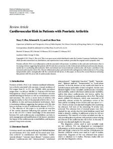
FIG. 1. (a) The hands of a patient with asymmetric polyarthritis showing shortening of third left digit, swelling of second left metacarpophalangeal and right fifth metacarpophalangeal joints, (b) The radiographs of the same patient's hands show 'pencil and cup' changes in third left distal interphalangeal, fifth right proximal interphalangeal and erosive changes in several metacarpophangeal joints and both wrists.
132
D. D. Gladman and others
(a)
Downloaded from by guest on November 25, 2014
FIG. 2. (a) The hands of a patient with psoriatic arthritis show significant distal interphalangeal joint disease as well as skin and nail changes. Based on clinical features he might have been included in the distal group, (b) Radiographs of both hands and wrists show significant damage in distal interphalangeal joints, but in addition, erosive changes are noted in several metacarpophalangeal joints as well as in the right wrists, where the ulnar styloid is tapered.
Psoriatic Arthritis (PSA) - An Analysis of 220 Patients
133
(a) Downloaded from by guest on November 25, 2014
(b)
FIG. 3. (a) The hands of a patient with distal involvement only, (b) Matching radiographs confirming the distal distribution.
134
D. D. Gladman and others
Downloaded from by guest on November 25, 2014
FIG. 4. (a) Symmetric polyarthritis, rheumatoid arthritis-like, with significant clinical deformity and damage. (b) The radiographs demonstrate that the damage is seen not only in the wrists, metacarpophalangeal and proximal interphalangeal joints but also in the distal interphalangeal joints bilaterally.
Psoriatic Arthritis (PSA) - An Analysis of 220 Patients
135
The last four drugs were prescribed primarily for skin disease, and there was no correlation between the severity of the arthritis and their use. We cannot comment on the effect of treatment on the course of the disease at this time. Laboratory features Results of the laboratory investigation are shown in Table 3. Anemia and leukocytosis are commonly associated with psoriasis and psoriatic arthritis. The elevated sedimentation rate noted in almost half of the patients represented both active arthritis and active skin disease. The frequency of positive rheumatoid factor was similar to the incidence of 13 per cent found in a group of 101 patients with psoriasis uncomplicated by arthritis [26], The highest titer of rheumatoid factor (1:640) was seen in a patient with spondylo-arthropathy, and another with distal joint disease and arthritis mutilans; neither had features of rheumatoid arthritis. Antinuclear factor was detected in 10 per cent of the patients, again a frequency similar to that seen in patients with uncomplicated psoriasis [26]. The highest titer of antinuclear factor was 1:160, Hypergammaglobulinemia which was observed in 11 per cent of the patients, was always nonspecifically polyclonal. Radiological manifestations
Psoriatic arthritis subsets Grading the pattern of arthritis at onset was based on patient history, and where available, previous records, The assignment of psoriatic arthritis pattern at initial assessment was based on both clinical and radiological findings at the first visit to the clinic. Table 4 compares the frequency of the various patterns at onset to that found at first assessment. Polyarthritis
TABLE 3. Laboratory and radiological features in psoriatic arthritis
Anemia (Hb450//mol/l) Females 14% Males 32% Rheumatoid factor (> 1:160) 9% Antinuclear factor (>l:40) 10% Hypergammaglobulinemia (> 16 g/1) 11% Erosive changes on radiographs 67% Stage IV changes (>5 joints) 16% Sacroiliitis (grade 2 or greater) 27% Syndesmophytes Classic 11% Paramarginal 15%
Downloaded from by guest on November 25, 2014
Radiographs revealed erosive disease in 67 per cent of the patients, with stage 4 changes (ankylosis and/or joint destruction) occurring in 30 per cent (Figs. l-4(b)). Sixteen per cent of patients had more than five joints at stage 4. Sacro-iliitis of grade 2 or more was detected in 59 (27 per cent). Eleven per cent of the patients had classic syndesmophytes (Fig. 5 panel a) while 33 patients (15 per cent) had paramarginal syndesmophytes (Fig. 5 panel b).
D. D. Gladman and others 116
c
c a
ca Q.
-O
x: a.
Downloaded from by guest on November 25, 2014
00
Psoriatic Arthritis (PSA) - An Analysis of 220 Patients
137
TABLE 4. Psoriatic arthritis pattern in 220 patients At onset
Pattern Distal Oligoarthritis Polyarthritis Back alone Back+distal Back-1-oligoarthritis Back+polyarthritis
20 22 41 3 4 3 7
First assessment Total
A'
S*
12 14 40 2 4 7 21
56 80 53 71 73 41
44 20 47 29 27 59
* A=asymmetric distribution; S=symmetric distribution (occurring in identical joints in both sides of the body). Figures are percentages.
TABLE 5. Psoriatic arthritis subsets
No. of patients F/M
Age onset (skin) (years) Age onset (joints) (years) Skin first (%) Joints first (%) Simultaneous onset (%) Family history (%) Simultaneous flares (%) FC m/IV (%) Iritis (%) Nail lesions (%) Sacroiliac pain (%) Back pain (%) Neck and back (%) Distal interphalangeal joints affected (%) Dactylitis (%)
Distal
Oligoarthritis
Polyarthritis
Back disease
Back+ distal
Back+ oligo
Back+ poly
27
31
89
60/29
30 36 70 11 19 37 30 4 0 89 0 0 0 74
24 34 84 6 10 55 29 3 6 90 6 19 6 29
29 35 61 22 17 40 36 12 7 83 7 13 6 63
5 0/5 22 44 80 20 0 40 20 0 0 100 40 80 40 0
8 3/4 24 36 86 14 0 43 43 0 14 71 14 14 14 71
15 7/8 36 38 67 33 0 47 40 13 13 60 20 33 33 20
45
14/17
48
19
40
0
42
27
24
9/18
23/23 32 40 65 13 22 33 41 20 9 85 15 24 17 56
Downloaded from by guest on November 25, 2014
comprised the largest group both at onset and at initial assessment, occurring in more than 40 per cent of the patients. Fewer patients were grouped in the distal and oligoarthritis pattern at initial visit than at onset. This may, however, reflect the patient's inability to remember the type of onset, as in many cases there was a delay in referral to the clinic. More patients in the back+polyarthritis group were found at initial assessment compared with onset. This may represent an actual change in the pattern of arthritis, or, alternatively, it may signify asymptomatic back disease which was only diagnosed after radiological assessment. The patients were divided further into those with symmetric and those with asymmetric distribution. Patients with oligoarthritis with or without back disease, and patients with distal joint disease with an affected back tended to have asymmetric arthritis, while among patients with polyarthritis symmetry and asymmetry were almost equally distributed (Table 4).
138
D. D. Gladman and others
A comparison was carried out among the patients assigned to the seven different patterns of psoriatic arthritis (Table 5). There was a male preponderance only among patients with distal disease and back disease only, while in the other groups, the sex ratio was close to 1. Patients with back disease were older at the time of onset of their arthritis than those without (40 versus 35 years, p=0.02), and had a longer disease duration (12 versus 8 years, p=0.01). ARA functional class III or IV occurred only in patients with polyarthritis, probably representing the extent of disease. As has been previously noted, dactylitis was more common in patients with distal interphalangeal joint disease (p=0.001) than in the other categories. Distal interphalangeal joint disease, however, occurred just as frequently among patients with polyarthritis as it did in the other groups. Grade 4 radiological changes were observed more commonly in patients with distal disease and those with symmetric polyarthritis, suggesting that these forms were more likely to be associated with severe disease.
DISCUSSION
Downloaded from by guest on November 25, 2014
The concept of psoriatic arthritis as a separate disease, first proposed by Alibert in 1822, received further support by the classic studies of Wright, who established much of the groundwork of our current understanding of the disease. In 1956, Wright described 42 patients with psoriasis and erosive arthritis, and compared them to patients with classical rheumatoid arthritis and patients with psoriasis without joint disease [2]. This study provided evidence that psoriatic arthritis was a specific entity. In contrast to rheumatoid arthritis, psoriatic arthritis occurred as frequently in males as in females, was less often polyarticular at onset, and tended to be less severe than rheumatoid arthritis. In general there were fewer affected joints, fewer deformities such as ulnar deviation and only the rare occurrence of mutilating disease. This paper was followed by a description in 1958 of 118 patients with psoriasis and an erosive arthritis [3]. The majority of patients were found to have an arthritis indistinguishable from rheumatoid arthritis. Although no attempt had been made to exclude rheumatoid arthritis patients from this group, only 17 per cent of them were seropositive compared to 80 per cent of patients with definite rheumatoid arthritis. In this study, it was noted that deforming arthritis as well as arthritis mutilans was associated with an earlier age of onset of joint disease, and with sacroiliitis. In 1967 a second study of this group of patients was reported [15]. Again, the majority of patients (79 per cent) belonged to the group that was indistinguishable from rheumatoid arthritis, while 16 per cent belonged to the distal group, and 5 per cent constituted the 'deforming' group with spinal abnormalities and severe deforming peripheral joint disease. They concluded that only a small proportion of patients with psoriatic arthritis developed severe arthropathy, in spite of the frequency of polyarthritis 'indistinguishable from rheumatoid arthritis'. In a review paper in 1973 [5], Moll and Wright summarized their concept of psoriatic arthritis as an inflammatory arthritis associated with psoriasis, usually seronegative, and described the five clinical patterns. It is of note that while in previous studies by Wright, the group indistinguishable from rheumatoid arthritis was the most common, the 1973 paper described the 'symmetric polyarthritis' in 15 per cent of the patients while an 'asymmetric' group emerged as the most common, occurring in 70 per cent of their patients. Although initially noted by Wright, Little et al. [11] and Leonard et al. [12] also point out the association of arthritis with severe psoriasis. Both studies reported a much higher frequency of arthritis among patients with psoriasis than had been previously described. In his analysis of 30 patients, Leonard confirmed the distribution into five clinical patterns, while Little did not describe the clinical features in his patients.
Psoriatic Arthritis (PSA) -An Analysis of 220 Patients
139
TABLE 6. Comparison of reported series Gladman (1985) M/F
Age (years) Age onset (skin) (years) Age onset (joints) (years) Family history (%) Nail lesions (%) Skin