PROTEINURIA AND RENAL DISEASE A ROUNDTABLE DISCUSSION
Sponsored by an educational grant from IDEXX Laboratories
PROTEINURIA AND RENAL DISEASE A ROUNDTABLE DISCUSSION Moderator: Roberta Relford, DVM, PhD, DACVP, DACVIM IDEXX Laboratories Dallas, Texas Participants: Scott A. Brown, VMD, PhD, DACVIM Department of Physiology & Pharmacology College of Veterinary Medicine University of Georgia Athens, Ga. Jonathan Elliott, MA, PhD, Vet MB, Cert SAC, DECVP&T, MRCVS
Department of Veterinary Basic Sciences Royal Veterinary College London, U.K. Fred Metzger, DVM, DABVP (canine and feline) Metzger Animal Hospital State College, Pa.
Dr. Roberta Relford: Our discussion today is about kidney disease in dogs and cats. Kidney disease has received a lot of attention in the literature recently, particularly the role of proteinuria in the detection, characterization, and prognosis of kidney disease. Because of the importance of proteinuria, the American College of Veterinary Internal Medicine (ACVIM) organized a focus group to produce a consensus statement on proteinuria to be published this spring. The following roundtable discussion is important because it addresses some of the confusion that exists about proteinuria utilization. The participants include several internationally recognized experts as well as members of the ACVIM focus group. Let’s begin with the current, accepted prevalence of acute and chronic kidney disease in dogs and cats.
George E. Lees, DVM, MS, DACVIM
Department of Small Animal Clinical Sciences College of Veterinary Medicine Texas A&M University College Station, Texas Sherry Sanderson, DVM, PhD, DACVIM, DACVN Department of Physiology & Pharmacology College of Veterinary Medicine University of Georgia Athens, Ga.
and one in five elderly dogs will develop some kind of kidney disease.
chronic kidney failure—at least in referral practices.2,3
Dr. Fred Metzger: Renal disease is certainly a common clinical presentation in private practice, but I’m interested to learn how many cases we currently miss. We’re a little better at picking up acute renal failure than chronic renal failure, but we miss a lot of cases because of the way we test for renal failure.
Dr. George Lees: About one in three cats and one in five dogs will develop kidney disease in their lifetimes. Historically, acute renal failure surfaces when the animal becomes ill, and practitioners seldom misdiagnose it. The International Renal Interest Society (IRIS) has developed a good staging system for chronic kidney disease. There are four stages: I, II, III, and IV, differentiated by plasma or serum creatinine concentrations. Late stage III and stage IV (i.e., moderately and severely azotemic) animals are typically symptomatic. The animals we are fuzziest about are in stages I and II (i.e., nonazotemic or mildly azotemic animals). We need studies to develop a better understanding of the numbers of animals in stages I and II and the clinical significance of their kidney disease. We must guess right now.
Dr. Scott Brown: In 1999, Dr. Elizabeth Lund reported that kidney disease was the No. 6 reason cats were presented to veterinarians—in the top six with gastrointestinal and dermatologic problems—and No. 10 in dogs.1 Metzger: That number goes way up when we look at the population of older cats.
Prevalence Dr. Sherry Sanderson: According to recent literature, one in three elderly cats
Gregory F. Grauer, DVM, MS, DACVIM Department of Clinical Sciences College of Veterinary Medicine Kansas State University Manhattan, Kan.
Brown: Absolutely. One in three senior cats is a number commonly cited with
2
A ROUNDTABLE DISCUSSION
Metzger: When our practice instituted a senior care program including diagnostic testing of asymptomatic patients seven or eight years ago, the number of patients diagnosed with renal disease increased substantially. I know we could be helping a lot more animals with better testing for renal disease. Dr. Jonathan Elliott: In our London practice, we diagnose about 60 new chronic kidney disease cases in cats a year. But we have a high index of suspicion because we have many senior clients with older cats. About 30% to 40% of these new cases are diagnosed without the owners noticing clinical signs. We’ve detected them with screening tests rather than waiting to test until they present with clinical signs. Some of these cats remain stable and never show clinical signs of chronic kidney disease. These are not stage I cats; they’re stage II according to the classification system. About half of them will die because of another problem without having shown clinical signs of kidney disease. Dr. Greg Grauer: A semiepidemiologic Morris Animal Foundation study that polled dog and cat owners about health concerns and causes of pets’ death revealed that for both species, renal disease was the No. 2 natural cause of death. We’re conducting a longitudinal study at Kansas State—similar to ones being done at North Carolina State and Colorado State—where we monitor staff- and student-owned dogs over a four-year period. We have about a 20% incidence of low-level proteinuria in both species. The bulk of these dogs and cats are persistently proteinuric. But that certainly doesn’t mean that all of those animals are going to progress to overt renal failure. Brown: I liken it to an iceberg. Traditionally, in veterinary medicine, we see the tip of the iceberg—those patients with clinical signs in late stage III and early stage IV in the IRIS classification system. If you screen select populations such as senior cats, as Dr. Elliott suggested, there is no question that kidney disease is much more common. But we need markers to help identify other patients. Proteinuria may well prove to be a good marker. Urine
screening and critical evaluation of serum creatinine levels will help in this regard. Rather than simply determining if creatinine is outside of a broad normal range, sequential serum creatinine determinations allow you to follow adult animals so you can look for trends. And viewing a high normal creatinine concentration with suspicion may cause veterinarians to critically evaluate urinalyses and other ancillary tests. We all agree that kidney disease is common, but it’s probably much more common than we think. Identifying stage I or II animals is a challenge that urinescreening tests facilitate.
S OUND B YTE -
D R . M ETZGER
“When our practice instituted a senior care program including diagnostic testing of asymptomatic patients seven or eight years ago, the number of patients diagnosed with renal disease increased substantially.” Lees: We’re trying to learn more about the bottom of the iceberg. Few studies have been performed, and they involve animal subjects in only the double digits—less than 100 in any one study where researchers have looked microscopically at kidney tissue from seemingly healthy dogs. However, they found that up to 80% of middleaged or older animals have kidney lesions and the majority of the lesions were observed in the glomeruli.4,5 Brown: The bottom 80% of the iceberg contains many animals that won’t progress to clinically apparent uremia as Dr. Elliott described—but some will. We don’t know yet how to predict progression. Metzger: I’m interested in learning more about early detection and intervention. This
3
disease is very emotional for clients and veterinarians. When we diagnose renal failure by detecting azotemia, the patients have progressed to stages III and IV. Because these are usually older patients, owners and team members are emotionally bonded to them.
Prevalence and age groups Relford: Senior patients, as most of you indicate, have the highest incidence of kidney disease, so is there a reason to monitor younger animals? Since most patients are identified when they’re in stage III and later, this suggests that undetected pathologic changes were occurring when they were younger. Is there a way to detect these patients earlier and alter their outcomes? Additionally, should certain at-risk patients be monitored? Brown: In surveys of dogs, there have usually been two peaks of higher prevalence. One occurs at an earlier age and is related to congenital or hereditary disease. This is different than what we’ve been talking about so far: acquired disease occurring later in an animal’s life. Hereditary or congenital kidney disease also progresses and may be identified by serum and urine screening tests, but in surveys, it’s seen in dogs about 1 to 3 years of age. Lees: The principal inheritable disease in cats is polycystic kidney disease, in which renal failure occurs in middle age. Juvenile onset (i.e., less than 3 years of age) of chronic renal failure is not common in cats. But some dog breeds have familial renal diseases that cause juvenile onset renal failure and are, therefore, at risk. Veterinarians caring for those animals should monitor them carefully. Metzger: Mandatory preanesthetic testing has been a huge wake-up call in our practice. With blood and urine tests, such as blood urea nitrogen (BUN), creatinine, and a complete urinalysis, we’re now diagnosing renal failure in pets much earlier. About 80% of us perform preanesthetic testing. It might not always be the right testing, but some renal tests are in those profiles. All patients—especially breeds predisposed to renal disease like Persian cats
PROTEINURIA AND RENAL DISEASE and Wheaton terriers—should be thoroughly evaluated before anesthesia. Before the advent of preanesthetic testing, I didn’t think renal disease was present in young patients. Lees: When you start testing young animals before routine spays and neuters, you need a different frame of reference than in older animals. A serum creatinine value of 1.3 mg/dl in a 5-month-old dog is alarmingly abnormal. Metzger: That’s a great point. Age-specific ranges are important too. Elliott: The average age of diagnosis of kidney disease in cats is about 12 years. But we do diagnose kidney disease in younger cats and not just in specific breeds. We see cats presenting with kidney disease at 8, 5, and even 3 years of age. These cats don’t seem to have a different clinical form of the disease than older cats—when we examine their kidney tissue, they apparently have the same type of end-stage lesions. The initiating cause, however, cannot be ascertained at this late stage. Relford: That brings us to our next question. Are there certain kidney disease categories that are more prevalent (e.g., glomerular vs. tubular)? What categories should veterinarians look for?
Types of kidney disease Sanderson: Classically, we thought most renal failures in dogs involved tubular interstitial disease, but a histopathologic study found that there is a lot more glomerular disease that we weren’t recognizing. Previously, we may have underestimated the prevalence of glomerular disease as the underlying cause of renal disease in dogs. A recent study found that seven of 15 dogs with end-stage renal failure had glomerular nephritis as the underlying cause of the renal disease rather than tubular interstitial disease.6 An earlier study also showed similar results.7 Lees: One principle of kidney disease pathology is that it all ends up in the same place regardless of its initial cause. The bulk is interstitial kidney disease. Renal
function correlates best with changes in the tubulointerstitial compartment in all types of kidney disease—whether it starts out as tubular or glomerular disease. Because kidney disease is historically diagnosed in late stage III and stage IV animals on the basis of clinical signs, clinicians only find the end-stage wreckage and it always looks the same. One of the potential benefits of screening seemingly healthy animals and detecting renal disease early is that it will not only help our patients, but we’ll also get a better idea of the inciting causes. We’ve been handicapped by not looking for disease at a time when we could determine the cause.
S OUND B YTE -
D R . L EES
“One of the potential benefits of screening seemingly healthy animals and detecting renal disease early is that it will not only help our patients, but we’ll also get a better idea of the inciting causes.” A wonderful example is the inherited kidney disease in cocker spaniels. This was the first inherited kidney disease that veterinarians recognized in dogs beginning in the 1940s. It was described in the veterinary literature for about 40 years as a renal cortical hypoplasia. We did electron microscopy, found some changes unique to that disease, and then did studies in dogs by collecting serial biopsies at the beginning, middle, and end. We were able to unravel how the disease progresses by looking at it from the beginning. If you only look at the end using light microscopy, as veterinarians did for 50 years, you can tell they’ve got it, but you can’t tell why.
Common causes of kidney disease Relford: What are the common causes of kidney disease? We’ve already talked
4
about the less common congenital or familial breed predispositions in young pets. Grauer: Dr. Lees and Dr. Sanderson have alluded to studies that suggest glomerular disease is associated with end-stage renal disease in dogs—perhaps to a greater extent than we once thought. In my opinion, however, there aren’t enough longitudinal studies (i.e., studies with serial renal biopsies that would facilitate primary disease process determination) in dogs with spontaneous kidney disease. These types of studies are invasive and expensive and, therefore, difficult to perform in clientowned animals. Based on the information we have today, it does appear that glomerular disease is an important cause of end-stage renal disease in dogs. What is the primary cause in cats? Elliott: I’d love to be able to take biopsies of cats when they have normal kidney function and when they have stage II kidney disease, but I’d have a hard time convincing cat owners to allow me to do that. Glomerular events are going to influence tubular pathology. Proteinuria is an indicator of renal pathology or renal disease and is a predictor of survival—proteinuric cats with kidney disease tend to progress more rapidly. We don’t know enough about it at this stage. We’re diagnosing cats at the end stage and seeing the tubulointerstitial fibrosis that’s resulted from the initial disease process, which is no longer evident. Lees: The kidneys are composed of four types of structures: vessels, glomeruli, tubules, and interstitium. Let’s discuss which components are the likely targets for initial insult. Vascular disease is not common in dogs or cats compared with people, who have a fair amount of primary cardiovascular disease. The glomeruli are an important target of the initial renal insult in dogs and possibly cats—far greater than we’ve appreciated in the past. Even so, glomeruli are injured secondary to diseases in other organ systems most of the time—the glomeruli are sentinels that often indicate problems elsewhere in the body. There are huge microvascular glomerular areas available to be injured. The tubules are the initial target of most diseases that present as acute renal
A ROUNDTABLE DISCUSSION
failure; leptospirosis and toxic nephropathies are examples. However, diseases that initially attack the tubulointerstitium sometimes do cause chronic renal failure. Pyelonephritis, which occurs when a bacterial bladder infection works its way up to the kidneys, is an example. Metzger: Clients ask, “What caused this?” I often tell clients with older pets that it’s just age. Maybe that’s true, but maybe it’s not. In Pennsylvania, I’m concerned about Lyme disease in relation to renal disease in dogs. But is hypokalemia in cats a cause of renal disease or a result? And what about hypercalcemia? Brown: Clearly, a variety of infectious diseases and cancers lead to immune complex formation that potentially causes glomerular inflammation and subsequent deterioration of renal function. And hypercalcemia damages the kidney through mineralization, fibrosis, and other secondary changes. These two conditions, immune complex formation and renal mineralization, are two primary causes of kidney disease. The link between hypokalemia and kidney disease is quite complex. Chronic kidney disease in cats tends to be a potassium-losing disease. Even with supplementation, our feline patients are often hypokalemic. And there are important consequences of hypokalemia, such as muscular weakness. A really interesting question is whether hypokalemia may be contributing to the deterioration of renal function and blood pressure elevation. Results of some laboratory studies in rodents and clinical trials in people suggest that blood pressure elevation is more likely to occur in association with low serum potassium levels. Elliott: We’ve noticed that low potassium levels tend to occur in cats with high blood pressure. The potassium remains low even if you supplement the cats. We’re quite interested in why that’s the case. It may give us an indication of the endocrine changes that surround hypertension. Hypokalemia may be a consequence of the disease causing hypertension and, therefore, a marker. Low serum potassium may actually have some influence on blood-pressure control. We con-
ducted a study where we supplemented potassium and then looked at the effects on blood pressure in cats with chronic kidney disease that weren’t severely hypertensive. We found no detectable influence on blood pressure. Brown: We conducted a similar laboratory study with precise direct measures of blood pressure and observed only small blood-pressure reductions with potassium supplementation in azotemic cats. I wonder if some therapies, such as salt restriction or amlodipine administration, activate the renin-angiotensin-aldosterone system in cats, which could enhance urinary loss of potassium. Elliott: We found that plasma renin activities increase when you decrease blood pressure with amlodipine treatment. Aldosterone production doesn’t change, which is strange. You’d expect aldosterone to increase in those animals. We’re investigating whether there is some dysfunction in aldosterone secretion control in these cats. Brown: Dr. Lees discussed how the tubulointerstitial compartment is commonly affected in kidney disease. Basically, any time there is kidney disease, the lesions that correlate best with disease severity are those in the tubulointerstitium— regardless of which compartment the disease attacks. But in addition, in every kidney disease, there will be significant lesions in the glomerulus that contribute to glomerular dysfunction, decreased glomerular filtration rate (GFR), and increased protein loss across the glomerulus. So there are two inevitabilities: 1) No matter what happens, the interstitial compartment is damaged, and 2) no matter where the disease originates, all compartments are affected— particularly the glomerulus. Relford: If one area is affected, then eventually all four are affected or, at least, the tubulointerstitium is affected. Are these fair statements? Brown: The two compartments that we know are affected eventually in every kidney disease case are the tubulointerstitium and the glomerulus in both dogs and cats.
5
Relford: And even though we may not know what initiated the disease, we need to learn what is causing it to progress. This can only be done with early detection and monitoring with treatment. Grauer: Nephrolithiasis, ureterolithiasis, ascending infections, hyperphosphatemia, acidosis, hypertension, and proteinuria are all potentially treatable causes of continued renal function deterioration once failure has been diagnosed. Elliott: With the exception of nephrolithiasis and ureterolithiasis in cats, we generally view those conditions as complications rather than initiating causes. We could argue whether hypertension comes first, but many of those conditions are a consequence of later kidney disease stages. Brown: I agree with your list, Dr. Grauer. Most of them occur as part of the milieu of what happens in kidney disease. Uremic syndrome is a term that is commonly used. Hypertension is potentially one kidney disease complication that may also be a cause of kidney disease. But we know remarkably less than we should about the causes. If one in three geriatric cats has chronic kidney disease, we had better find ways of detecting it earlier and figuring out what the primary causes are.
Clinical signs Relford: What are the common clinical signs that veterinarians should be looking for in the different stages of kidney disease? And what about animals that show no clinical signs? Grauer: It’s easy to diagnose overt stage III or IV renal failure, that is, azotemia or uremia with persistent urine concentration deficits. We need to focus on diagnosing patients earlier when there may be no clinical signs, beginning with annual or semiannual physical exams for mature and senior pets. Dr. Lees has wonderful data that show you can have significant declines in renal excretory function, and yet still have creatinine levels well within the normal limits simply because that normal range is so wide. Plus you have compensation occurring with nephron loss. Looking at the trends even within that normal range, you
PROTEINURIA AND RENAL DISEASE can see greater than a 50% reduction in excretory function. You may also see trends in urine specific gravity, creatinine, phosphorus, packed cell volume, proteinuria, or body weight over time. Lees: Of the four stages, stages I and II are, by definition, absent of clinical signs. So if by clinical signs, we mean something the client or veterinarian would observe and report—we are limiting ourselves to diagnosing late stage III and stage IV disease. The disease is insidious, so the signs are insidious. Changes in urinary habits (including frequency and amounts), water consumption, appetite, and body condition are a few signs. Weight loss can be hard to pin down; it’s a fairly early, subtle sign. The exception would be the acutely ill animal— whether it’s a decompensation of chronic renal failure or acute renal failure crisis. Grauer: But when you put it all together over time and start to see declines in urine specific gravity, increases in proteinuria and creatinine concentration, and declines in body weight, it should make you take a closer look. Metzger: It sounds like an easy question: What are the clinical signs? But it’s complicated. I think the “ain’t doing right” animal is a perfect candidate for chronic renal failure. Cat owners might not notice polyuriapolydipsia (PU-PD), anorexia, and lethargy, for example. You would think it would be easy to tell if an animal is drinking and urinating more or eating less. If there are multiple animals living in the same home, it’s not that easy unless you are looking for it. That’s why I’m so interested in senior care programs and testing animals before they are clinical.
head, there are steps you can take to evaluate patients. Brown: I agree. By definition, there are no uremic signs in stages I and II. By uremic signs, I mean such things as vomiting, lethargy, weakness, and anorexia associated with marked azotemia as seen in late IRIS stage III or stage IV. But there is further complexity as there are two other sets of clinical signs that can occur— albeit less frequently. First, there are disease-specific signs. For example, if the animal has pyelonephritis, it may not be uremic, but it may have renal pain observed by owners or veterinarians as reluctance to handling or inappetence. Here, clearly, urinalysis is the key to disease identification. The other type of early preuremic clinical sign is hypertension. Animals with stage I or stage II chronic kidney disease might exhibit systemic hypertension. In these animals, retinal abnormalities or other sequelae of systemic hypertension might appear before the uremic signs. Because hypertension increases protein traffic across the glomerulus, many of these animals will be proteinuric. Here, urinalyses, blood pressure measurements, and ophthalmic exams are the key to disease identification. Lees: How do we define a clinical sign? I don’t hear a heart murmur until I use a stethoscope, but to me, it’s a clinical sign. The same might be said for screening urinalyses for proteinuria. If we define clinical signs to include abnormalities that we can detect using tools—stethoscopes, blood pressure cuffs, urinalyses—then we end up with a different set of signs.
Diagnostic tests Elliott: Cats don’t show clinical signs until the latter stages of many different diseases. So screening is important. Retrospectively, the owners will often say, “Well, yes, I noticed that.” Lees: It’s partly what you are looking for— a product of your index of suspicion. Some clients are astute judges of their animals’ well-being; others never notice. So, yes, it’s crucial to have tools available so that once the light bulb goes on in your
Relford: If veterinarians suspect kidney disease, what are some diagnostic tests they should consider? Lees: There is nothing more important than a complete urinalysis. Urinalysis is the beginning and the fulcrum around which everything else should hinge— there should never be a clinical investigation involving screening tests, such as complete blood counts (CBCs) or chemistry profiles, without a urinalysis as well.
6
Brown: We’re trying to identify animals with early kidney disease. Late stage III and stage IV animals are relatively easy to diagnose if they have severe azotemia in the face of a low urine specific gravity. But the only thing on the serum chemistry panel that is going to be helpful in those early patients is the serum creatinine determination. Here, either a high normal serum creatinine or a serum creatinine that has been inching up within the normal range is helpful. In IRIS stage II, a slightly elevated serum creatinine is helpful as well. Historical studies in both dogs and cats show that in order for creatinine to increase out of the normal range, three out of every four nephrons in the kidneys must be destroyed, so we’re talking about severe disease at that stage. As a profession, we should begin to look at serial creatinine levels, measure them routinely, and compare current to previous values in senior patients. A high normal value should make a veterinarian suspicious even if earlier values aren’t available. But generally, you won’t see any obvious abnormalities on the CBC and serum chemistry panel in stage I and stage II. Elliott: Body condition score is important when you’re interpreting creatinine because the animal’s muscle mass will make a big difference. We see a lot of old skinny cats that have normal creatinine levels. It’s quite abnormal if the creatinine level is at the top end of the reference range. Grauer: The urinalysis is a must if you’re going to interpret the serum chemistry profile. I think serial serum chemistry and urinalysis assessments are absolutely critical. Sanderson: Pretreatment urine samples are ideal. They ensure you’re not interpreting therapy’s effects on top of everything else. Lees: Additionally, it’s important to interpret the various laboratory test results in light of one another and the patient’s status. Clinical judgment is what the client pays you to bring to the table. Do the numbers make sense in the context of this patient? Sanderson: If you’re looking for glomerular disease, you may not see any changes
A ROUNDTABLE DISCUSSION
on the initial blood work. You may be missing glomerular disease if you don’t look at the urine. Lees: Historically, veterinarians have not always been good at getting a clean urine specimen. Somehow it’s easier to do a venipuncture than it is to get a urine sample. If you’re not doing a urinalysis, you’re missing the single biggest window into the problem. Brown: One of our faculty members at the University of Georgia always refers to urine as liquid gold, meaning it’s critical to evaluate the urine if we want to identify these patients in the early stages. Even in the absence of a creatinine change, a low specific gravity should raise a veterinarian’s index of suspicion. With the dipstick evaluation, you’re looking for proteinuria and other abnormalities that may indicate kidney disease. You must evaluate the sediment in order to identify urinary tract infections (UTIs) or ongoing renal damage. Lees: Let’s not forget that in these same patients, lower urinary tract disease is fairly common as well. Twenty percent of dogs, female dogs particularly, will have bacterial UTIs. Stone disease is fairly common. So a complete urinalysis that allows you to examine the sediment for pus, blood, and so on is screening for not only kidney disease but other common urinary tract diseases that are important to the animal’s health as well. We’ve already talked about ascending infections and urolithiasis damaging the kidneys. But again, these are the tip of the iceberg of a larger set of diseases that you will not find regularly unless you look. A number of years ago, we did studies on UTI. For an 18-month period, we carefully evaluated all the dogs that we diagnosed with UTIs in our teaching hospital. Exactly half (19 out of 38 patients) had uncomplicated bacterial UTIs that were initially detected only by urinalysis (asymptomatic UTIs). Elliott: In my experience, it’s almost 100% in cats. We don’t see lower urinary tract signs when they have poor concentrating ability and bacterial infections. Those pa-
tients are not showing the increased frequency or dysuria. Sanderson: It’s unfortunate that some people think that cats don’t get UTIs. Young, healthy cats are not at great risk, but add renal failure to the equation and a fair number of them do get concurrent UTIs. You cannot always rely on the urinalysis to diagnose UTIs; there may not be a significant inflammatory response. If you’re trying to rule out an infection, the gold standard is actually the culture. Lees: Especially when there is concurrent renal failure.
S OUND B YTE -
Proteinuria Relford: What are some ways of evaluating proteinuria?
D R . B ROWN
“One of our faculty members at the University of Georgia always refers to urine as liquid gold, meaning it’s critical to evaluate the urine if we want to identify these patients in the early stages.” Relford: Your minimum database should include a CBC, serum chemistry panel, and a complete urinalysis with specific gravity, dipstick chemistry analysis, and a sediment examination. If you suspect renal disease, there should be a culture. Metzger: I think we’d all agree that cystocentesis is the way to go, but it can be a problem in practice. If a practice owns an ultrasound machine, it’s a wonderful way to use it. At our practice, we’ll do an ultrasound urine collection for a minimum of $38. You can almost always get a cystocentesis if you have an ultrasound. You can also do a quick screen of the bladder and kidneys for urolithiasis, neoplasia, and other abnormalities. Elliott: Another practical point: If you advise the owner not to feed the cat in the morn-
7
ing, you have a much better chance of getting a urine sample because cats often urinate after they’ve eaten. We’re able to get samples from about 75% of the cats that have fasted and haven’t urinated in their boxes on the way to the clinic. Detecting a full bladder on palpation suggests that the cat is polyuric; a urine specific gravity will help with the interpretation, particularly if the patient fasted overnight. If it has a small bladder and the owners confirm it has not urinated that morning, then chances are it’s not polyuric.
Grauer: I think we’d all agree that proteinuria, with some caveats (i.e., kidney localization and persistence), is a sign of kidney disease. The standard assessment of protein in the urine, the dipstick, is unfortunately not a reliable or sensitive test. It’s nonspecific in cats—in other words, there is a high percentage of false-positive results. Thorough analyses for proteinuria should combine the standard dipstick analysis with additional testing. Relford: What are some recommendations for additional proteinuria testing? Grauer: In academic settings, the sulfosalicylic acid turbidimetric test is a good test, but it hasn’t caught on with practitioners. Lees: The sulfosalicylic acid test is a good, simple test. It’s a lot easier than several other procedures veterinarians learn to do and become adept at. We’ve gone from no laboratory testing within recent memory to some laboratory testing. Generations of veterinarians were raised on doing a urinalysis with a dipstick. The protein pad on the dipstick will often change color in moderately to highly concentrated urine from dogs or cats even when there is no protein. These falsepositive reactions occur because of the pH and concentration of the urine and the limited buffer capacity of the reagent test strip. So we have a whole generation of veterinarians finding trace and 1+ positive color changes in animals that they knew were perfectly normal. They thought it was
PROTEINURIA AND RENAL DISEASE protein and said to themselves, “A little bit of proteinuria is normal in cats and dogs.” The truth: The level of proteinuria that would cause the dipstick to turn color is never normal if it’s a true positive. We’ve desensitized ourselves while learning to do lab tests. We only think about it being proteinuria if it is 4+, which means we need to change our frame of reference. Grauer: So a positive dipstick should be followed up with a confirmatory test? Lees: Yes. Grauer: If you’re interested in detecting a low level of protein in the urine, the dipstick is not sensitive enough. We’re going to get a lot of false positives, but we may also see some false negatives with a low proteinuria level. The sulfosalicylic acid, urine microalbumin assay, and urine protein–creatinine ratio tests are much more sensitive than the dipstick at detecting these lower concentrations of protein in the urine. Relford: What about quantitation with the urine protein–creatinine ratio? Elliott: The standard dipstick tests for protein are of little help when screening cat urine because there are so many false positives. There are two possible approaches: 1) run a urine protein–creatinine ratio test initially and forget the screening test or 2) screen with the microalbuminuria test (if this is negative, you can be confident the urine protein–creatinine ratio will be so low that it’s not worth quantifying). Any positive on the microalbuminuria test should be followed up by a urine protein-creatinine ratio test in my opinion. Either of these two approaches is appropriate provided the urine sediment has been carefully examined for signs of active inflammation. In our experience, if the microalbuminuria assay is negative in a cat, then the urine protein–creatinine ratio is likely less than 0.2. Grauer: What is the clinical significance of low-level proteinuria? And what do clinicians do with that information? Lees: That low level of proteinuria alone signifies that the animal is in the bottom
part of the iceberg—a small, abnormal amount of proteinuria that shouldn’t be there. We don’t know the proportion of those animals that will ultimately be diagnosed with end-stage renal disease. If we think they’re at risk, we need serial evaluations to detect trends in a timely manner—if the protein amount rises, the kinds of protein change, or the creatinine begins to creep up. Ideally we notice when the creatinine goes from 1.2 to 1.5 mg/dl— not when it leaps from 2.5 to 4 mg/dl.
S OUND B YTE -
D R . G RAUER
“If you’re interested in detecting a low level of protein in the urine, the dipstick is not sensitive enough.” Elliott: It’s going to be difficult to educate practitioners because there will be animals with transient proteinuria that test positive. The next time you test them, the protein level is below the threshold and they test negative. It should be interpreted as transient proteinuria in those instances, not false-positives. Grauer: It was abnormal at the time but transient proteinuria is probably associated with little consequence. Persistence is the key to pathology.
Localization Relford: Once you’ve determined that proteinuria exists, it’s important to determine the source or location of the protein— whether it’s prerenal, renal, or postrenal in origin. How should veterinarians localize the source of the proteinuria? Lees: There is no urine test that says where the protein originated. You rely on clinical judgment and other information to make a reasonable assessment of the likely source of the protein. Healthy kidneys produce urine that contains negligible amounts of protein. Protein often enters the
8
urine in its course through the excretory pathway through blood or exudation from the walls of the urinary tract. Sometimes, depending on how the sample is collected, protein is added later. We call that postrenal proteinuria—either urinary or extraurinary. The key finding in this situation is usually the coincident presence of red blood cells and positive dipstick reactions for blood or white blood cells, which is why it’s crucial to examine the sediment. Studies that use the commercially available dipsticks with leukocyte esterase assays have shown them to be unreliable in dogs and cats. So you must examine the urine sediment microscopically for the presence of red or white blood cells—or both. If they are present, the proteinuria is explained by the primary hemorrhagic or inflammatory disease process and becomes an incidental finding. Determine whether there is an infection—do a culture. Do a survey for stones or, in an older animal, determine if the proteinuria is associated with a urinary tract tumor. Grauer: With postrenal proteinuria, there would be no need to do a microalbuminuria assay, a urine protein–creatinine ratio, or a sulfosalicylic acid test. Brown: We’re using the same nomenclature for protein in the urine that we use for azotemia, which is prerenal, renal, and postrenal. Prerenal refers to extraurinary causes, such as Bence Jones proteinuria of multiple myeloma. Postrenal refers to conditions of the lower urinary tract. With renal and postrenal being the most common, veterinarians should perform the set of evaluations exactly as you proposed any time proteinuria is identified. Is it prerenal, renal, or postrenal? It’s critical because each is addressed in a completely different manner diagnostically and therapeutically. Elliott: How you’ve collected the urine samples matters as well. If you collected samples by cystocentesis, then you’ve ruled out one part of the urinary tract— the urethra—as a source of additional protein. You may iatrogenically cause hematuria by taking a sample by cystocentesis, but the studies suggest it’s not sufficient to cause significant proteinuria on the urine protein–creatinine ratio test.
A ROUNDTABLE DISCUSSION
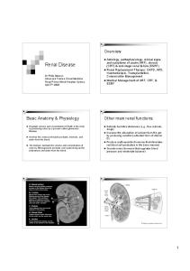
Urine Protein–Creatinine Ratio Diagnostic Protocol
History and physical exam
CBC, serum chemistry profile, urinalysis (examine urine sediment and specific gravity)
Active sediment * Pollakiuria Azotemia
Prerenal
Active sediment Pollakiuria Azotemia
Active sediment Pollakiuria Azotemia
If prerenal ruled out
Hemoglobinuria Myoglobinuria Bence Jones proteinuria (myeloma)
If postrenal ruled out
Renal
Postrenal
Urinary tract infection Stones Tumors
Urine protein–creatinine ratio
Azotemia
0.5 canine 0.4 feline Within reference range Monitor, investigate, and treat **
Investigate azotemia
Kidney disease with proteinuria Azotemia
0.5 to 1.0 to 2.0 canine and feline Monitor, investigate, and treat
Repeat urine protein–creatinine ratio in two weeks
Investigate for potential causes of immune-mediated glomerulonephritis or interstitial nephritis
Work up for proteinuria
Investigate = determine underlying cause, if any. Consider ancillary testing as needed (radiology, ultrasonography, blood pressure evaluation, infectious disease testing, endocrine testing, and autoimmune panel). Treat = consider the following treatment options when appropriate: dietary changes, ACE inhibitor therapy, phosphate binders, and fluid therapy.
* Active sediment = cells, casts, and bacteria ** Monitor = recheck to determine trends. Recheck should include renal panel (albumin, BUN, calcium, creatinine, phosphorus, and total protein), urine protein–creatinine ratio, CBC, and electrolytes.
9
PROTEINURIA AND RENAL DISEASE So if it appears on the dipstick as microscopic hematuria but you can’t see urine discoloration, then it’s unlikely to alter the urine protein–creatinine result. The situation is different with acute or chronic urinary tract inflammation, which results in protein exudation and white blood cells in the sediment. Urine microscopy is important in these cases. If you have exudation, the white cell content will exceed the red cell content. If you just nicked a blood vessel while taking the sample, then you’ll have many more red cells. Grauer: Renal proteinuria caused by inflammatory diseases of the kidney is another example of proteinuria typically associated with active sediment where quantitation of the protein is not indicated. Examples of kidney tissue inflammation that can result in renal proteinuria include pyelonephritis and renal urolithiasis. Proteinuria associated with kidney inflammation is classically associated with an active sediment, whereas glomerular and tubular proteinuria are typically associated with inactive sediments.
Persistence Relford: So if a patient has proteinuria, the first step is to determine if it’s from the lower urinary tract or from the kidneys. If it’s from the kidneys, the next step is to determine if the proteinuria is persistent. How should veterinarians determine persistence? What would be their diagnostic plan? Lees: Persistence means that you find it repeatedly. When you take a snapshot of a train on a track, you can’t tell whether it’s going 100 miles per hour or has been parked there for years. You need several different observations with some sense of time in between to know whether things are changing or staying the same. Relford: When should the animal be retested? In six months? Six weeks? Lees: It depends on the clinical context. The standard would be about two to four weeks in apparently healthy animals. If you’re dealing with a sick animal, the time frame may need to be pushed up a little. If you get a positive protein and think it might
be transient, you want to learn if it’s repeatable. Somewhere within two to four weeks would be a good recommendation.
Grauer: What change in the urine protein– creatinine ratio do you consider significant vs. just day-to-day variability?
Lees: If I see it twice, then there is something to worry about, and I’m going to continue evaluating this patient.
Lees: We’re analyzing data on that subject. We have dogs with clinically stable glomerular proteinuria that are in stage I (i.e., they aren’t azotemic). We’ve done daily urine protein–creatinine ratios for three to five consecutive days in these dogs and looked at how much the values varied from day to day. It takes at least a 30% change in the urine protein–creatinine ratio for the difference to exceed ordinary day-to-day variation. If a urine protein–creatinine ratio is 3, then 3.2 a week or two later, the animal’s level of proteinuria probably hasn’t changed. But if it increases to 5 or 6, it almost certainly reflects a meaningful change. It’s the same principle if it decreases. If it decreases from 3 to 2.5, it’s borderline, but if it decreases from 3 to 2 or less, it’s likely a genuine change. This also applies when using proteinuria magnitude changes as treatment response indicators to guide further treatment decisions.
Elliott: It would be similar to how we approach a cat with marginally high blood pressure. I would want to see it in two weeks to see if its blood pressure is still above our threshold.
Monitoring, investigation, and intervention Relford: So we’ve localized the proteinuria to the kidney and identified that it’s persistent. Let’s discuss the ACVIM consensus statement and guidelines for monitoring, investigating, and intervention. Lees: “Monitor, investigate, and intervene” became our mantra while working on the consensus statement. Monitoring means doing the same tests over and over again (i.e., serial evaluations to look for worrisome trends over time). Investigate means performing additional tests, which are usually focused on discovering a potentially treatable underlying cause. Intervene means treat with medication, diet, exercise, or other lifestyle changes. So let’s say we’ve determined that the patient has persistent renal proteinuria. The key is magnitude, and we especially want to know about changes in magnitude over time. For microalbuminuria, we can use the semiquantitative amounts (i.e., low, medium, and high positives) indicated by the commercially available point-of-care test. However, we mainly use serial urine protein–creatinine ratios to evaluate proteinuria magnitude because they are useful for measuring any level of proteinuria. We want to monitor the proteinuria and the patient. With regard to renal function, we want to monitor the serum creatinine concentration. But don’t lose sight of the patient and any changes in weight or body condition. The three key questions to answer regarding monitoring are: How is the urine protein–creatinine ratio doing? How is the serum creatinine doing? How is the patient doing?
10
Elliott: You’re dealing with lower level proteinuria in cats—urine protein–creatinine ratios of about 0.4 to 1 in most proteinuric cats. We’d classify nonproteinuric cats as those with ratios of less than 0.2. Urine protein–creatinine ratios between 0.2 and 0.4 could reflect normal variability. The ratio would need to double in a cat before we’d say it’s a significant change. Lees: Another consideration that’s received little attention in veterinary medicine—the urine protein–creatinine ratio is influenced by not only the protein in the urine but by the creatinine in the urine. If less creatinine is produced in the body, the urine creatinine will decrease. You’re dividing the protein by a smaller number and, therefore, getting a higher ratio. I think that’s why urine protein–creatinine ratios are often high in puppies even though there is not much protein in the urine; puppies don’t have as much creatinine in their urine. Grauer: The key to monitoring is looking for trends over time. Lees: Yes. It’s the serial evaluation of crucial variables over time. Monitoring can tell
A ROUNDTABLE DISCUSSION
you to investigate immediately because the trend is worsening.
Monitoring frequency Relford: Before moving to investigation, let’s discuss monitoring frequency. How often should you monitor patients’ conditions? Grauer: If your screening test was negative and you’re dealing with a mature animal with no hereditary risk for disease, I’d recommend annual or semiannual monitoring.
the cats that progress? About one-third of these animals show a nice linear progression. Our initial studies suggest that urine protein behaves the same as the plasma creatinine concentration—jumping to higher values when the plasma creatinine concentration jumps. I was quite hopeful that the proteinuria preceded and predicted those jumps in plasma creatinine but this does not appear to be the case.
S OUND B YTE -
D R . E LLIOTT
Lees: That’s the frequency for screening apparently healthy animals. How frequently should patients with persistent renal proteinuria be monitored? Grauer: The magnitude of the positive result and the patient’s condition will drive how zealous you need to be. Brown: Dr. Grauer, let’s say you have a nonazotemic animal with persistent renal proteinuria. How frequently would you want to measure serial serum creatinine levels and to reevaluate the urine protein– creatinine ratio? Grauer: Again, I think it depends on the magnitude. If the magnitude causes me to intervene, I’d reassess that patient monthly. If the magnitude doesn’t require intervention but I want to see if it’s persistent, I’d also reassess monthly. Lees: A monthly interval is a good recommendation to begin monitoring. If you reassess after a month and it hasn’t changed, it’s reasonable to extend the recheck intervals. For example, you could double the recheck interval with every good recheck. Rather than imposing one schedule, I’m guided by what I learn. Some animals are reasonably stable for extended periods of time; then, without any outward manifestation, they abruptly change. But you’ve done the best you can by monitoring them carefully. Elliott: That is one of the issues we’re trying to address in longitudinal studies: What can we use to predict those abrupt decreases in kidney function? Or jumps in plasma creatinine concentration or weight loss, which we see in about two-thirds of
dose or add other drugs. With renal disease, once the patient’s magnitude of proteinuria is in, or progresses to, the treatable range, monitoring intervals should be dictated by your need to assess response to treatment. Grauer: I like the idea of the monthly approach that doubles if the patient is stable. When you first detect and confirm abnormal proteinuria but you don’t know if it’s transient or persistent, would you wait a month to reevaluate? Lees: No, I’d wait two weeks in that instance.
“The degree of proteinuria when the animal is first diagnosed with renal disease is helpful in predicting survival time, according to our studies. It’s a gradation; the lower the protein level in the urine, the longer the cat is going to survive.” The degree of proteinuria when the animal is first diagnosed with renal disease is helpful in predicting survival time, according to our studies. It’s a gradation; the lower the protein level in the urine, the longer the cat is going to survive. That’s fine in a population study, but not very useful in the individual patient. Veterinarians want the cut-off point— when should they start worrying about proteinuria? We say it’s linear across the whole range and the lower it is, the better the cat’s prognosis. We need to monitor it along with the parameters you’d normally monitor in the azotemic animal. Lees: Monitoring also happens during therapy—to monitor how the patient is doing and to evaluate treatment efficacy. You can use a blood pressure analogy. If you have high blood pressure, you give medicine to lower it, but you still need to check to see whether it went down. If you didn’t reach your target, you change the
11
Brown: We seem to agree that two to four weeks is a good recheck interval for patients with proteinuria. One month is advisable for the otherwise healthy patient with persistent renal proteinuria. I would prefer not to go beyond the sixmonth interval if it’s a senior pet. I think we, as veterinarians, tend to wait too long in the face of stability and the lack of clinical abnormalities. Elliott: You need to consider what interval is acceptable to owners and doctors. Pet owners are not paying for the rechecks involved in the research we’re doing in the United Kingdom. Even so, rechecks every six weeks, in our experience, seem acceptable. If I take blood samples more frequently, many owners with cats that appear outwardly stable will question it. Metzger: One of the possible pitfalls of testing is whether you can use a free catch sample. Is that an acceptable sample for the urine protein–creatinine ratio or microalbuminuria test? Often when I tell owners that I need another sample, they’ll say they’d prefer to drop the sample off. Grauer: Several studies have examined multiple collection techniques. As long as we rule out proteinuria caused by a postrenal component or an inflammatory renal component with a sediment exam, sure. Metzger: Great, I think you’d see excellent compliance then.
PROTEINURIA AND RENAL DISEASE Lees: Voided samples are fine if they’re collected properly. We routinely use midstream, voided samples for proteinuria assessment in our male research dogs. Catching a clean midstream sample in a small cup is easy with these Labradorsized dogs. It’s much harder to get good midstream samples from females. We routinely use cystocentesis to obtain urine samples from the female dogs we study. Grauer: Are you suggesting that a midstream, voided sample obviates the need for a sediment exam? Lees: No, not at all. Proteinuria should be assessed in the context of a complete urinalysis, but catching a clean midstream sample during voiding can be satisfactory. Even if the genitalia harbor some secretions or debris, contamination should be negligible after the first washout.
the role of renal biopsies (i.e., examining renal tissue to diagnose the cause of the primary renal lesions). Renal biopsies are only as good as the people performing them. Good pathologic evaluation of kidney tissue requires expertise. In human medicine, renal biopsies are evaluated by nephropathologists—pathologists who specialize in renal disease. At some point, veterinary medicine will bring that level of expertise to renal biopsies. I liken it to dermatopathology. It used to be that we’d do a skin biopsy, send it in, and they’d say, “It’s dermatitis.” But now we know that if you look in the right place in the right way with the right approach, you see things that are indeed useful. Now people do skin biopsies correctly and helpful information comes back from dermatopathology services.
S OUND B YTE -
D R . R ELFORD
Investigation Relford: We’ve discussed performing several diagnostic tests to determine that the patient has renal proteinuria, but let’s expand on the ACVIM recommendations for investigation. Lees: Basically, the idea is to do new tests. “Tests” can even mean new physical exams and thorough history reviews. Examine pets like you’ve never seen them before to make sure you’re not missing anything. Ask a colleague to look at the patient. Learn what drugs are being administered—not just medication you’ve prescribed, but health foods, nutraceuticals, or other supplements. Get them off unnecessary or harmful drugs. Because so much of the kidney disease that occurs in dogs and cats is secondary to other phenomena, we’re really looking for some underlying disease that might affect the kidneys, such as active periodontal disease or inflammatory bowel disease. Exhaustive searches can be daunting. The other crucial issue is whether you should investigate the kidney disease directly with testing. Minimally invasive ultrasound evaluations and radiographs answer questions about the kidney’s size, the presence of stones, and so on. As we progress, one of the key issues will be
“The investigation phase involves two areas. One phase constitutes identifying the underlying cause of the kidney disease. The second phase involves kidney function evaluation.” Today, with renal disease, we’re in that “it’s dermatitis” phase. One of our challenges is to investigate kidney disease in a way that’s productive for the patient and the client. It’s coming, but not here yet. One of the reasons we perform kidney biopsies less often is because the biopsy reports would say kidney disease, but they rarely taught us anything. With early diagnosis, the view through the pathologic window into kidney disease is more interesting and useful. We still need to examine biopsies in more sophisticated and helpful ways. But we possess the ability to evaluate the patient as a whole and to find underlying, treatable systemic diseases—whether they are
12
inflammatory, infectious, noninfectious, or degenerative. Relford: So the investigation phase involves two areas. One phase constitutes identifying the underlying cause of the kidney disease—whether it’s the initial cause or the cause of the current kidney function deterioration. The second phase involves the kidney function evaluation. Are there some special conditions you’d look for in cats? Elliott: Hyperthyroidism is common in cats and is occasionally problematic to diagnose. Hyperthyroidism causes proteinuria at levels we can detect with microalbuminuria tests but also at levels significantly greater than urine protein–creatinine ratio thresholds. We often see urine protein– creatinine ratios between 0.5 and 1 in cats with hyperthyroidism. You should rule that out with your initial screen. But occasionally, you see cats with hyperthyroidism and normal T4 concentrations initially that you diagnose as hyperthyroid when you retest. Hypertension is also an issue in cats. The higher the blood pressure, the more likely the cat is to have significant proteinuria. Blood pressure measurement is something I’d recommend in the initial evaluations of aging nonazotemic proteinuric cats. Proteinuria will actually resolve as you treat the hyperthyroidism in most cases. We’ve done prospective studies involving hyperthyroid cats with proteinuria and examined their response to treatment. Whether they become azotemic or not, the proteinuria decreases with hyperthyroidism treatment. We’d classify hyperthyroidism as a risk factor for hyperfiltration and potential kidney damage.
Intervention Relford: Let’s now shift our discussion to intervention. Dr. Brown, what interventions should the veterinarian consider with proteinuric patients? Brown: We’re somewhat limited in terms of intervention, but there are some therapies that we know are antiproteinuric. Veterinarians may ask themselves, “Should proteinuria be treated? Is that the target of therapy?”
A ROUNDTABLE DISCUSSION
Mild renal proteinuria, sometimes called microalbuminuria, has been studied extensively in rodents and people. In people, it’s clearly a marker for two diseases. The first is early stage kidney disease. The second is cardiovascular disease where mild renal proteinuria seems to be an indicator of generalized endothelial or vascular dysfunction with myocardial infarction or stroke as potential sequelae. Based on a substantial amount of data, even mild renal proteinuria is a target for therapy in people. The identification of proteinuria and therapies that reduce proteinuria are valued. Lees: The worst outcomes in people with chronic renal disease often are not endstage renal failure but fatal cardiovascular events. In people, kidney disease and proteinuria are risk factors for serious cardiovascular disease. Brown: One reason for excitement is that proteinuria may be a marker that allows us to assess the efficacy of renal failure treatments. Drs. Elliott and Syme reported that the magnitude of proteinuria is a predictor of mortality in cats8 and a recent University of Minnesota study suggests that the level of proteinuria (measured by the urine protein– creatinine ratio) predicts mortality as well.9 Lees: That’s the level of proteinuria at the time of renal failure diagnosis. Brown: Correct. And we have antiproteinuric therapies that seem beneficial. These are interventions that can lower the magnitude of renal proteinuria. The most common are dietary protein restriction, dietary fish oil supplementation, and interventions that interfere with the renin-angiotensinaldosterone system. In veterinary medicine, that latter group includes angiotensinconverting enzyme (ACE) inhibitors, such as benazepril or enalapril. In terms of renoprotection, the interventions that are supported by studies are ACE inhibitors and dietary fish oil supplementation. In dogs, for example, there is evidence that ACE inhibitors decrease proteinuria and renal lesion magnitude concurrently.10 In addition, a multicenter trial that Dr. Grauer led indicated that ACE inhibitors are antiproteinuric and potentially beneficial in spontaneous glomerular disease.11
Fish oil supplementation is also antiproteinuric and renoprotective in dogs,12 but there is no evidence yet of fish oil’s benefits in chronic kidney disease. Ongoing clinical trials in which Dr. Sanderson is involved are evaluating the potential benefits of long-term fish oil supplementation in dogs with spontaneous chronic kidney disease. There is reason for excitement. Lees: I’m a “work in stages” sort of a person. My first impulse is to fix underlying concurrent conditions—whether it’s periodontal disease, skin problems, cancer, or infectious disease—and then reevaluate and decide whether to add in additional therapies (e.g., renoprotective diets or drugs). Elliott: So we wouldn’t put our hyperthyroid cats on ACE inhibitors. We’d treat the hyperthyroidism. Lees: The interventions we’ve talked about decrease proteinuria and injury to the kidneys. I’ll use the term renoprotective. Each of these diet-related therapies—whether it’s adjusting fish oil, fatty acid, or protein content—affect the patient and the kidneys beyond the proteinuria. ACE inhibitors have an antiproteinuric effect as well as a number of other beneficial effects on the kidney. Brown: Everybody has their own way of approaching these patients, but in summary, there are three different types of available therapy. I’d call the first, and the most important, disease-specific therapy. If a cat has hyperthyroidism, you treat the hyperthyroidism. If a dog has pyelonephritis, you treat the pyelonephritis. We may not find the problem, but we certainly will not find it if we don’t look for it. If we’re fortunate enough to find the disease, it becomes the most important therapy to institute. The second is renoprotective therapy (i.e., therapy designed to prevent chronic kidney disease progression). For example, this could be ACE inhibitors, dietary protein restriction, and fish oil supplementation in dogs. This approach is generally effective because it influences multiple mechanisms in several renal diseases— proteinuric and nonproteinuric. The third category is symptomatic therapy. Here we may be alleviating signs of
13
uremia. Systemic hypertension, for example, is one sign of chronic kidney disease that needs to be managed in stages I to IV. Symptomatic therapy is not for stage III and IV animals only.
The role of diet Relford: In talking about intervention, diet has come up several times. Dr. Sanderson, can you comment on nutrition in regard to renal disease and proteinuria? Sanderson: Diet is still one of our main treatments for renal disease. There is a lot of confusion right now surrounding diet and its role in slowing the progression of renal failure in animals. I get calls from practitioners who think that we don’t use special diets to treat renal failure anymore. That is not true. In renal failure models, phosphorus restriction has been shown to slow disease progression. Omega-3 fatty acids in diets have beneficial effects that relate to metabolites. The omega-6 fatty acids produce more inflammation than omega-3 fatty acids. When changing protein levels, we may not be changing the outcome of renal failure itself, but we’re hoping to lessen some of the extrarenal signs. Protein restriction is still indicated in these cases. When animal protein is metabolized, it creates acid metabolites that need to be excreted by kidneys that are already having difficulties regulating the animal’s acid base status. But you don’t want to go to the other extreme and create protein malnutrition by severely restricting protein. We try to have a balance. Most renal diets will include B vitamin supplementation because of the loss of water-soluble vitamins. The potassium level in the feline renal diets is higher than in maintenance diets. And if you add certain kinds of fiber into the diets, you can create alternative routes for urea excretion. Normally, the kidneys would excrete urea. But if you add fiber sources in the colon, the bacteria will metabolize them and use urea as a source of nitrogen—lessening some of the urea that is ultimately excreted by the kidneys. Another consideration is canned vs. dry diets. It’s beneficial for cats and dogs to eat canned diets because they’re 60% to 70% water, and cats, especially, are not big drinkers. But some clients may not be
PROTEINURIA AND RENAL DISEASE able to afford canned food. So adding additional water to diets will also be helpful. There are a lot of therapeutic options available. The five main pet food companies all make renal diets now. Grauer: Although it may be difficult to ferret out the actual influences of the various diet manipulations, research shows that appropriate dietary therapy in naturally occurring disease can significantly affect patient longevity. Sanderson: I definitely agree. Maintenance diets were compared with renal failure diets in the research you mentioned.13 But was it phosphorus restriction, phosphorus and protein restriction, omega-3 fatty acid supplementation, or a combination of variables in the renal diet that were beneficial and increased patient longevity? There is a lot we need to learn. There haven’t been many studies in patients with naturally occurring renal disease—the gold standard. I hope that in the years to come we’ll acquire more information to help clarify these issues. Lees: The more protein you feed to a proteinuric subject, the more protein appears in the urine. Studies in people and rodents show it has a direct effect on the permselectivity of the glomerular filtration barrier. The diet’s protein can cause the damaged glomerulus to be even more permeable to protein. Such studies have not been repeated in dogs or cats. But there is a physiologic rationale for limiting protein and adding ACE inhibitors on top of protein restriction in proteinuric patients. I wouldn’t opt for drugs instead of diet. I would change the diet, then add in drugs as needed. Grauer: Dr. Elliott, did you study the addition of a phosphate binder in your research? Elliott: The goal was to control parathyroid hormone secretion in the group that would accept phosphorus restriction. It was an uncontrolled study.14 Grauer: But you saw almost a threefold increase in lifespan, did you not? Elliott: Yes, the difference was quite staggering. But as I mentioned, it was an uncontrolled study; the cats were self-
selecting. The cats that wouldn’t eat the diet at the beginning were fed standard maintenance diets. But there were no significant differences between anything we measured at entry (e.g., plasma creatinine, phosphorus, potassium, packed cell volume, urine specific gravity, and body weight) between the two groups.
S OUND B YTE -
D R . S ANDERSON
“There are advantages to introducing renal diets during the early renal failure stages.” Brown: It was important and very well conducted. The results of that study as well as Jacob’s study13 imply that dietary intervention can have a powerful influence in cats and dogs with chronic kidney disease. But the right diet in stage I may be different from the right diet in stage IV. In dogs, which nutrients are more important? The data are much stronger for fish oil supplementation and phosphorus restriction than protein reduction in slowing progression from stage II and stage III. I think we all agree protein restriction is essential in reducing uremia in stage IV. Dr. Elliott, in what stage of kidney disease were your cats when you found benefits to restricting phosphorus and controlling parathyroid hormone secretion? Elliott: They were in late stage II and early stage III. There was an effect in both stages. Lees: We all recognize the importance of controlling serum phosphorus through dietary phosphorus restriction with intestinal phosphate binders as needed. And the restricted dietary protein in renal diets is rational even if it has no other effect than allowing us to limit dietary phosphorus for stage II and III patients, which we know is crucial.
14
Elliott: Some cats with kidney failure are able to regulate their phosphorus concentrations with a standard maintenance diet. The goal should be to keep the serum phosphorus concentration at the lower end of the reference range. Metzger: I begin with phosphorus restriction or a phosphate binder (aluminum hydroxide) before they are hyperphosphatemic. A lot of clinicians wait until patients are azotemic with phosphorus at 16 mg/dl before starting phosphorus restriction. Sanderson: There are advantages to introducing renal diets during the early renal failure stages. If animals have uremic crises and you put the food in front of them, it’s probably the last thing they want to see again. But if they’re still feeling good when the diet is introduced, they may be more likely to eat it on a long-term basis. Elliott: We occasionally see cats become hypercalcemic with phosphorus restriction. They don’t seem to do as well clinically. It reverses when we add more phosphorus to their diets. Not all treatments suit all animals with kidney disease—diets need to be tailored to the individual animal’s needs. Metzger: How about the nonclinical, nonazotemic animal with persistent proteinuria? If I have a senior patient with persistent proteinuria, should I initiate a renal diet? Sanderson: Yes, there are some indications for renal diets in the nonazotemic proteinuric patient. If you have a choice between early-stage renal diets, which tend to have a little more protein, and advanced-stage diets, go with the earlystage diet in the beginning. The benefits are the omega-3 fatty acids and the protein itself. You want to keep up with what they’re losing, but you don’t want to give them so much that more and more protein is leaked into the kidneys and causes damage. Mild dietary protein restriction is warranted in those cases. Renal diets are indicated in both chronic renal failure and glomerular disease. Grauer: We’re trying to expand our therapeutic window for proteinuria and renal
A ROUNDTABLE DISCUSSION
disease, but here is the conundrum: We don’t have prospective controlled randomized clinical trial data showing we can influence the outcome of nonazotemic proteinuric disease in its early stages with any type of treatment. There was a multicenter clinical trial with enalapril showing that the drug decreased proteinuria and systolic blood pressure and prevented serum creatinine increases, but the study involved spontaneous disease in nonazotemic proteinuric dogs with urine protein–creatinine ratios of 3 or more to begin with.
urine protein–creatinine ratio of less than 2 in a dog that has good renal function otherwise, I’m not going to intervene. I’ll watch it. If it’s higher—say a 4 to 6 urine protein–creatinine ratio, or a 2 that’s risen to a 4—I make the decision to treat. Even if the serum creatinine is fine and the dog looks fine, I’ll recommend a renal diet and see how it affects the urine protein–creatinine ratio. Then, I’ll carefully add enalapril or benazepril in stages and increase the dose until I get the effect I want.
Lees: But they did receive a renal diet as part of their treatment.
Brown: The ACVIM proteinuria consensus panel recommendations are consistent with our discussions here today. If the urine protein–creatinine ratio is persistently abnormal and the protein is renal in origin but less than 1 in stable, nonazotemic animals, the panel recommends monitoring. If it’s between 1 and 2, we investigate the kidneys and the patient to determine potential causes. A ratio above 2 means we should monitor and investigate but it’s now appropriate to intervene. That leaves a large group of animals to monitor, so we’ll need to convince clients to comply. With a nonazotemic patient with a urine protein–creatinine ratio that exceeds 2, intervention could include fish oil supplementation, dietary protein restriction, and ACE inhibitors. We don’t have evidence that suggests that fatty acids, special diets, or ACE inhibitors are routinely needed for animals with lower urine protein–creatinine ratios, such as 0.6 or 0.7—the panel would recommend monitoring these nonazotemic patients.
Grauer: Yes, in addition to the enalapril and low-dose aspirin. Metzger: What forms do omega-3 fatty acids come in? Brown: We tend to think of fish oil as a primary source of omega-3 polyunsaturated fatty acids but there are other sources. Omega-3 fatty acids are often incorporated into renal diets for cats and dogs, though I’m not aware of evidence that shows their benefits in cats. Clients can buy capsules at pet stores, health food stores, or grocery stores. Metzger: What would be the dose, even if it were anecdotal? Brown: It has been proposed that a 1-g gel cap of concentrated omega-3 polyunsaturated fatty acids be added per 250 kcal of diet. That’s probably as close as we can come to a dose. One problem is that each commercial diet has different levels and types of polyunsaturated fatty acids and the ideal dosage is thus hard to predict.
Grauer: With this exception: If you see a urine protein–creatinine ratio of 0.6 in an animal with a concurrent underlying disease, you should treat the underlying disease.
Conclusion Lees: So which patients do we treat? If it’s stable, low-level proteinuria, such as a
Relford: We hope you’ve found the discussion on kidney disease, proteinuria, and
the ACVIM’s recommendations for monitoring, investigation, and intervention useful. The consensus panel’s full recommendations will be published in the May/June 2005 issue of the Journal of Veterinary Internal Medicine.
References 1. Lund E, Armstrong P, Kirk C, et al. Health status and population characteristics of dogs and cats examined at private veterinary practices in the United States. J Am Vet Med Assoc 1999;214:1336-1342. 2. Polzin DJ, Osborne CA, Adams LG, et al. Medical management of feline chronic renal failure. In: Kirk RW, Bonagura JD, eds. Current Veterinary Therapy XI. Philadelphia, Pa: WB Saunders Co, 1992:848-853. 3. Krawiec D, Gelberg H. Chronic renal disease in cats. In: Kirk RW, ed. Current Veterinary Therapy X. Philadelphia, Pa: WB Saunders Co, 1989:1170-1173. 4. Rouse BT, Lewis RJ. Canine glomerulonephritis: prevalence in dogs submitted at random for euthanasia. Can J Comp Med 1975;39:365-370. 5. Short RP, Lobetti RG, Nesbit JW. Renal pathology in working dogs in the South African National Defence Force. J S Afr Vet Assoc 1999;70:158-160. 6. Mathews KA, Holmberg DL, Miller CW. Kidney transplantation in dogs with naturally occurring endstage renal disease. J Am Anim Hosp Assoc 2000;36: 294-301. 7. MacDougall DF, Cook T, Steward AP, et al. Canine chronic renal disease: Prevalence and types of glomerulonephritis in the dog. Kidney Int 1986;29:1144-1151. 8. Syme HM, Elliott J. Relation of survival time and urinary protein excretion in cats with renal failure and/or hypertension. J Vet Int Med 2003;17:405A. 9. Jacob F, Polzin DJ, Osborne CA, et al. Evaluation of the association between initial proteinuria and morbidity rate or death in dogs with naturally occurring chronic renal failure. J Am Vet Med Assoc 2005; 226:393-400. 10. Brown SA, Finco DR, Brown CA, et al. Evaluation of the effects of inhibition of angiotensin-converting enzyme with enalapril in dogs with induced chronic renal insufficiency. Am J Vet Res 2003;64:321-327. 11. Grauer G, Greco D, Gretzy D, et al. Effects of enalapril treatment versus placebo as a treatment for canine idiopathic glomerulonephritis. J Vet Intern Med 2000;14:526-533. 12. Brown SA, Brown CA, Crowell WA, et al. Effects of dietary polyunsaturated fatty acid supplementation in early renal insufficiency in dogs. J Lab Clin Med 2000;135:275-286. 13. Jacob F, Polzin DJ, Osborne CA, et al. Clinical evaluation of dietary modification for treatment of spontaneous chronic renal failure in dogs. J Am Vet Med Assoc 2002;220:1163-1170. 14. Elliott J, Rawlings JM, Markwell PJ, et al. Survival of cats with naturally occurring chronic renal failure: effect of dietary management. J Small Anim Pract 2000;41:235-242.
Cover art by Kip Carter. The views in this publication represent those of the participants and do not necessarily reflect those of IDEXX Laboratories or the publisher. © 2005 Advanstar Communications Inc. All rights reserved. Published by Advanstar Veterinary Healthcare Communications, Lenexa, Kan. Printed in the United States of America.
15
09-65318-00