original article
Annals of Oncology 19: 970–976, 2008 doi:10.1093/annonc/mdm595 Published online 13 February 2008
Prognostic significance of serial determinations of lactate dehydrogenase (LDH) in the follow-up of patients with myelodysplastic syndromes F. Wimazal1, W. R. Sperr1, M. Kundi2, A. Vales1, C. Fonatsch3, R. Thalhammer-Scherrer4, I. Schwarzinger4 & P. Valent1* 1 4
Department of Internal Medicine I, Division of Hematology and Hemostaseology; 2Institute of Environmental Health; 3Department of Medical Genetics; Clinical Institute of Medical and Chemical Laboratory Diagnostics, Medical University of Vienna, Vienna, Austria
Received 14 September 2007; revised 14 November 2007; accepted 13 December 2007
original article
Background: Early recognition of disease progression in low-risk myelodysplastic syndromes (MDS) is an important decision point concerning intensive therapies. In a screen program searching for dynamic prognostic determinants, we have identified lactate dehydrogenase (LDH) as a most suitable follow-up parameter. Patients and methods: LDH levels were serially determined in 221 patients with de novo MDS (median age 70 years, range 24–94). The increase in LDH was correlated with survival and acute myeloid leukemia (AML) evolution. Results: Confirming previous data, an elevated LDH at diagnosis was found to be associated with an increased probability of AML evolution and decreased probability of survival (P < 0.05). In the follow-up, we found that in patients who progressed (to higher IPSS category or AML), LDH levels were significantly higher in the two 3-month period preceding progression compared with the initial two 3-month period (P < 0.005). In a subgroup of patients, the increase in LDH was accompanied or followed by other signs of disease progression, such as occurrence of thrombocytopenia or appearance of circulating blasts. In multivariate analyses, the LDH increase was found to be an independent prognostic variable. Conclusions: LDH is an interesting follow-up parameter in MDS, which may assist in early recognition of disease progression and thus help in risk stratification and patient selection for interventional therapies. Key words: IPSS, LDH, MDS, prognosis
introduction Myelodysplastic syndromes (MDS) represent a heterogenous group of myeloid neoplasms characterized by disturbed maturation of myeloid cells, bone marrow (BM) failure, and enhanced risk to transform to secondary acute myeloid leukemia (AML) [1–4]. Traditionally, MDS are categorized according to the cytological features and karyotypes. A most useful classification system has been the proposal of the French–American–British (FAB) study group [5, 6]. However, in each FAB group, the prognosis and clinical picture, however, vary among patients. Whereas some patients transform to leukemia or die of complications of BM failure within short time, other patients survive for years without major hematological problems [1–4]. More recently, the World Health Organization (WHO) has worked out an updated proposal for the classification of MDS [7, 8]. In this *Correspondence to: Dr P. Valent, Department of Internal Medicine I, Division of Hematology and Hemostaseology, Medical University of Vienna, Wa¨hringer Gu¨rtel 18-20, A-1090 Vienna, Austria. Tel: +43-1-40400-6085; Fax: +43-1-40400-4030; E-mail:
[email protected]
classification, several new MDS categories have been defined, whereas the FAB categories refractory anemia with excess of blasts in transformation (RAEB-T) (now defined as AML) and chronic myelomonocytic leukemia (CMML) (now within the myelodysplastic/myeloproliferative overlap syndromes) were deleted [7, 8]. During the past decades, a number of attempts have been made to establish useful MDS scoring systems that can indicate the prognosis concerning survival and AML evolution [9–15]. These systems have been developed on the basis of multiple prognostically important parameters such as the percentage of BM blasts or the number of cytopenias. In 1997, the ‘International Prognostic Scoring System’ (IPSS) has been established [14]. This system is based on the percentage of BM blasts, number of cytopenias, and chromosomal findings [14]. Apart from IPSSbased variables, also other prognostic parameters, have been identified however. Likewise, transfusion dependence has recently been identified as a prognostic variable in MDS [15]. A number of previous and more recent studies have shown that an elevated serum lactate dehydrogenase (LDH) is associated with a poor prognosis in MDS [12, 16–18].
ª The Author 2008. Published by Oxford University Press on behalf of the European Society for Medical Oncology. All rights reserved. For permissions, please email:
[email protected]
original article
Annals of Oncology
In most scoring systems presented so far, prognostic variables including LDH were used as ‘static’ variables determined at the time of diagnosis. The dynamics of the disease, however, may also be of great importance, especially when considering ‘decision points’ in treatment algorithms such as stem-cell transplantation (SCT). Such dynamic variables should be simple tests that can be applied serially in all MDS patients without extensive (invasive) evaluation. The aim of the present study was to determine the prognostic value of LDH as a dynamic variable in MDS. For historical reason and proper comparison, patients with all FAB categories were included in this retrospective analysis.
patients and methods patients A total number of 316 consecutive patients with de novo MDS seen at our department were screened. In 221 patients (102 females, 119 males; female/ male ratio = 1 : 1.2), LDH levels were determined serially. The median observation time was 17.1 months (range 3.3–145.4 months) in these 221 patients and was 20.2 months (range 3.4–145.4 months) in patients without death and without AML development. The patients’ characteristics and diagnoses are shown in Table 1. The median age was 70 years (range 24–94). According to FAB criteria [5, 6], 62 patients (28%) had refractory anemia (RA), 46 (21%) refractory anemia with ringed sideroblasts (RARS), 48 (22%) refractory anemia with excess of blasts (RAEB), 36 (16%) RAEB-T, and 29 (13%) CMML. Long-term follow-up data with LDH levels, measured over at least 13 months, were available in 114 patients. The median observation time in all these 114 patients was 29.5 (range 13.5–145.4) months and was 29.0 (range 13.5–145.4) months in those without death or AML development. Data were analyzed retrospectively. Informed consent was obtained in each case. Patients with secondary MDS were excluded. Most patients were treated with supportive therapy. In a group of high-risk patients, polychemotherapy was initiated (n = 35). In two patients, SCT was carried out. Hydroxyurea was used as palliative cytoreductive drug.
prognostic parameters at diagnosis and scoring systems At the time of diagnosis, the following parameters were analyzed and recorded: percentage of BM blasts (BM smears), cytogenetics, complete blood counts and differential counts, and serum chemistry including LDH. During the follow-up, the BM was reexamined in case of suspected progression. Transformation to AML was defined by the presence of ‡30% blast cells in BM smears or differential blood counts. In 173 of 221 patients (78%), karyograms were available. Karyotypes were established using
standard techniques [19] and classified according to the International System for Human Cytogenetic Nomenclature (ISCN) [20]. Sixteen patients with CMML were not included in IPSS calculations because no karyotype was available or/and white blood counts (WBC) were >12 · 109/l (proliferative type). A total of 163 patients were analyzed using the IPSS. Serum LDH activity levels were determined by a commercial assay (Boehringer Mannheim, Mannheim, Germany) as described [17].
serial determinations of LDH levels and other disease-related parameters LDH levels were determined at diagnosis and at routine follow-up visits. Patients were seen at least four times per year. For calculations, LDH levels from 3-months periods were employed. In those in whom the LDH level was measured more than 1 time in a 3-month period, the median LDH level from these investigations were used for statistical calculations. Along with LDH, the following follow-up parameters were determined: hemoglobin, platelets, WBC, absolute neutrophil counts (ANC), monocyte counts, and percentage of peripheral blast cells. These variables were analyzed and compared with LDH levels in the follow-up.
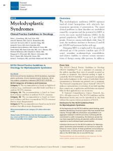
analysis of survival and disease progression The product-limit method of Kaplan and Meier [21] was applied to determine the probability of survival, event-free survival, or disease progression (AML evolution or progression from low- to high-risk MDS = change in FAB category). The significance of differences in survival and AML evolution between the patients’ groups was calculated by log-rank test. To analyze the significance of differences of initial LDH levels in FAB groups and IPSS groups, the Kruskal–Wallis test (variance analysis for nonparametric data) was applied. Differences were considered significant when P was 240 U/l) amounted to 26.8 months, and thus was significantly shorter when compared to patients with normal LDH (44.6 months; P < 0.05). Confirming previous data [15–17], significant differences in LDH levels were found when the various FAB and various IPSS groups were compared. The highest LDH levels were found in the high-risk group with a median LDH level of 254 U/l (Table 2). By contrast, the median LDH level in low-risk patients amounted to 176 U/l. LDH as a prognostic follow-up parameter in patients with MDS In a first step, we compared LDH levels in patients with and without progression (to a ‘higher grade MDS category’ or to AML) in 114 patients. We found that in patients with progression, median LDH levels increased to higher levels in the follow-up compared with patients without progression (Figure 1). When comparing the first two 3-months periods (at diagnosis) with the last two 3-months periods preceding
972 | Wimazal et al.
Figure 1. Lactate dehydrogenase (LDH) levels in myelodysplastic syndromes (MDS) patients according to disease progression. MDS patients (n = 221) were divided into those with progression into a higher disease category or acute myeloid leukemia (h–h) and those without progression (e–e). LDH levels were measured in 3-months periods as described in the text. As visible, patients with disease progression were found to develop higher LDH levels during their follow-up compared with patients without progression (P < 0.05).
progression, the increase in LDH was found to be statistically significant (P < 0.05) (Figure 2A). In contrast, no significant increase in LDH was observed when comparing the first two 3-months periods with the last two 3-months periods in the follow-up in patients without progression (Figure 2B). Similar results were obtained when MDS patients were analyzed after excluding CMML and RAEB-T (WHO-based sample; not shown). To determine the optimal cut-off level for an increase in LDH indicating prognostic significance, LDH levels were analyzed in the follow-up in patients with or without progression: variations in LDH levels were computed as a function of the distance in time between measurements, and the upper 90th percentile values of LDH differences were determined. In these calculations, the optimal prognostic ‘LDH increase cut-off’ indicating progression of the disease was found to be 70 U/l. In 48 of the 114 patients with a long-term LDH follow-up, an increase in the LDH level by at least 70 U/l was documented
Volume 19 | No. 5 | May 2008
Annals of Oncology
original article
Figure 2. Lactate dehydrogenase (LDH) levels in myelodysplastic syndromes (MDS) patients with or without disease progression. LDH levels were serially determined in 3-months periods in the follow-up in 114 patients with MDS. In patients with disease progression (A), the last two 3-months periods before progression were compared with the two initial (first two) 3-months periods after diagnosis. As visible, LDH levels were significantly higher shortly before progression compared with diagnosis in these patients (P < 0.05). For control purpose, the last two 3-months periods in the follow-up were compared with the two initial (first two) 3-months periods in patients who did not show a progression (B). Here, no differences in LDH levels were found (P > 0.05). Whiskers represent the minimum and maximum LDH levels, the box indicates the 25%–75% quantil levels, and the horizontal line within the box shows median LDH levels. The P values are also shown.
during the observation time. The median time interval between diagnosis and a documented increase in LDH (by at least 70 U/l) was 15 months. An increase in LDH by at least 70 U/l was found to be associated to a significantly higher probability of disease progression and a significantly reduced probability of survival when compared to patients without increasing LDH in the follow-up (P < 0.05) (Figure 3). In particular, the median AML-free survival in patients with increasing LDH was 23.8 months and was not reached in patients without increasing LDH (by at least 70 U/l). Similarly, the median survival in patients with increasing LDH was 35.8 months and thus significantly lower as compared with survival in patients without increasing LDH (79.8 months; P < 0.01). Whereas progression to a more advanced MDS category or to AML was diagnosed in 29 of 48 patients (60%) with increasing LDH, only 5 of 66 patients (8%) without an increase in LDH showed a progression to a higher grade MDS or AML (P < 0.05). Typical examples for the clinical course in MDS patients in whom an increase in the LDH level was recorded over time are depicted in Figure 4. In almost all of these cases, the increase in LDH was indicative of disease progression. Hence, applying the criterion for an LDH increase defined above sensitivity for disease progression was found to be 85% and specificity was found to be 76%. Overall, the predictive value of an LDH increase is 60%, and the negative predictive value is 92%.
comparison between LDH and other follow-up parameters In a next step, we asked whether the increase in LDH in transforming patients is accompanied by other signs of disease progression. For this purpose, we compared the dynamics in LDH levels with that of WBC, ANC, monocytes, platelets, and percentage of circulating blasts in the follow-up. In these
Volume 19 | No. 5 | May 2008
investigations, we found that platelets often decrease shortly before or at the time of documented progression, often in association with an increase in LDH (Figure 5). In many of these patients, circulating blasts appeared in the blood (Figure 5). Thus, by statistical analyses, the occurrence of thrombocytopenia was identified as an additional prognostic follow-up parameter. However, in some of these patients, the increase in LDH preceded the decrease of platelets, and in other cases, constant thrombocytopenia was already present before progression and before the increase in LDH was noted. Interestingly, the other parameters tested (WBC, ANC, and monocytes) showed no significant changes in the follow-up, and thus were not found to be of prognostic value. As assessed by multivariate analysis, the increase in LDH was found to be an independent predictive variable concerning disease progression.
predictive value of the IPSS at diagnosis compared with the LDH in the follow-up To determine the role of the LDH as a follow-up parameter in relationship to the IPSS, patients were split into three groups: (i) MDS without progression and without LDH increase, (ii) patients with stable LDH over at least 1 year, who then showed an increase in LDH and disease progression, and (iii) patients with a rapid increase in LDH and rapid progression (within 1 year). When comparing these three groups concerning the (distribution of the) IPSS, it was found that most group (i) patients were within the low-risk IPSS category. Interestingly, a majority of patients with progression and increase in LDH were within the INT-2 or high-risk group (Table 3). However, it was also found that several patients with disease progression in whom progression was also indicated by an increase in LDH were in the low or INT-1 categories (Table 3). These data
doi:10.1093/annonc/mdm595 | 973
original article
Annals of Oncology
with an increasing LDH, whereas no progression was found in five of these patients (14.7%). In 15 patients (13.2%), the increase in LDH was found to precede documented progression, in six patients (5.3%), LDH increased at the time of progression, and in eight patients (7%), the increase in LDH was found after progression had been documented. In 19 patients (16.7%), an increase in LDH but no progression was found, and in 61 patients (53.5%) no increase in LDH and no progression was seen.
discussion
Figure 3. Influence of increasing lactate dehydrogenase (LDH) on survival. A total number of 114 patients with myelodysplastic syndromes (MDS) were examined. Acute myeloid leukemia (AML)-free survival (A), survival (B), and event free survival (C) were determined in patients without increasing LDH (no LDH increase) and those with an increase in LDH by at least 70 U/l. Survival, AML-free survival, and event free survival were calculated by the product-limit method of Kaplan and Meier [21]. The differences in survival and AML-free survival were found to be significant (P < 0.05). Tic marks represent censored patients. For calculating event-free survival, events were defined as either progression to a more advanced type of MDS or to AML or death without progression.
suggest that the IPSS may predict disease progression to a degree confirming previous studies [14], but that in several of these patients, monitoring of LDH in the follow-up may better predict transformation. Finally, we compared the numbers of patients in whom LDH and progression were compared. In this assessment, progression of MDS was detectable in 29 of 34 patients (85%)
974 | Wimazal et al.
A number of prognostic variables predicting survival and/or AML evolution in MDS have been identified [9–18]. Among well-established factors are the percentage of blasts, number of cytopenias, age, karyotype, and transfusion dependence [9–18]. The LDH has also been described as a prognostic variable [12, 16–18]. However, whereas the prognostic value of LDH at diagnosis is well established, little is known about the value of LDH as a marker of disease monitoring. We have serially determined LDH levels in a cohort of MDS patients in order to determine its prognostic value as a dynamic parameter. The results of our study show that an increase in LDH over time is associated with a higher probability of AML evolution and a reduced probability of survival. On the basis of these results, we recommend to use LDH as a prognostic follow-up parameter in MDS. A number of previous data have shown that the LDH at diagnosis is of prognostic significance in MDS [12, 16–18], which could be confirmed in the present study. In fact, an elevated LDH at diagnosis was found to be associated with a reduced probability of survival and an increased probability of AML evolution. To determine the prognostic value of an increasing LDH in the follow-up, a statistical model comparing LDH levels determined in 3-month intervals was applied. Using this approach, we were able to show that an increase in LDH over time is associated with disease progression. In fact, in most patients in whom an increase in LDH was recorded, progression to AML or to high-grade MDS was seen. In addition, we were able to show that an increase in LDH in the follow-up is associated with a significantly reduced survival. As assessed by statistical evaluation, an increase in LDH by at least 70 U/l was defined as the optimal prognostic cut-off in these calculations. A number of different factors may contribute to an increase in LDH. Apart from a transient increase in LDH seen in infectious (viral) diseases or hemolysis, patients with MDS may also develop a paroxysmal nucturnal hemoglobinuria (PNH) subclone or hepatopathy (iron overload) with increase in LDH [22, 23]. In our patients, we were also able to identify a few patients with a PNH subclone or hepatopathy. In two patients with increasing LDH in the follow-up, no signs of MDS progression and no PNH subclone or hepathopathy, however, were found. All in all, MDSrelated and unrelated factors must be considered when using the LDH as a follow-up parameter in MDS. Several different mechanisms may underly an increase in LDH in progressing MDS. One possible factor may be the increased turnover and degradation of myeloid cells in the BM, spleen, and other tissues. Another reason may be ineffective
Volume 19 | No. 5 | May 2008
Annals of Oncology
original article
Figure 4. Follow-up of individual patients with myelodysplastic syndromes (MDS). Examples of patients with MDS in whom an increase in lactate dehydrogenase (LDH) was detected in the follow-up (A–C) or no increase in LDH was detected (D). As visible, an increase in LDH was found to be associated with disease progression.
Figure 5. Comparison of lactate dehydrogenase (LDH) with other disease-related follow-up parameters. Case example of an myelodysplastic syndrome patient in whom progression of disease was accompanied (preceded) by an increase in LDH, decrease in platelet counts, and an increase in circulating blasts. The absolute neutrophil counts (ANC) are also shown.
hematopoiesis. Additional cofactors may be an infiltration of the liver and spleen by immature myeloid cells or iron overload. The exact biochemical basis of an increasing LDH in these patients remains unknown. A number of different prognostic factors indicate the probability of survival and AML evolution in MDS [9–18,
Volume 19 | No. 5 | May 2008
24–29]. We therefore asked whether an increase in LDH in progressive MDS is also accompanied by other signs of disease progression and found that disease progression is also associated with occurrence of thrombocytopenia or appearence of circulating blasts. An interesting observation was that in several cases, the increase in LDH may precede the decrease in platelets or the occurrence of blasts. In multivariate analyses, the increase in LDH and the occurrence of peripheral blasts were found to be independent prognostic parameters. Whether the increase in LDH was also accompanied by cytogenetic evolution, which may also be indicative of disease progression [24–26] was not assessed in this study. As more and more therapeutic approaches become available for MDS patients and since more and more (younger) patients are diagnosed at an early disease phase, it is of importance to determine the exact risk profile in these patients and to predict survival and AML evolution at diagnosis and in the follow-up [27–30]. The results of our study show that LDH levels may serve as a useful follow-up parameter. Notably, on the basis of our data, we recommend to carry out LDH determinations in patients with MDS in the follow-up in clinical practice. An increase in LDH in these patients should lead to a thorough reevaluation of the disease status with a reexamination of the BM. This may be of particular interest for patients who have low-risk MDS but would be eligible for intensive therapies and SCT. In fact, recent data suggest that such low risk MDS
doi:10.1093/annonc/mdm595 | 975
original article
Annals of Oncology
Table 3. Distribution of IPSS categories in MDS patients with progression that was indicated (accompanied) by an increase in LDH in the follow-up Definition
Group i Group ii
Distribution of IPSS groups, patients = n Low INT-1 INT-2 High
Stable during observation 29 Stable >1 year, then 1 progression Group iii Progression within 1 year 2
16 1
4 5
0 3
4
3
3
Patients with MDS in whom LDH was serially determined for at least 6 months were separated into three groups, i.e. (i) patients with no increase in LDH and no progression, (ii) patients with a stable LDH for at least 1 year who later showed an increase in LDH and disease progression, and (iii) patients with a rapid increase in LDH and rapid disease progression (within the first year of observation). In these three groups, patients were subdivided according to the IPSS. The differences in the percentage of patients belonging to a distinct IPSS type in the various groups (i), (ii), and (iii) were calculated by chi-square test and were found to be significant (P < 0.05). IPSS, International Prognostic Scoring System; MDS, myelodysplastic syndromes; LDH, lactate dehydrogenase.
10.
11.
12.
13. 14.
15.
16.
17. 18.
patients should be considered for SCT as soon as diseaseprogression occurs, but not when the course of their disease remains stable [27, 28]. LDH assessments in the follow-up could be a most helpful predictive parameter in these cases. In conclusion, LDH should be recommended as a follow-up parameter in patients with MDS, especially when disease progression is an important decision point for treatment. The exact value of LDH measurements in the follow-up in patients with MDS should be determined in prospective clinical studies.
19.
20. 21. 22.
funding Medical University of Vienna.
23.
references 1. Cazzola M, Riccardi A. Myelodysplastic syndromes: biologic and clinical aspects. Haematologica 1986; 71: 147–159. 2. Alessandrino EP, Amadori S, Cazzola M et al. Myelodysplastic syndromes: recent advances. Haematologica 2001; 86: 1124–1157. 3. Hofmann WK, Koeffler HP. Myelodysplastic syndrome. Annu Rev Med 2005; 56: 1–16. 4. Bennett JM, Komrokji RS. The myelodysplastic syndromes: diagnosis, molecular biology and risk assessment. Hematology 2005; 10: 258–269. 5. Bennett JM, Catovsky D, Daniel MT et al. Proposals for the classification of the myelodysplastic syndromes. Br J Haematol 1982; 51: 189–199. 6. Bennett JM, Catovsky D, Daniel MT et al. The chronic myeloid leukaemias: guidelines for distinguishing chronic granulocytic, atypical chronic myeloid, and chronic myelomonocytic leukaemia. Proposals by the French-American-British Cooperative Leukaemia Group. Br J Haematol 1994; 87: 746–754. 7. Brunning RD, Bennett JM, Flandrin G et al. Myelodysplastic syndromes. In Jaffe ES, Harris NL, Stein H, Vardiman JW (eds): Tumours of Haematopoietic and Lymphoid Tissues, Pathology & Genetics, World Health Organization Classification of Tumours. Lyon, France: IARC Press 2001; 61–73. 8. Bennett JM. A comparative review of classification systems in myelodysplastic syndromes (MDS). Semin Oncol 2005; 32: 3–10. 9. Morel P, Hebbar M, Lai JL et al. Cytogenetic analysis has strong independent prognostic value in de novo myelodysplastic syndromes and can be incorporated
976 | Wimazal et al.
24.
25.
26.
27.
28.
29. 30.
in a new scoring system: a report on 408 cases. Leukemia 1993; 7: 1315–1323. Sanz GF, Sanz MA, Vallespi T et al. Two regression models and a scoring system for predicting survival and planning treatment in myelodysplastic syndromes: a multivariate analysis of prognostic factors in 370 patients. Blood 1989; 74: 395–408. Toyama K, Ohyashiki K, Yoshida Y et al. Clinical implications of chromosomal abnormalities in 401 patients with myelodysplastic syndromes: a multicentric study in Japan. Leukemia 1993; 7: 499–508. Aul C, Gattermann N, Heyll A et al. Primary myelodysplastic syndromes: analysis of prognostic factors in 235 patients and proposals for an improved scoring system. Leukemia 1992; 6: 52–59. Mufti GJ, Stevens JR, Oscier DG et al. Myelodysplastic syndromes: a scoring system with prognostic significance. Br J Haematol 1985; 59: 425–433. Greenberg P, Cox C, LeBeau MM et al. International scoring system for evaluating prognosis in myelodysplastic syndromes. Blood 1997; 89: 2079–2088. Malcovati L, Germing U, Ku¨ndgen A et al. Time-dependent prognostic scoring system for predicting survival and leukemic evolution in myelodysplastic syndromes. J Clin Oncol 2007; 25: 3503–3510. Aul C, Gattermann N, Germing U et al. Risk assessment in primary myelodysplastic syndromes: validation of the Du¨sseldorf score. Leukemia 1994; 8: 1906–1913. Wimazal F, Sperr WR, Kundi M et al. Prognostic value of lactate dehydrogenase activity in myelodysplastic syndromes. Leuk Res 2001; 25: 287–294. Germing U, Hildebrandt B, Pfeilstocker M et al. Refinement of the international prognostic scoring system (IPSS) by including LDH as an additional prognostic variable to improve risk assessment in patients with primary myelodysplastic syndromes (MDS). Leukemia 2005; 19: 2223–2231. Fonatsch C, Gudat H, Lengfelder E et al. Correlation of cytogenetic findings with clinical features in 18 patients with inv(3)(q21q26) or t(3;3)(q21;q26). Leukemia 1994; 8: 1318–1326. ISCN. An international system for human cytogenetic nomenclature. Cytogenet Cell Genet 1981; 31: 5. Kaplan EL, Meier P. Nonparametric estimation from incomplete observations. J Am Stat Assoc 1958; 53: 157. Iwanaga M, Furukawa K, Amenomori T et al. Paroxysmal nocturnal haemoglobinuria clones in patients with myelodysplastic syndromes. Br J Haematol 1998; 102: 465–474. Oelschlaegel U, Besson I, Arnoulet C et al. A standardized flow cytometric method for screening paroxysmal nocturnal haemoglobinuria (PNH) measuring CD55 and CD59 expression on erythrocytes and granulocytes. Clin Lab Haematol 2001; 23: 81–90. Tomonaga M, Tomonaga Y, Kusano M, Ichimaru M. Sequential karyotypic evolutions and bone marrow aplasia preceding acute myelomonocytic transformation from myelodysplastic syndrome. Br J Haematol 1984; 58: 53–60. Tricot G, Boogaerts MA, De Wolf-Peeters C et al. The myelodysplastic syndromes: different evolution patterns based on sequential morphological and cytogenetic investigations. Br J Haematol 1985; 59: 659–670. Haase D, Fonatsch C, Freund M. Karyotype instability in myelodysplastic syndromes–a specific step in pathogenesis preceding clonal chromosome anomalies. Leuk Lymphoma 1992; 8: 221–228. Cutler CS, Lee SJ, Greenberg P et al. A decision analysis of allogeneic bone marrow transplantation for the myelodysplastic syndromes: delayed transplantation for low-risk myelodysplasia is associated with improved outcome. Blood 2004; 104: 579–585. Deeg HJ. Optimization of transplant regimens for patients with myelodysplastic syndrome (MDS). Hematology Am Soc Hematol Educ Program 2005; 1: 167–173. List A, Dewald G, Bennett J et al. Lenalidomide in the myelodysplastic syndrome with chromosome 5q deletion. N Engl J Med 2006; 355: 1456–1465. Valent P, Horny HP, Bennett JM et al. Definitions and standards in the diagnosis and treatment of the myelodysplastic syndromes: consensus statements and report from a working conference. Leuk Res 2007; 31: 727–736.
Volume 19 | No. 5 | May 2008