Mucinous Neoplasms of the Appendix and Peritoneum Nicole C. Panarelli, MD; Rhonda K. Yantiss, MD
Context.—Appendiceal mucinous neoplasms are considNered enigmatic tumors of unpredictable biologic potential. Their importance lies in their potential to spread to the peritoneum and viscera in the form of gelatinous mucin deposits. Extra-appendiceal spread of these tumors is the most common etiology of pseudomyxoma peritonei, which is a descriptive term encompassing a number of neoplastic and nonneoplastic peritoneal disorders. Many studies aimed at evaluating the biologic importance of appendiceal mucinous neoplasms and pseudomyxoma peritonei have employed inconsistent histologic criteria for their diagnosis and descriptive terminology for their classification. As a result, appendiceal mucinous neoplasms and associated peritoneal disease represents one of the most confusing and controversial areas in gastrointestinal pathology. rimary appendiceal tumors are found in less than 2% of P surgically removed appendices, but still pose a variety of diagnostic and therapeutic challenges. In particular, the classification and management of appendiceal mucinous neoplasms has proven problematic for both clinicians and pathologists. These tumors characteristically cause cystic dilatation of the appendix owing to accumulation of copious gelatinous material in the lumen (Figure 1, A and B). They may disseminate throughout the peritoneal cavity in the form of gelatinous deposits, termed pseudomyxoma peritonei (Figure 1, C and D). Many controversies surrounding appendiceal neoplasms and pseudomyxoma peritonei stem from the use of inconsistent histologic criteria for their diagnosis and philosophic differences of opinion regarding their pathogenesis. Several investigators have proposed classification schemes that employ similar terminology to describe lesions of variable biologic potential or that use dissimilar terms to describe the same entity. Moreover, an origin in the appendix is not suspected before surgery in many cases because patients present with ovarian or peritoneal tumors that are managed by gynecologists, rather than general or colorectal surgeons. These different clinical Accepted for publication March 11, 2011. From the Department of Pathology and Laboratory Medicine, Weill Cornell Medical College, New York, New York. The authors have no relevant financial interest in the products or companies described in this article. Presented in part at the Tutorial on Pathology of the GI Tract, Pancreas, and Liver; Westin Diplomat Resort & Spa; November 15–19, 2010; Hollywood, Florida. Reprints: Rhonda K. Yantiss, MD, Weill Cornell Medical College, 1300 York Ave, New York, NY 10065 (e-mail:
[email protected]. edu). Arch Pathol Lab Med—Vol 135, October 2011
Objectives.—To summarize the literature regarding the biologic potential of appendiceal mucinous neoplasms and pseudomyxoma peritonei and to discuss the similarities and differences between proposed systems for their classification. Data Sources.—Literature review and case-derived material. Conclusions.—Many studies have contributed to an increased understanding of the natural progression of mucinous neoplasms of the appendix and peritoneum, and the adoption of a uniform reporting system, as advocated by the American Joint Committee on Cancer and the World Health Organization, will facilitate clear communication among pathologists and clinical colleagues. (Arch Pathol Lab Med. 2011;135:1261–1268; doi: 10. 5858/arpa.2011-0034-RA)
groups are usually familiar with some, but not all, of the terminology used to describe mucinous tumors of the appendix and peritoneum. As a result, management strategies across different institutions, or even among clinicians at the same institution, are not standardized. The purpose of this review is to provide a comprehensive discussion of the biologic potential of, and proposed classification schemes for, appendiceal mucinous neoplasms and pseudomyxoma peritonei. WHAT IS PSEUDOMYXOMA PERITONEI? Pseudomyxoma peritonei is a term used to describe mucinous ascites or mucin deposits within the peritoneal cavity. Pseudomyxoma peritonei consists of organizing pools of mucin within peritoneal fat or on the serosal surfaces of the viscera, which contain variable numbers of neoplastic epithelial cells. Most cases reflect dissemination of an appendiceal mucinous neoplasm, in which case, mucin pools that contain scant strips and clusters of lowgrade neoplastic epithelial cells are typical (Figure 2, D), similar to those present in the appendiceal mucosa (Figure 2, A and B). However, pseudomyxoma peritonei is not synonymous with a neoplasm nor are all neoplastic cases derived from the appendix. Ruptured diverticula of the appendix and colon may spill mucin into the peritoneal cavity, and mucin-producing carcinomas of the colon, pancreas, and other organs may disseminate throughout the peritoneum in the form of gelatinous ascites. Unfortunately, much of the earlier literature is of limited value in assessing the biologic potential of appendiceal mucinous neoplasms and pseudomyxoma peritonei. Most reports before 2000 did not distinguish between neoplastic and nonneoplastic etiologies for pseudomyxoma peritonei, appendiceal and nonappendiceal neoplasms, or
Mucinous Neoplasms of the Appendix and Peritoneum—Panarelli & Yantiss
1261
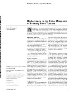
Figure 1. Mucinous neoplasms often cause appendiceal distention (A) because of massive accumulation of extracellular mucin within the lumen (B). The neoplasms may spread to the serosal surfaces of the viscera (C) or form gelatinous tumor deposits in the omentum (D).
cytologically low- and high-grade tumors in the peritoneal cavity.1 In our practice, we generally avoid using pseudomyxoma peritonei as a diagnostic term and limit its inclusion in pathology reports to a comment aimed at facilitating communication with clinicians. ORIGIN OF PSEUDOMYXOMA PERITONEI The combined results of several studies published in theearly 1990s implicated the appendix as a likely site of origin in most of nearly 100 patients with pseudomyxoma peritonei but also suggested that some cases may be derivedfrom either the ovary or peritoneum. Prayson etal2 evaluated a series of 10 men and 9 women with pseudomyxoma peritonei and found that 94% of patients had appendiceal mucinous neoplasms, including all of the women with ovarian tumors. Seidman et al3 found a high frequency of bilateral ovarian mucinous tumors, or rightsided ovarian mucinous tumors in unilateral cases, among women with concomitant mucinous tumors of the appendix, leading them to conclude that pseudomyxoma peritonei may be derived from either site or represent a primary peritoneal neoplasm. Young et al4 studied 22 appendiceal mucinous neoplasms associated with mucinous ovarian tumors and noted that tumors in both sites 1262
Arch Pathol Lab Med—Vol 135, October 2011
were histologically similar. They also found ovarian disease to be bilateral in most cases, whereas unilateral ovarian tumors showed a right-sided predominance. Unlike Seidman et al,3 however, they concluded that these features suggested a primary appendiceal origin with secondary ovarian involvement. Molecular data further support the notion that pseudomyxoma peritonei is usually derived from the appendix, rather than the ovary or peritoneum. Cautrecasas et al5 analyzed KRAS mutational status in 6 synchronous mucinous tumors of the appendix and ovary, 5 (83%) of which showed identical KRAS mutations in codon 12 in tumors from both sites. Chuaqui et al6 analyzed loss of heterozygosity on chromosomes 5 and 17 in synchronous appendiceal and ovarian mucinous neoplasms obtained from 12 patients. They found either loss of heterozygosity at the same locus or no loss of heterozygosity in paired tumors in 75% of cases. Subsequent studies have shown progressive loss of heterozygosity in ovarian mucinous tumors, when compared with synchronous appendiceal neoplasms, leading most investigators to conclude that most pseudomyxoma peritonei cases are derived from appendiceal mucinous neoplasms, whereas an ovarian origin is considered a rare occurrence.7,8
Mucinous Neoplasms of the Appendix and Peritoneum—Panarelli & Yantiss
Figure 2. Mucinous neoplasms typically display a circumferential growth pattern in the appendiceal mucosa with a variable papillary architecture (A). The tumor cells contain abundant cytoplasmic mucin and enlarged, hyperchromatic nuclei that are basally located, with minimal cytologic atypia (B). Peritoneal mucin deposits contain scant strips and clusters of mucin-containing epithelial cells (C) that are cytologically low grade, similar to the neoplastic cells present in the appendix (D) (hematoxylin-eosin, original magnifications 340 [A and C] and 3400 [B and D]).
PERIAPPENDICEAL MUCIN DEPOSITS (LOCALIZED PSEUDOMYXOMA PERITONEI) Not uncommonly, surgical pathologists encounter appendiceal resection specimens with mucin deposits on the appendiceal serosa, within the mesoappendix, or limited to the right lower quadrant (Figure 3, A and B). Extra-appendiceal mucin deposits limited to the periappendiceal area are usually devoid of neoplastic epithelial cells (acellular), although one may infrequently observe strips or clusters of neoplastic mucinous epithelial cells in periappendiceal mucin, similar to cases with widespread peritoneal disease (Figure 3, C and D). Yantiss et al9 evaluated the prognostic implications of peritoneal mucin localized to the right lower quadrant in 65 patients with appendiceal mucinous neoplasms. Fifty patients (77%) had acellular extra-appendiceal mucin, and 15 (23%) had periappendiceal mucin deposits that contained scant mucinous epithelium with low-grade cytologic features. At follow-up, 48 (96%) patients without extra-appendiceal epithelium were disease free (mean, 52 months), whereas 33% of patients with any neoplastic epithelium outside the appendix developed widespread peritoneal tumor deposits, including one patient who died of disease (mean, Arch Pathol Lab Med—Vol 135, October 2011
38 months). Notably, 2 patients with acellular periappendiceal mucin developed disseminated peritoneal disease, but neither of those appendices was submitted entirely for histologic examination. These results emphasize the importance of adequate sampling in determining the prognosis of patients with appendiceal mucinous neoplasms and indicate that tumors without extra-appendiceal neoplastic epithelium are associated with an excellent prognosis. CLASSIFICATION OF APPENDICEAL MUCINOUS NEOPLASMS AND PSEUDOMYXOMA PERITONEI The proposed classification schemes for appendiceal mucinous neoplasms and pseudomyxoma peritonei reflect differences of opinion regarding the fundamental nature of mucin deposits in the peritoneum and comparisons between them are summarized in the Table. Some investigators think that peritoneal mucin deposits are the consequence of appendiceal rupture and spillage of mucin and ‘‘adenomatous’’ epithelium into the peritoneal cavity, rather than carcinoma. They argue that appendiceal mucinous tumors frequently lack infiltrating, malignant glands and desmoplastic stroma and note that
Mucinous Neoplasms of the Appendix and Peritoneum—Panarelli & Yantiss
1263
Figure 3. Appendiceal mucinous neoplasms may show areas of mucin extrusion through the wall (A) or organizing mucin on the serosal surface (B). The mucin deposits contain inflammatory cells and granulation tissue in most cases (C), but occasional tumors are associated with extraappendiceal neoplastic epithelium, similar to that seen in patients with pseudomyxoma peritonei (D) (hematoxylin-eosin, original magnifications 320 [A], 340 [B], 3400 [C], and 3600 [D]).
extra-appendiceal epithelium usually shows bland cytologic features insufficient for a diagnosis of malignancy. On the other hand, there are compelling reasons why the American Joint Committee on Cancer, World Health Organization, and most other investigators consider pseudomyxoma peritonei to be carcinoma.10,11 First, some carcinomas are extremely well differentiated with minimal atypical cytologic features, so low-grade cytology does not exclude a diagnosis of malignancy. Second, parenchymal invasion of solid organs is common in patients with pseudomyxoma peritonei and is generally considered a feature of malignancy. Third, some cancers, particularly mucinous or colloidal carcinomas may invade tissues with a broad, pushing growth pattern, so one need not see infiltrating glands or desmoplasia to make a diagnosis of malignancy. Finally and most important, mucinous neoplasms that spread beyond the appendix are associated with progressive mucinous ascites, disease recurrence, and death in at least half of patients.12–14 In a study of 184 epithelial noncarcinoid appendiceal tumors, Carr et al13 found that mucinous neoplasms limited to the appendix pursued an indolent course, whereas those with extra-appendiceal spread were associated with decreased survival. These authors proposed 1264
Arch Pathol Lab Med—Vol 135, October 2011
that appendiceal mucinous neoplasms be classified as adenomas (or cystadenomas), tumors of uncertain malignant potential, and carcinoma. Adenomas were defined by the presence of mucinous neoplasia confined to the mucosa with intact muscularis mucosae, whereas tumors that showed mural invasion or proliferation of neoplastic epithelium outside the appendix were considered carcinomas. Neoplasms with pushing, but not infiltrative, mural invasion or acellular mucin pools on the serosa were considered to be of uncertain malignant potential. Misdraji et al15 later analyzed the histologic features of 107 appendiceal mucinous tumors, which they classified as either low- or high-grade based on cytoarchitectural features. Tumors with low-grade cytology were termed low-grade appendiceal mucinous neoplasms. High-grade lesions limited to the appendiceal mucosa were termed noninvasive mucinous cystadenocarcinomas, whereas peritoneal deposits containing overtly malignant cells were labeled as invasive mucinous adenocarcinomas. All patients with low-grade neoplasms confined to the appendiceal mucosa were free of disease at last followup (median, 6 years), whereas 67% of patients with peritoneal involvement experienced disease recurrence or death from disease. Patients with overtly malignant
Mucinous Neoplasms of the Appendix and Peritoneum—Panarelli & Yantiss
Comparisons Among Classification Schemes for Appendiceal Mucinous Neoplasms and Pseudomyxoma Peritonei Source, y Carr and Sobin,11 2010
Misdraji et al,15 2003
Pai and Longacre,17 2009
Ronnett et al,1 1995
Bradley et al,12 2006
AJCC and WHO10,11 2010
Tumor confined to appendix Limited to mucosa Low-grade cytology High-grade cytology Positive surgical margin Neoplastic epithelium in appendix wall Tumor beyond appendix Low-grade epithelium in peritoneal mucin High-grade epithelium in peritoneal mucin
Adenoma Adenoma Adenoma Uncertain malignant potential
Low-grade appendiceal mucinous neoplasm Noninvasive mucinous cystadenocarcinoma Low-grade appendiceal mucinous neoplasm Low-grade appendiceal mucinous neoplasm
Adenoma
NA
NA
Adenoma
Adenoma
NA
NA
Adenoma
Uncertain malignant potential Uncertain malignant potential
NA
NA
Adenoma
NA
NA
Invasive Mucinous Adenocarcinoma
Disseminated peritoneal adenomucinosis
Low-grade Low-grade mucinous mucinous carcinoma adenocarcinoma peritonei High-grade High-grade mucinous mucinous carcinoma adenocarcinoma peritonei
Invasive mucinous Low-grade adenocarcinoma appendiceal mucinous neoplasm
High-risk for recurrence
Invasive mucinous Invasive mucinous adenocarcinoma adenocarcinoma
Invasive mucinous Peritoneal adenocarcinoma mucinous carcinomatosis
Abbreviations: AJCC, American Joint Committee on Cancer; NA, not applicable; WHO, World Health Organization.
peritoneal disease had 3- and 5-year survival rates of 90% and 44%, respectively, compared with 100% and 86%, respectively, among patients with low-grade peritoneal disease. The authors failed to identify any pathologic features in the appendiceal component that reliably predicted the presence or absence of pseudomyxoma peritonei and suggested that all low-grade tumors be labeled low-grade appendiceal mucinous neoplasm, regardless of the extent of disease, which would be documented in the pathology report. Two years later, Pai and Longacre16 proposed an alternative classification scheme that built on that suggested by Carr et al.13 They recognized lesions confined to the appendiceal mucosa as adenomas but expanded the definition of mucinous tumors of uncertain malignant potential to include those tumors with mucosal disease at the proximal margin, those with mucin and epithelium in the appendiceal wall without obvious destructive invasion, and those in which the diagnosis of extra-appendiceal epithelium was in doubt. Mucin pools containing scant, low-grade epithelium outside the appendix were classified as mucinous neoplasms of low malignant potential, whereas those with destructive invasion were considered to represent adenocarcinomas. This same group later analyzed 116 mucinous appendiceal neoplasms and modified their original classification scheme to a 4-tiered system.17 Low-grade tumors confined to the appendiceal mucosa were classified as adenomas, whereas similarly low-grade tumors with acellular extraappendiceal mucin were considered to be of low risk for recurrence. High-grade tumors in the peritoneal cavity were classified as adenocarcinomas, similar to their prior Arch Pathol Lab Med—Vol 135, October 2011
proposal. Cytologically low-grade tumors with extraappendiceal epithelium (previously termed mucinous neoplasms of low malignant potential) recurred in 21 of 27 cases (78%) and, thus, were reclassified as low-grade mucinous neoplasms with high-risk of recurrence. All patients with invasive carcinoma and who had available follow-up data died of disease with significantly decreased overall survival rates compared with patients with low-grade extra-appendiceal disease. Other authors have devised classification schemes for pseudomyxoma peritonei based on the cytologic appearance of peritoneal epithelium. Ronnett et al1 considered pseudomyxoma peritonei with low-grade cytologic features to represent passive spread of adenomatous epithelium into the peritoneum, secondary to appendiceal rupture, and, thus, termed such cases disseminated peritoneal adenomucinosis (Figure 4, A and B). They consider mucinous tumors of the peritoneum with more-abundant, cytologically malignant epithelium to represent peritoneal mucinous carcinomatosis (Figure 4, E and F), and cases with both low- and high-grade features were designated peritoneal mucinous carcinomatosis, intermediate grade (Figure 4, C and D). The authors applied this classification scheme to 109 multifocal peritoneal mucinous tumors derived from the appendix, colon, or small intestine and compared survival rates among the 3 groups. All 65 cases of disseminated peritoneal adenomucinosis were derived from the appendix and were associated with 5- and 10-year survival rates of 75% and 68%, respectively. Both intermediate and high-grade mucinous carcinomas were more frequently derived from nonappendiceal primary tumors and pursued an aggressive clinical course with 5- and
Mucinous Neoplasms of the Appendix and Peritoneum—Panarelli & Yantiss
1265
Figure 4. Mucin deposits in the peritoneal cavity classified as disseminated peritoneal adenomucinosis (A and B), peritoneal mucinous carcinomatosis–intermediate grade (C and D), and peritoneal mucinous carcinomatosis (E and F). Disseminated peritoneal adenomucinous contains paucicellular mucin pools (A) with scant strips of low-grade neoplastic epithelium (B). Peritoneal mucinous carcinomatosis displays mucin pools with abundant epithelium (E) that show overtly malignant cytologic features (F). Peritoneal mucinous carcinomatosis–intermediate grade is less cellular than peritoneal mucinous carcinomatosis (C), but the degree of cytologic atypia exceeds that of disseminated peritoneal adenomucinosis (D) (hematoxylin-eosin, original magnifications 3100 [A], 3600 [B and F], 340 [C], 3200 [D], and 320 [E]).
10-year survival rates of 50% and 21% for intermediategrade peritoneal mucinous carcinomatosis and 14% and 3% for high-grade peritoneal mucinous carcinomatosis, respectively.1,14 Although these results suggest histologic 1266
Arch Pathol Lab Med—Vol 135, October 2011
grade is important in predicting behavior among peritoneal mucinous tumors, inclusion of intestinal carcinomas in the analysis of high-grade tumors makes it difficult to interpret the data.
Mucinous Neoplasms of the Appendix and Peritoneum—Panarelli & Yantiss
Bradley et al12 better demonstrated the importance of cytologic grade in predicting behavior of pseudomyxoma peritonei in a later study. These authors evaluated the natural history of 101 patients with appendiceal mucinous tumors and peritoneal disease, all of whom were treated in a similar fashion at a single institution. These authors used a 2-tiered system to classify pseudomyxoma peritonei. Low- and intermediate-grade tumors were considered to be low-grade mucinous carcinoma peritonei, whereas cases with severe cytologic atypia were classified as highgrade mucinous carcinoma peritonei. The authors found that low-grade mucinous carcinoma peritonei was associated with a significantly better 5-year survival rate (63%) than was high-grade mucinous carcinoma peritonei (38%). The combined results of all of these studies can be summarized into several major points. First, tumors limited to the appendiceal mucosa have no potential for aggressive disease when completely resected, regardless of the degree of dysplasia, and, therefore, it is appropriate to classify them as adenomas or cystadenomas. Appendiceal mucinous neoplasms associated with acellular mucin limited to the appendiceal wall and/or mesoappendix are also cured by excision, in most cases and, thus, we consider these tumors to be adenomas as well. However, all neoplasms that are classified as adenomas should be completely submitted for histologic evaluation to exclude the possibility of extra-appendiceal epithelium. Clinicians should also be informed of the remote possibility that a small amount of extra-appendiceal epithelium may not be detected by sampling of tumors with acellular periappendiceal mucin and thus, clinical follow-up should probably be considered. Any proliferation of neoplastic epithelium beyond the muscularis mucosae is at risk for peritoneal dissemination and should be graded and staged as a carcinoma, as described in the World Health Organization classification and staging guidelines put forth by the American Joint Committee on Cancer.10,11 The World Health Organization regards any neoplastic epithelial proliferation confined to the appendiceal mucosa as an adenoma. Appendiceal mucinous tumors with extra-appendiceal neoplastic epithelium are classified as mucinous adenocarcinomas and subcategorized as lowor high-grade because increasingly severe cytoarchitectural atypia is associated with poorer outcome.11 The American Joint Committee on Cancer staging guidelines assign tumor (T) stage for appendiceal mucinous neoplasms similar to those used for colonic adenocarcinoma, with the exception that T4a denotes both serosal involvement and extra-appendiceal disease limited to the right lower quadrant. Mucinous deposits beyond the right lower quadrant (pseudomyxoma peritonei) are considered to represent metastatic disease and are denoted as M1a.10 Confusing terminology, such as low-grade appendiceal mucinous neoplasm in the peritoneum, disseminated peritoneal adenomucinosis, and mucinous neoplasm at high-risk for recurrence, should probably be avoided to facilitate clear communication with our clinical colleagues. TREATMENT OF PSEUDOMYXOMA PERITONEI Treatment of pseudomyxoma peritonei has historically consisted of surgical debulking of gross disease, with or without various forms of chemotherapy. Recent years have seen a trend toward more aggressive management strategies that include multiple surgeries, complete Arch Pathol Lab Med—Vol 135, October 2011
peritonectomy, intraoperative intraperitoneal chemotherapy, and additional cycles of postoperative chemotherapy. Miner et al18 retrospectively analyzed the relative contribution of histologic features of peritoneal disease, patient characteristics, and extent of treatment in 97 patients who underwent extensive debulking surgery, which achieved complete cytoreduction in 53% of cases. They found that low-grade cytology was independently associated with disease-free survival and that 90% of patients who achieved 10-year survival had tumors of low-histologic grade. Outcome data from other studies using these aggressive treatment strategies have also shown improved survival among patients with low-grade peritoneal disease compared with survival rates of those with highgrade tumors, which are probably not amenable to surgical management.19–22 Thus, therapeutic decisions largely rest on the distinction between low-and highgrade peritoneal disease. SUMMARY AND CONCLUSIONS Appendiceal mucinous neoplasms represent a relatively homogeneous group of neoplasms that pursue a predictable clinical course based on tumor stage and grade. Those confined to the appendiceal mucosa are cured by excision, whereas any proliferation of neoplastic epithelium beyond the mucosa places the patient at risk for peritoneal dissemination. The histologic grade of peritoneal disease is extremely important. Patients with low-grade tumors may benefit from aggressive management, consisting of a combination of chemotherapy and cytoreductive surgery, whereas those with high-grade tumors probably do not benefit from aggressive debulking but may be better served by systemic chemotherapy. Decisions regarding clinical management require clear communication among treating physicians, so adoption of a uniform reporting system for appendiceal mucinous neoplasms with peritoneal metastases by the World Health Organization and American Joint Committee on Cancer represents a major advancement in the field. References 1. Ronnett BM, Zahn CM, Kurman RJ, Kass ME, Sugarbaker PH, Shmookler BM. Disseminated peritoneal adenomucinosis and peritoneal mucinous carcinomatosis: a clinicopathologic analysis of 109 cases with emphasis on distinguishing pathologic features, site of origin, prognosis, and relationship to ‘‘pseudomyxoma peritonei.’’ Am J Surg Pathol. 1995;19(12):1390–1408. 2. Prayson RA, Hart WR, Petras RE. Pseudomyxoma peritonei: a clinicopathologic study of 19 cases with emphasis on site of origin and nature of associated ovarian tumors. Am J Surg Pathol. 1994(6);18:591–603. 3. Seidman JD, Elsayed AM, Sobin L, Tavassoli FA. Association of mucinous tumors of the ovary and appendix: a clinicopathologic study of 25 cases. Am J Surg Pathol. 1993;17(1):22–34. 4. Young RH, Gilks CB, Scully RE. Mucinous tumors of the appendix associated with mucinous tumors of the ovary and pseudomyxoma peritonei: a clinicopathologic analysis of 22 cases supporting an origin in the appendix. Am J Surg Pathol. 1991;15(5):415–429. 5. Cautrecasas M, Matias-Guiu X, Prat J. Synchronous mucinous tumors of the appendix and the ovary associated with pseudomyxoma peritonei: a clinicopathologic study of six cases with comparative analysis of c-Ki-ras mutations. Am J Surg Pathol. 1996;20(6):739–746. 6. Chuaqui RF, Zhuang Z, Emmert-Buck MR, et al. Genetic analysis of synchronous mucinous tumors of the ovary and appendix. Hum Pathol. 1996; 27(2):165–171. 7. Young R. From Krukenberg to today—the ever present problems posed by metastatic tumors of the ovary, part I: historical perspective, general principles, mucinous tumors including Krukenberg tumor. Adv Anat Pathol. 2006;13(5):205–227. 8. Young RH. Pseudomyxoma peritonei and selected other aspects of the spread of appendiceal neoplasms. Semin Diagn Pathol. 2004;21(2):134–150. 9. Yantiss RK, Shia J, Klimstra DS, Hahn HP, Odze RD, Misdraji J. Prognostic significance of localized extra-appendiceal mucin deposition in appendiceal mucinous neoplasms. Am J Surg Pathol. 2009;33(2):248–255. 10. Edge SB, Byrd DR, Compton CC, Fritz AG, Greene FL, Trotti A, eds. AJCC Cancer Staging Manual. 7th ed. New York, NY: Springer; 2010:133–141.
Mucinous Neoplasms of the Appendix and Peritoneum—Panarelli & Yantiss
1267
11. Carr NJ, Sobin LH. Tumours of the appendix. In: Bosman FT, Carneiro F, Hruban RH, Theise ND, eds. WHO Classification of Tumours of the Digestive System. 4th ed. Lyon, France: IARC Press; 2010:122–125. World Health Organization Classification of Tumours; vol 3. 12. Bradley RF, Stewart JH, Russell GB, Levine EA, Geisinger KR. Pseudomyxoma peritonei of appendiceal origin: a clinicopathologic analysis of 101 patients uniformly treated at a single institution, with literature review. Am J Surg Pathol. 2006;30(5):551–559. 13. Carr NJ, McCarthy WF, Sobin LH. Epithelial noncarcinoid tumors and tumor-like lesions of the appendix: a clinicopathologic study of 184 patients with a multivariate analysis of prognostic factors. Cancer. 1995;75(3):757–768. 14. Ronnett BM, Yan H, Kurman RJ, Shmookler BM, Wu L, Sugarbaker PH. Patients with pseudomyxoma peritonei associated with disseminated peritoneal adenomucinosis have a significantly more favorable prognosis than patients with peritoneal mucinous carcinomatosis. Cancer. 2001;92(1):85–91. 15. Misdraji J, Yantiss RK, Graeme-Cook FM, Balis UJ, Young RH. Appendiceal mucinous neoplasms: a clinicopathologic analysis of 107 cases. Am J Surg Pathol. 2003;27(8):1089–1103. 16. Pai RK, Longacre TA. Appendiceal mucinous tumors and pseudomyxoma peritonei: histologic features, diagnostic problems, and proposed classification. Adv Anat Pathol. 2005;12(6):291–311.
1268
Arch Pathol Lab Med—Vol 135, October 2011
17. Pai RK, Beck AH, Norton JA, Longacre TA. Appendiceal mucinous neoplasms: clinicopathologic study of 116 cases with analysis of factors predicting recurrence. Am J Surg Pathol. 2009;33(10):1425–1439. 18. Miner TJ, Shia J, Jaques DP, Klimstra DS, Brennan MF, Coit DG. Long-term survival following treatment of pseudomyxoma peritonei: an analysis of surgical therapy. Ann Surg. 2005;241(2):300–308. 19. Baratti D, Kusamura S, Nonaka D, et al. Pseudomyxoma peritonei: clinical pathological and biological prognostic factors in patients treated with cytoreductive surgery and hyperthermic intraperitoneal chemotherapy (HIPEC). Ann Surg Oncol. 2008;15(2):526–534. 20. Baratti D, Kusamura S, Nonaka D, Cabras AD, Laterza B, Deraco M. Pseudomyxoma peritonei: biologic features are the dominant prognostic determinants after complete cytoreduction and hyperthermic intraperitoneal chemotherapy. Ann Surg. 2009;249(2):243–249. 21. Yan H, Pestieau SR, Shmookler BM, Sugarbaker PH. Histopathologic analysis in 46 patients with pseudomyxoma peritonei syndrome: failure versus success with a second-look operation. Mod Pathol. 2001;14(3):164– 171. 22. Sugarbaker PH. Cytoreductive surgery and peri-operative intraperitoneal chemotherapy as a curative approach to pseudomyxoma peritonei syndrome. Tumori. 2001;87(4):53–55.
Mucinous Neoplasms of the Appendix and Peritoneum—Panarelli & Yantiss