PRENATAL DIAGNOSIS IN WOMEN OF ADVANCED MATERNAL AGE
ISBN 90-9005585-1
Met dank aan Schering Nederland b.v. Cover: A group of women at Baartmansfontcin in the Southern Cape. From "Africa's vanishing art. The Rock Paintings of Tanzania" by Mary Leaky. Published by Hamish HamiltonJRainbird 1983.
Prenatal diagnosis in women of advanced maternal age Prenatale diagnostiek op leeftijdsindicatie
PROEFSCHRIFT TER VERKRDGING VAN DE GRAAD VAN DOCTOR AAN DE ERASMUS UNNERSITEIT ROTTERDAM OP GEZAG VAN DE RECTOR MAGNIFICUS PROF. DR. C.J. RUNVOS EN VOLGENS BESLUIT VAN HET COLLEGE VAN DEKANEN. DE OPENBARE VERDEDIGING ZAL PLAATSVINDEN OP WOENSDAG 11 NOVEMBER 1992 OM 15.45 UUR
DOOR
HELEN BRANDENBURG GEBOREN TE AMSTERDAM
1992
Pasmans Offsetdrukkerij b. v., 's-Gravenhage
Promotiecommissie:
Promotores:
Prof.Jhr.Dr.J.W. Wladimiroff Prof.Dr.E.S.Sachs
Overige !eden: Prof.Dr.N.J. Leschot Prof. C.H.Rodeck. M.D.
Human power is very limited and infinitely surpassed by the power of external causes. Spinoza. Ethica. c.1665.
Aan mijn ouders.
Contents
Chapter 1 Deimition of study objectives and general introduction to pregoancy and prenatal diagnosis in women of advanced maternal age. 1.1 1.2
1.2.1 1.2.2 1.2.3 1.2.4 1.2.5 1.3
Objectives of the study. General introduction to pregnancy and prenatal diagnosis in elderly
11
women. Effect of maternal age on complications of pregnancy.
11 11 12 12 13 16 16 17
Introductory remarks. Conception. Abortion risk. Cytogenetic studies. Congenital abnormalities. Obstetric complications. Closing remarks.
Chapter 2 Aspects of prenatal cytogenetic diagnosis in women of advanced maternal age.
2.1 2.2
Introductory remarks. Procedures: technical aspects and risks. Utilization of prenatal diagnosis in women of advanced maternal age in the southwest region of the Netherlands between 1984-1989.
Chorionic villus sampling: sampling approach. utilization and acceptability. 2.3.1 Sampling approach. Introductory remarks. 2.3.1.1 Transabdominal villus sampling in early second trimester: a safe sampling method for women of advanced age. (Prenat Diagn 1990:10:307-311). 2.3.1.2 Transcervical (TC) and transabdominal (TA) CVS for prenatal diagnosis in Rotterdam: experience with 3611 cases. (Prenat Diagn 1991:11:559-561) 2.3.2 Utilization. Introductory remarks. Effect of CVS on utilization of prenatal diagnosis in women of advanced maternal age. (Ciin Genet 1992:4:239-242). Acceptability. Introductory remarks.
19 20 24
2.3
7
28 28 28
29
33 36 36
36 42 42
2.3.3.1 Acceptance of chorionic villus sampling in the southwest region of
the Netherlands: A 5-year evaluation. (AmJ Med Genet 1991:41:236-238).
42
2.3.3.2 Prenatal diagnosis in advanced maternal age. Amniocentesis or
2.4
CVS. a patients' choice or lack of information? (Prenat diagn 1991:11 :685-690). Conclusions.
47 53
Chapter3 Abnormal cytogenetic resnJts following prenatal diagnosis in women of advanced maternal age.
Introductory remarks. 3.1
3.2 3.3
55
Reproductive behaviour and prenatal diagnosis following genetic termination of pregnancy in women of advanced maternal age.
(Accepted in Prenatal Diagnosis).
56
Continuation of pregnancy in the presence of a prenatally diagnosed chromosome abnormality. Conclusions.
61 62
Chapter4 Spontaneous fetaJ Joss after prenatal diagnosis in women of advanced maternal age. Introductory remarks.
63
4.1
Reproductive behaviour following spontaneous loss of a pregnancy after prenatal diagnosis.
4.2
Conclusions.
(Accepted in Clinical Genetics).
64 68
Chapter 5 Advanced maternal age and twin pregnancy. 5.1 5.2
5.3
Introductory remarks. The Rotterdam experience.
71 73
A quantitative estimation of the effect of prenatal diagnosis in dizygotic twin pregnancies in women of advanced maternal age.
(Submitted for publication). Conclusions.
75 81
8
Chapter 6 General conclusions.
83
References. Summary. Samenvatting. Acknowledgements. Curriculum vitae.
85
97 99 101 103
9
Chapter I
Definition of study objectives and general introduction to pregnancy and prenatal diagnosis in women of advanced maternal age.
1.1 Objectives of the study. Since the availability of prenatal diagnosis to women of advanced maternal age in the late sixties. certain changes have taken place world-wide. in the Netherlands
and in our department. Firstly. the number of elderly gravidas increased during the last ten years in the
Netherlands as in other western countries (Hansen. 1986: Utian and Kiwi. 1988: Fonteyn and Isada. 1988: Tas. 1990). Secondly. chorionic villus sampling (CVS) was introduced in our department in 1984. Another change occurred in 1984 when in the Netherlands the age limit for women who were entitled to have prenatal diagnosis was lowered from 38 to 36
years of age. Finally. from 1987 onwards in our department all CVS procedures for advanced maternal age were performed transabdominally as opposed to the transcervical approach between 1984-1987. The purpose of the present study was to determine the impact of these changes on prenatal diagnosis in women of advanced maternal age during the period 19841990. The following aspects were studied: 1. The uptake (utilization) of prenatal diagnosis in women of advanced maternal age in the Southwest region of the Netherlands.
2. The effect of the introduction of CVS on the utilization of prenatal diagnosis: which sampling approach should be adopted and the acceptability of CVS by the patient and the referring physician. 3. The effect of CVS on pregnancy termination and spontaneous fetal loss and subsequent reproductive behaviour. 4. The difference in prenatal diagnostic approach between singleton and twin pregnancies.
1.2 General introduction to pregnancy and prenatal diagnosis in elderly women.
Effect of maternal age on complications of pregnancy. Introductory remarks. During the last ten years an increasing number of women has been postponing
childbearing until their rnidthirties (Hansen. 1986: Utian and Kiwi. 1988: Fonteyn and lsada. 1988).
11
Not only has the absolute number in the age group of 35-45 years grown because of the post second world war baby boom. but also a higher percentage of these women has become pregnant (Davidson and Fukushima. 1985:· Urian and Kiwi. 1988). One reason for the increased tendency to delay pregnancy is that more women complete their training and establish a career before starting a family of their own (Daniels and Weingarten. !979; Kessler et a! .. 1980). Another reason for delayed parenthood is a history of infertiliry (Kessler et a! .. !980). Further. as a result of present medical care pregnancy at advanced maternal age is no longer subject to considerable risks (Kirz et al.. 1985). However. pregnancy outcome and maternal mortality are still less favourable in women over 35 years compared with younger women (Hansen. !986; Hogberg. 1986; Utian and Kiwi. !988). A brief review concerning pregnancy and fetal outcome in women of advanced maternal age will be presented in this introductory chapter in order to define the position of prenatal diagnosis. Fetal loss after prenatal diagnosis and genetic termination of pregnancy can only be judged when compared with the natural course of pregnancy in the elderly gravida.
1.2.1 Conception. Despite a regular menstrual period and no obvious explanations for reduced fertility. women beyond the age of 38 may have an infertility rate as high as 50% (Stein. 1985; Hansen. 1986). Likewise. in a series of women benveen 35 and 40 years of age receiving donor insemination. the conception rate was as low as 50% (Ceros et al.. 1982: van NoordZaadstra eta! .. 1990). The following explanations for the reduced fertility in elderly women have been put forward: l) uterine and ovarian dysfunction (luteal insufficiency) caused by an impaired circulation resulting in early. unnoticed abortions (Stein. 1985); 2) an increased number of pre-implantation abortions as a result of a higher incidence of chromosomal abnormalities (Simpson. 1990); 3) It has recently been demonstrated that pOor oOcyte quality plays an important role in age-related reduction in fertility (Navot eta! .. 1991).
1.2.2 Abortion fisk. Both the difficulty in conceiving and the risk of early abortion constitutes a problem for the elderly woman. After the age of 35 there is a steep increase in the percentage of spontaneous early abortions (McFadyen. 1985; Fonteyn and Isada. 1988). from approximately I 0-15% at the age of 35 to 25% at the age of 40 years (Stein. 1983; Hassold and Chiu. 1985). Similar data were found in our department (CohenOverbeek eta! .. 1990). The increased abortion risk for elderly gravidas reflects the impaired function of the uterus and ovaries as well as the increased rate of trisomic conceptions (see 1.2.3).
12
1.2.3 Cytogenetic studies. Aneuploidy of chromosomes, monosomy or trisomy. is caused by non-disjunction during meiosis or by anaphase lag, when a chromosome is lost during early mitosis of the zygote. The risk of aneuploid conceptions increases- with advancing
maternal age (Ferguson Smith and Yates. 1984: Hook et a! .. 1988: Morton eta! .. 1988). Down's syndrome with trisomy 21 in pregnancies of older mothers is usually caused by non-disjunction in maternal meiosis I (Stewart et a! .. 1988). The recurrence risk of trisomy 21 is 1-2%. irrespective of maternal age and should be added to a possible age factor. The role of paternal age in trisomy has long been subject to debate. Initially. the age of 55 served as the paternal age limit. beyond which prenatal diagnosis should
be offered (Stene eta! .. 1977). However. many later studies showed that the paternal age appeared to be of little. if any. influence (Hook and Cross. 1982: Ferguson Smith and Yates. 1984: Morton eta! .. 1988). The recurrence of trisomy 21 in a subsequent pregnancy should always be an indication for karyotyping both parents. preferably in more than one cell type, to detect mosaicism. There is a possibility of gonadal mosaicism as was found in the
ovaries of a woman whose first child displayed Down's syndrome. followed by three subsequent pregnancies with trisomy 21 (Sachs eta! .. 1990b). Down's syndrome can also be caused by an unbalanced Robertsonian transloca-
tion of chromosome 21. mostly to chromosome 14. Approximately one tltird of these are caused by a parental carrier. with 45 chromosomes, however. 95% of the Down's
syndromes are caused by non-disjunction (Pulliam and Huether. 1986). The unexpected finding of a balanced reciprocal translocation after prenatal diagnosis occurred in 0.23% in our centre. In nearly 70% one of the parents appe-
ared to be carrier (personal communication E.S.Sachs). A translocation (whether balanced or unbalanced) in the (fetal) index patient should always be followed by chromosome studies in the family. starting with the parents. to detect familial translocations and by counselling of carriers on their risk for an unbalanced translocation.
causing Down's syndrome. In general. the incidence of chromosomal aneuploidy depends on both gestation-
al and maternal age. (Fig I. table 1.2 and 3). The percentage of abnormal fetal karyotypes increases with advancing maternal age but decreases as pregnancy
advances because of spontaneous abortion of fetuses with aneuploidy (Hassold and Chiu. 1985: Hook. 1988 and 1989) (Fig 1). Early spontaneous abortions showed an abnormal fetal karyotype in 60-70% (Boue et a! .. 1975: Stein. 1985: Hassold and Chiu. 1985). A survey of nearly 3000 spontaneous abortions by Jacobs eta!. (1987) showed autosomal trisomy in 53% of abnormal karyotypes and both monosomy X and polyploidy in about 20%. Because of this natural selection of chromosomally abnormal fetuses. only few pregnancies with an abnormal karyotype will result in the birth of a live infant. Recurrent abortions may be caused by a parental carrier of a mosaic or a structurally balanced ano-
maly. For this reason couples should be karyotyped when they have had two or more spontaneous abortions, to detect these carriers and counsel them about their risks. In
a study by Sachs eta!. ( 1985) a carrier was detected in about 5% of couples. 13
%
l4r----------------------------------,
81--6 t---------
maternal age -
live birth
-+- amniocentesis
""""*- cvs
Fig. I. Percentage of abnormal fet:l.l karyotype at CVS, amniocentesis and at Jive birth relative to maternal age.
Table I. Incidence of chromosomal abnormalities per ye::rr of age in CVS at 10 weeks. Maternal age (years) 36 37
38 39 40 41 42 43 44 45
Down's syndrome
All chromosomal abnormalities.
%
%
0.6 0.7 1.0 1.3 1.8
1.1 1.5
2.0 2.6 3.4 4.5 5.9 7.7 10.1
2.4
3.2 4.2 5.6 7.5
J3.2
Based on Hook et at 1988: Hook and Cross. 1989.
14
Table 2. Incidence of chromosomal abnormalities per year of age at :mmiocentesis. Maternal age (years)
36 37 38 39 40 41 42 43 44
45
Down's syndrome
All chromosomal abnormalities
%
%
0.5
l.O
0.6 0.8
1.2
1.5 1.8
LO 1.3 l.7 2.2 2.7 3.5 4.4
2.3 2.9
3.7 4.5 5.0 6.2
Based on Ferguson Smith. 1983.
Table 3. Incidence of chromosomal abnormalities per year of age in live births. Maternal age (years)
%
36 37 38 39 40 41 42 43
0.7 0.8 0.9
44
4.2
45
5.4
1.2
1.6 2.0 2.5
3.3
Based on Hook. 1981.
Abnormal karyotypes with trisomy which are compatible with life after birth are trisomy 21 (Down's syndrome). trisomy 13 (Patau syndrome) and trisomy 18 (Edwards' syndrome). Autosomal trisomies are associated with congenital malformations and severe mental retardation. About half of all Down's syndrome infants display a congenital heart anomaly. which will shorten their life expectancy. The other two trisomies are usually assiociated with more serious malformations resulting in perinatal death. whilst only 10% of these children survive their first year. The anomalies caused by aneuploidy of X-chromosomes are less evident. Turner's syndrome (45.X) can be diagnosed at birth by short length and lymphedema of the hands and feet. whilst Klinefelter's syndrome (47.XXY) in most cases will only manifest itself at puberty or later by small testes and sterility. Cognitive development in Klinefelter boys is slightly impaired compared to the average range. Their IQ is on average 10 points lower compared to a control group. Speech and language skills are reduced. Most boys need special attention for learning difficulties (Robbertson et al .. l991). 15
In the triple X syndrome (47.XXX) cognitive skills are depressed with a significant lower IQ than controls. Furthermore, speech. language and educational progress are delayed (Robbertson et al .. l991). If a sex chromosome aneuploidy is diagnosed after prenatal testing counselling
of the parents must include information about the expected phenotype. fertility and psychosocial abilities and behaviour (Verp eta! .. 1988). In case of unexpected cytogenetic results in prenatal karyotyping it is often necessary to karyotype the parents to detect familial anomalies. Additional ultrasound examinations may support the diagnosis of an abnormal fetal karyotype.
1.2.4 Congenital abnonnalities.
Congenital abnormalities, unrelated to chromosomal disorders were only found slightly more often in children born to mothers over the age of 40 (Hay and Barbano. 1971: Stein. 1985). Heart defects show a relationship with maternal age for all parities. Clefts. syndactyly and reduction deforntities show a relationship with elderly primiparas only. All other catagories of malformations demonstrate a slightly raised incidence with advancing maternal age especially after the age of 40 (Hay and Barbano. 1972). Czeizel (1988) established a positive correlation between maternal age and the incidence of neural tube defects, cleft lip whether or not associated with cleft palate
and congenital inguinal heraia after birth. Other malformations (hypertrophic pyloric stenosis, ventricular septal defect and orthopedic malformations) showed no statistically significant relation with maternal age.
In 1982. Gillberg et a!. reported that fine motor-problems and visuo-perceptual dysfunction were significantly more common in children born to older women
(mean age 39.4 years) than in children born to younger mothers (mean age 27.9 years). However. a large population-based analysis in Canada could not demonstrate any association between the indicence of birth defects of unknown etiology and
advancing maternal age (Baird et al .• l991).
1.2.5 Obstetric complications.
It seems that in the majority of serious threats to the fetus in elderly mothers
(prematurity. stillbirth and low birth weight). hypertensive disorders must be held responsible (Grimes. 1981: Kirz et al .. 1985: Hansen eta! .. 1986: Fonteyn and Isada. 1988). The incidence of hypertensive disorders increases with age. Pregnancy increases the incidence of hypertensive disorders in elderly women 2-4 times (Utian and
Kiwi. 1988). In the study of Stein (1983) in which all elderly gravidas with pre-existing hypertension. diabetes. obesity and thrombophlebitis were excluded. no incre-
ased perinatal mortality could be established compared with younger gravidas. Also.
16
the increased maternal mortality rate in women over the age of 35 finds its origin in a higher incidence of pre-eclampsia and eclampsia (Hogberg. 1986). Complications of labour associated with advanced maternal age include abruptio placentae. placenta praevia and a raised caesarean section rate (Naeye. 1983; Hansen. !986: Martel et aL. 1987: Berkowitz et aL. 1990).
1.3 Closing remarks. Women of advanced maternal age without hypertensive disorders or other conditions that may effect the vascular system. are not at increased risk for pregnancy complications when compared with younger women. However, the elderly woman is less fertile, runs a higher risk of spontaneous early abortion and has an increased risk for chromosomally abnormal offspring. Since the number of women who postpone childbearing until their mid-thirties is growing, the medical profession must be prepared for more requests for assisted fertility. a rise in spontaneous abortion rate, more requests for prenatal diagnosis and a rise in chromosomally abnormal infants. Prenatal diagnosis offers the elderly pregnant woman the possibility of avoiding the delivery of a chromosomally abnormal infant.
17
18
Chapter 2
Aspects of prenatal cytogenetic diagnosis in women of advanced maternal age.
Introductory remarks. The earliest medical literature on a specific type of congenital mental retardation. now known as trisomy 2L dates from 1846 (Seguin). In 1866 John Langdon Down wrote his important book "Observations on an ethnic classification of idiots'. Because of the frequently observed round facies and the typical eyefold. these patients had features in common with the Mongolian people. Down therefore called these mentally retarded people Mongols. Fraser and Mitchell noticed in 1876 that a great number of these patients were born as the youngest of many brothers and sisters. They held exhaustion of the uterus responsible for Down's syndrome. In 1909 Shuttleworth noted that in large families maternal age was advanced and could be the cause of Down's syndrome. It was not until 1933 that Penrose actually proved the relationship between maternal age and the frequency of Down's syndrome. independent of parity. Two years after Tjio and Levan showed that the human cell has 46 chromosomes (1957). Lejeune et al. (1959) demonstrated an extra chromosome in cells of Down's syndrome patients~ later identified as chromosome 21. In 1966 Steele and Breg were the first to analyze chromosomes of cultured amniotic fluid cells. One year later Jacobson and Barter (1967) reported on amniocentesis for prenatal detection of chromosomal abnormalities. The first publication on prenatal diagnosis of Down's syndrome originates from Valenti eta!. (1969). In the early seventies amniocentesis for prenatal diagnosis starts to play a role of clinical importance. the majority of procedures being done for advanced maternal age. For almost fifteen years amniocentesis was the procedure of choice for prenatal karyotyping. In 1983 chorionic villus sampling (CVS) and karyoryping of uncultured chorionic villi appeared to be an alternative to amniocentesis (Simoni et al .. 1983). The advantages were obvious~ the procedure is carried-out early in pregnancy (4-8 weeks earlier compared with amniocentesis) and the results are rapidly available. providing a diagnosis at 10-11 weeks compared with 18-19 weeks after amniocentesis. This time difference has major consequences in terms of early termination of pregnancy and reduced maternal anxiety. Since the mid-eighties CVS has become an important method in prenatal diagnosis for women with high genetic risks but also for women of advanced maternal age. In this chapter attention is given to clinical aspects of prenatal diagnosis in women of advanced maternal age. First we will deal with the technical aspects and risks of the two procedures. amniocentesis and chorionic villus sampling (2.1). The uptake rate of prenatal diagnosis in women of advanced maternal age in our
19
region is presented in 2.2. Factors that influence the utilization of the facilities for prenatal diagnosis are discussed. Sub-chapter 2.3 focuses on chorionic villus sampling. Sampling approach (transcervical or transabdominal). utilization and acceptability of CVS are discussed.
2.1 Procedures; technical aspects and risks. Amniocentesis.
Today. notwitbstanding tbe widespread use of chorionic villus sampling (CVS). amniocentesis remains an important tool in obtaining cells for fetal karyotyping. During the early years of prenatal diagnosis most gynaecologists used a static Bmode ultrasound scanner to identify the most suitable place for tapping amniotic fluid (Gerbie and Shkolmik. 1975: Hill eta! .. 1982: Librach eta! .. 1984). Witb tbe arrival of tbe real-time ultrasound technique it became possible to perform amniocentesis under continuous ultrasound guidance (Benacerraf and Frigoletto. 1983: Jeanty eta! .. 1983). A 20 or 22 gauge needle witb stylet is widely used. Between 1020 ml of anmiotic fluid is aspirated to obtain sufficient anmiotic fluid cells for karyotyping. Aspiration of 20 ml amniotic fluid represents 7-12.5% of the total volume at 16 weeks (Finegan. 1984). In our centre amniocentesis is performed as an outpatient procedure. No local anaestbesia is used. The results are known witbin 12-16 days. depending on tbe quality and growtb rate oftbe cultured fibroblasts. The risks of amniocentesis to the fetus have been well studied during the past twenty years. Fetal Joss rates after tbe procedure differ between tbe various centres. A procedure related fetal loss rate of 1.5% is reported in early studies (Working Party on Amniocentesis. 1978: Hill eta! .. 1982). Later studies show lower abortion rates of approximately 0.5-1% (Sachs eta! .. 1982: O"Brien. 1984: Leschot eta!.. 1985: Taboret a! .• 1986). In 1991 tbe fetal loss rate in our centre was 0.3% (1268 amniocenteses) before 28 weeks of pregnancy. Other complications associated with amniocentesis include loss of amniotic fluid. post partum respiratory problems and mild orthopedic abnormalities (Working Party on Amniocentesis, 1978) Amniotic fluid leakage after amniocentesis varies between 0.2% and 1.7% (Hanson eta! .. 1985: Taboret a! .. 1986). The incidence of unexpected respiratory difficulties at birtb was 1.15% in tbe anmiocentesis group and 0.42% in a control group (p=0.006) (Working Party on Amniocentesis. 1978). This finding was explained by tbe possibility of mild oligohydramnios appearing after amniocentesis which could cause hypoplasia of tbe fetal lungs. Mild orthopedic abnormalities were found in tbe same study in 2.2% of tbe amniocentesis group compared with to 1.1% in a control group. Other studies could not confmn tbe increased risk for respiratory and orthopedic problems (NICHD Amniocentesis Registry. 1976: Simpson eta! .. 1976: Crandall eta! .. 1980). Fetal injury during amniocentesis is extremely rare. It was seen only once in a group of 1745 women who underwent anmiocentesis (Hanson eta! .. 1985). none
20
were seen in the study of Tabor et al. (1986). who evaluated 4606 children after amniocentesis. The Working Party on Amniocentesis reports a similar incidence of needle-like scars in the study group and in a control group. An early report from our centre reported no fetal punctures (Niermeijer et al .. 1976). Until now no serious needle injuries were reported in our centre other than a few questionable skin scars in a series of more than 17000 amniocenteses. The risk of Rhesus sensitization after amniocentesis in Rhesus negative mothers can be reduced. or even eliminated by the administration of a low dose of anti·D gamma-globulin (Brandenburg et al .. 1989).
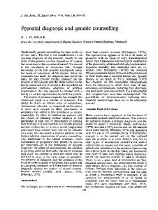
Chorionic villus sampling. Chorionic villus sampling (CVS) was introduced in our centre in 1983. Since then the number of procedures has increased each year (Brandenburg et al .. 1991a). Two different techniques of chorionic villus sampling can be used, the transcervical and the transabdominal approach. Transcervical (TC) CVS is performed between 911 weeks. transabdominal (TA) CVS is carried out after 11 weeks of pregnancy. For both methods continuous ultrasound guidance is mandatory. A diagram of both methods is given in fig. 1 and fig. 2. A minimum of 10 mg chorionic tissue is required for karyotyping. The results are known within 5-10 days.
0
0
monitor
uterus ~~!~~~~~placenta yolk sac
~
amnion ~f--chorion
syringe with catheter
Fig. I transcervical chorionic villus sampling.
21
monitor
;::;~~~~;==:yolk sac ~ placenta uterus
Fig. 2 transabdominal chorionic villus sampling.
Discrepancies between direct chorionic villi chromosome studies and fetal cells are caused by cell lines of non-fetal origin, present as a mosaic or even in all cells (Simoni eta!., 1985: Leschot et aL 1987: Sachs eta!., 1990a). Mosaicism is reported in 1-2% of samples (Leschot et aL 1989). Mosaic cell lines of trisomy 3, 7, 16 are always of non-fetal origin and 45.X cells in more than 90%. The recognition of false mosaics, which represent the great majority. is possible by examining cultures of the samples. In most cases mosaicism is confined to the cytotrophoblast (Crane and Cheung, 1988: Sachs et a! .. 1990a). Contamination by maternal cells in case of cell culture of an XX fetus should always be considered. additional amniocentesis is only rarely necessary. Fetal loss rates after CVS are higher than after amniocentesis since CVS is performed 4-6 weeks earlier in pregnancy. Older women have a high spontaneous abortion rate in early pregnancy which affects the post-procedme fetal loss rate (Jahoda et aL 1987). Various studies have been performed to estimate the procedure related fetal loss after CVS. Some studies report an excess abortion risk of CVS compared with amniocentesis between 3% and 5% (Hogge et al., 1985: Brambati et aL 1987: MRC Working Party on the evaluation of CVS, 1991) However, in the latter (a multi centre study). data from centres that only had limited experience with the procedure were included.
22
Several studies report a similar abortion rate after CVS as after amniocentesis (Crane eta! .. 1988: Green eta! .. 1988: Canadian Collaborative CVS-Amniocentesis Clinical Trial Group. 1989: Rhoads eta! .. 1989). Factors that influence the fetal loss rate after CVS include the experience of the operator (Brambati et a! .. 1987). the sampling method (Philip et a! .. 1991) and maternal age (Jahoda eta! .. 1987. 1989). A recent review of literature concluded that transcervical CVS had a 1. 7% higher total fetal loss than amniocentesis. whereas the fetal loss rate of transabdominal CVS was the same as after amniocentesis (Philip et a! .. 1991). Jahoda eta!. (1987. 1989) stressed the role of maternal age in estimating the abortion risk after CVS. Women over 36 years had a fetal loss rate up to 28 weeks of6.1% compared to 3.1% in younger women. CVS for women of advanced maternal age is not performed in the Rotterdam centre until 12 weeks of pregnancy because of a higher spontaneous abortion rate before that time (Cohen-Overbeek eta! .. 1990). It appears that the risk of obstetric complications after 20 weeks of pregnancy does not differ between a 1st trimester CVS group and a 2nd trimester amniocentesis group (MRC Working Party on the evaluation of chorion villus sampling, 1991: Rhoads eta! .. 1989). The rate of congenital abnormalities was also similar in both groups. This was confirmed by the study of Kaplan et a!. (1990). Recently. an association between CVS and limb reduction deformities was suggested. Five of the infants born to 289 women who underwent early CVS displayed a limb abnormality (Firth eta! .. 1991). CVS in these cases was performed very early in gestation (56-66 days) during the phenocritical stage of development. Monni et al.(l991) reported an incidence of transverse limb reductions of 0.07%. In our centre an overall incidence of 0.075% was established. The incidence was 0.14% in the group that was sampled before 11 weeks and 0.04% in the group that was sampled after 11 weeks (Jahoda et a! .. in press). By performing TACVS in advanced maternal age at 12 weeks a low abortion rate is achieved and the risk to the fetus is minimized. A genetic termination of pregnancy can then still be performed as an out-patient procedure.
23
2.2 Utilization of prenatal diagnosis in women of advanced maternal age in the southwest region of the Netherlands between 1984-1989. Introduction.
The uptake rate of prenatal diagnosis in women of advanced maternal age in our region was initially studied between 1978 and 1984 and reported in 1985 (Thomassen-Brepols. 1985). At that time amniocentesis was the only method of prenatal diagnosis for women of advanced maternal age. Late in 1983 the technique of chorionic villus sampling was introduced in our centre directly after its clinical applicability was demonstrated (Simoni eta!.. 1983; Ward et al .. 1983). In the Netherlands the age limit for advanced maternal age was reduced from 38 to 36 years in 1984. From 1985 onwards a substantial number of 36 and 37-year-old women visited our centre for prenatal diagnosis. Both the introduction of CVS and the reduction of the age limit might effect the utilization of prenatal diagnosis. Furthermore. during the past ten years medical information has become more accessible to the public. Increased awareness of the higher risk of chromosomal abnormalities in women of advanced maternal age and therefore a growing uptake of prenatal diagnosis was expected. In addition. an increasing number of women postpone childbearing because of their career (Kessler et al.. 1980; Holloway and Brock, 1988). This will result in a rise in the number of older women who may use prenatal diagnostic facilities. In this study the following questions were addressed: (i) has the pregnancy rate in women of advanced maternal age gone up in our region during the last six years: (ii) has the uptake rate for prenatal diagnosis in this age group increased during the same period: (iii) can a relationship be established betvveen maternal age and uptake rates.
Material and Methods.
The centre for prenatal diagnosis in Rotterdam covers the Southwest region of the Netherlands with approximately 45.000 births per year. Advanced maternal age was defined as 36 years or older at the gestational age of 20 weeks. The number of births provided by the Central Bureau for Statistics (CBS) was given per maternal year of age at the time of delivery. It follows that not all women who were 36 years at the time of delivery were entitled to have prenatal diagnosis. Of the women who were 36 years at that time only (52-20)/52 (62%) were included in the smdy. All women of advanced maternal age who lived in the Rotterdam Region and underwent prenatal diagnosis at our centre between January 1984 and January 1990 entered the study. The following data were collected: maternal age. date and type of procedure and place of residence. Uptake rate graphs were constructed from these data. Data analysis was performed by logistic regression.
24
Results.
The total number of women of 36 years and older at a gestational age of 20 weeks is given in Table l. An overall increase from 1668 women in 1984 to 2264 women in 1989 is seen. This rise occurred in all age groups with the exception of the group of women of 46 years and older. Table 2 shows the increase in percentage of deliveries in elderly women in our region during the study period. A statistically significant increase of utilization of prenatal diagnosis was established for women of 36 and 37 years of age. This resulted in a significant increase in the overall uptake rate (p 37 weeks
1545*
(97.6)
1466*
(96.0)
Perinatal death
(0.95)
15
(0.9)
14
~ 1 IUFD in twins at 31 weeks.
*including six pairs of twins.
Table 2. Relation between gestational age at TC- and TA-CVS and abortion rate for 2362 mothers aged 36 years or more and in 1079 younger mothers. Gestational age at CVS
> 12 weeks Fetal loss
< 12 weeks FeW loss
Mothers aged < 36 years
Mothers aged > 36 years
%
Type of CVS
N
%
TC TA
18
2
(2.7) (1.8)
5
(1.7)
TC TA
62 11
(6.2) (5.8)
29
(2.4)
35
N
2.3.2 Utilization. Introductory remarks.
With the introduction of CVS in early pregnancy, a first trimester abortion can be carried-out in case of an abnormal result. This has major consequences since an
early abortion is less traumatic than a mid-trimester termination. Furthermore, a first trimester abortion is to be preferred for economic as well as logistic reasons compared with a termination in the second trimester which requires hospitalization of
several days. It is, therefore, important to be informed about the utilization of CVS as well as the effect that CVS has on the uptake of prenatal diagnosis. The hypothesis that more women will make use of prenatal diagnostic facilities because an early abortion is more acceptable than mid-trimester termination will be tested in this subchapter.
Effect of chorionic villus sampling on utilization of prenatal diagnosis in women of advanced maternal age. Helen Brandenburg'-'-· Coen G .Gho'. Milena G.J.Jahoda'-'-· Theo Stijnen', Hans Bakker', Jury W.Wiadimiroff '2 • Departments of Obstetrics and Gynecology. ~Clinical Genetics and JBiostatistics. Academic Hospital-Dijkzigt. Dr.Molewaterplein 40. 3015 GD Rotterdam. The Netherlands 1
Published in Clinical Genetics 1992;41:239-242. Reprinted with permission from Munksgaard International Publishers Ltd, Copenhagen.
The effect of the introduction of chorionic v~llus sampling on the utilization rate of prenatal diagnosis in advanced maternal age was studied during the period I
January 1985- I January 1991. On the first of January 1984. the age limit for prenatal diagnosis in The Netherlands was lowered from 38 to 36 years of age, but it lasted until 1985 before women of 36 and 37 years made use of the facilities for prenatal diagnosis. The overall uptake rate during the studied period increased significant-
ly, but only because of the increased uptake rate in the group 36 and 37 years.ln the maternal age group of 42 years and older, an uptake rate as low as 15.9% was established. This was mainly determined by the relatively high percentage (73.0%) of women from ethnic minorities in this age group. The number of CVS procedures increased significantly during the study period. but the utilization rate was not influ. enced. since the number of amniocenteses decreased accordingly. An increase in
acceptability of prenatal diagnosis by women of advanced maternal age due to early testing and early termination of pregnancy could not be substantiated in the present
study.
36
Key words : advanced maternal age - chorionic villus sampling - prenatal diagnosis uptake rate. In the mid-eighties. two major changes involving prenatal diagnosis took place in The Netherlands. Firstly, in 1984 chorionic villus sampling (CVS) became available for clinical use, soon after the publications in 1983 by Ward et a!. and Simoni et a!. It was expected that since this test was performed earlier in pregnancy and results were obtained faster than after amniocentesis, prenatal diagnosis would become acceptable to more women of advanced maternal age. A higher uptake rate was therefore anticipated. Secondly. in the same year the age limit for prenatal diagnosis in women of advanced maternal age was reduced from 38 to 36 years of age. In this study the following questions were addressed: a) Has there been an increase in the uptake rate for prenatal diagnosis since 1985; b) Has a shift occurred from amniocentesis towards CVS during the same period; c) What was the uptake for prenatal diagnosis relative to advancing maternal age; d) Did a relationship exist between maternal age and the nature of the procedure (CVS or amniocentesis)?
Material and methods. In the Netherlands, approximately 35% of all confinements take place at home, under the care of a midwife or general practitioner (Kleiverda eta! .• 1990). All these deliveries involve low risk patients. All births in primigravidas over 35 years of age and in multigravidas over 40 years of age take place in a hospital. Around 80% of all women of advanced maternal age in Rotterdam including suburbs deliver in one of the hospitals in the Rotterdam region. All women of advanced maternal age who delivered in one of the six District hospitals or the University Hospital in Rotterdam between first Januaty 1985 and flrst Januaty 1991 were included in the study'. Advanced maternal age was defined as 36 years or more at twenty weeks of gestation. A total of 2045 records was analysed. The calendar year and maternal age at the time of the procedure and the nature of the procedure were evaluated retrospectively for both women who undenvent prenatal diagnosis and those who did not. Uptake rates were calculated for all women together I?er year of maternal age and per calendar year, using the ordinary X::-test and the X'-test for trend proportions (Armitage and Berry, 1987).
1 During the years 1989 and 1990 data from three District hospitals plus the University Hospital were studied. Three hospitals denied the disposal of information because of changed -laws on patients privacy.
37
Results. Fig. 1 shows the uptake rate for prenatal diagnosis between 1985-1990. There is a statistically significant increase (p
.----------- --- --- ------ ---- -- . --· . -.. -------- . --------- --
~
0
"S
~
E
" Q
0.25
.0.00
+------,---,------,-------,---.-------.--, 0
20
40
60 80 time in months
100
120
140
Fig. 1. Cumulative incidence of subsequent pregnancies following a genetic termination. 1 = after CVS: 2 = after amniocentesis
Figure 1 gives the cumulative incidence of subsequent pregnancies after genetic termination related to CVS and amniocentesis. No significant difference could be established between the two procedures. Figures 2 and 3 demonstrate that the cumulative incidence of subsequent pregnancies is significantly lower in older women (P