Postnatal Growth of Broilers in Response to In Ovo Administration of Chicken Growth Hormone H. KOCAMIS,* Y. N. YENI,† D. C. KIRKPATRICK-KELLER,* and J. KILLEFER*,1 *Division of Animal and Veterinary Sciences, West Virginia University, Morgantown, West Virginia 26506-6108, and †Bone and Joint Center, Henry Ford Hospital, Detroit, Michigan 48202 ABSTRACT The effect of in ovo administration of chicken growth hormone (cGH) on growth rate and efficiency of gain, organ, and long bone growth of 42-d-old broiler chickens was investigated. Eggs were injected once with 100 mL vehicle (0.03 M NaHCO3, 0.15 M NaCl, pH 8.3) per embryo or vehicle containing 100 ng cGH/100 mL per embryo (n = 630 eggs total) on one of the following Days: 1, 4, or 7 through 18 of embryogenesis. There was no significant difference in hatchability between control and cGH treatment groups on any given injection day. Cumulative feed conversion of all treatment groups was improved relative to their respective control groups (P < 0.05). In ovo administration of cGH on Day 15 or 16 of incubation increased body weights (P < 0.01) of female broilers. On the other hand, body weights of male broilers were significantly increased by treatment on Day 1 (P < 0.04). Breast
weights of female broilers from treatment groups Day 15 or 16 were increased (P < 0.01, P < 0.05, respectively). Liver weights of female broilers from treatment groups Day 1 and 15 were increased (P < 0.05, P < 0.01, respectively). In contrast, in ovo administration of cGH on Day 11 of incubation increased liver weights of male broilers (P < 0.03). There was no significant difference between control and treatment groups, in terms of heart or leg weights, or in Warner-Bratzler shear force of Pectoralis profundus muscle. Hydroxyproline concentration and cross-sectional area of female broiler tibias from treatment groups Day 11 or Day 16 were increased (P < 0.05), and ultimate breaking strength (stress) of tibias from the same groups was reduced (P < 0.05). In ovo administration of cGH altered growth and tissue development of broiler chickens in a time by sex dependent fashion.
(Key words: in ovo, growth hormone, feed efficiency, muscle, bone) 1999 Poultry Science 78:1219–1226
Growth hormone (GH) is required for normal development of chickens. Serum growth hormone is first detectable on Day 12 of embryonic development but remains under 10 ng/mL until Day 20 of incubation (Kikuchi et al., 1991). Growth hormone receptors and signal transduction mechanisms are developed during the latter embryonic stages, starting on about Day 12 (Kuhn et al., 1986; Berghman et al., 1989; Porter et al., 1995). The circulating concentration of GH has been positively correlated with growth rate in early posthatch chickens (Burke and Marks, 1982). Moreover, plasma GH decreases as growth rate declines with posthatch age (Vasilatos-Younken and Zarkower, 1987; VasilatosYounken et al., 1990). On the other hand, slower growing chickens display higher circulating concentrations of GH than faster growing chickens (Burke and
Marks, 1982; Stewart and Washburn, 1983; Goddard et al., 1988). When growth in chickens is retarded by hypophysectomy or when endogenous GH is low, growth can be stimulated by posthatch administration of mammalian GH (King and Scanes, 1986). Additionally, growth rate of intact birds was reduced by antisera against chicken GH (Scanes, 1987). Although there are some exceptions (Myers and Peterson, 1974; Scanes et al., 1975; Hargis et al., 1989), mammalian GH generally has been ineffective in stimulating growth rate, efficiency of gain, or organ growth in the normal growing chicken during either early or late posthatch periods (Scanes et al., 1975; Tojo et al., 1978). Furthermore, chicken (c) GH (pituitary derived or recombinant) does not substantially or consistently improve growth performance variables in the early posthatch period when administered by injection or chronic infusion (Scanes et al., 1986; Bowen
Received for publication September 24, 1998. Accepted for publication March 24, 1999. 1To whom correspondence should be addressed:
[email protected]
Abbreviation Key: c = chicken; GH = growth hormone; IGF = insulin-like growth factor; IGFBP = insulin-like growth factor binding proteins.
INTRODUCTION
1219
1220
KOCAMIS ET AL.
et al., 1987; McGuinness and Cogburn, 1988; Peebles et al., 1988; Cogburn et al., 1989). Growth hormone is the most important hormone for normal postnatal longitudinal bone growth. Exogenous GH increased long (predominantly cortical bone) bone growth in normal (Jorgensen et al., 1991; Andreassen and Oxlund, 1996), dwarf (Wright et al., 1995; Martinez et al., 1996), ovariectomized (Andreassen and Oxlund, 1996; Noland et al., 1996), and hypophysectomized rats (Schiltz et al., 1992), whereas no significant effect on cancellous bone mass was seen (Jorgensen et al., 1991). Furthermore, these results were supported in GHtransgenic mice with a very high concentration of GH (Ohlsson et al., 1996) and in old female monkeys injected with 100 mg GH/kg per d for 7 wk (Sass et al., 1997). In the chicken, there is evidence that exogenous mammalian GH stimulates tibial growth of chicks during embryonic development (Hsieh et al., 1952; Blumenthal et al., 1954). Additionally, Hargis et al. (1989) demonstrated that in ovo administration of ovine GH on Day 11 of embryonic development increased subsequent shank length of 7-wk-old male broilers. Although systemic GH administration increased circulating concentrations of other hormones that influence bone such as insulin-like growth factor (IGF)-I and the active vitamin D metabolite (Goff et al., 1990; Klindt et al., 1996), GH stimulated bone formation via direct interaction with bone tissue (Baker et al., 1992; Hedner et al., 1996). Growth hormone stimulated the proliferation of osteoblasts and the differentiated function of these cells, such as type I collagen synthesis, osteocalcin expression, and alkaline phosphatase activity in primary isolated rat (Ernst and Froesch, 1988) and chicken osteosarcoma cell culture (Slootweg et al., 1988). Increased cortical bone growth often has been associated with increased mechanical strength of whole bone as a function of both the mineral and the matrix (primarily type I collagen fibrils) constituents (Martin and Ishida, 1989; Shah et al., 1995). In ovo injections of either ovine or porcine GH increased posthatch growth in male broilers (Hargis and Pardue, 1989) and ovine GH improved feed efficiency in male broilers (Hargis et al., 1989). However, whether exogenous cGH in ovo has any influence on chicken embryonic or later posthatch growth has remained unresolved. Additionally, differences between chicken and mammalian GH sequences (Leung et al., 1984) may result in variations in bioactivity. Therefore, the objective of this work was to administer cGH in ovo and then measure its effect on growth, feed efficiency, and biomechanical properties of cortical bone development of chickens.
MATERIALS AND METHODS
Hatching Eggs Fertilized eggs (Hubbard × Hubbard) were obtained from the West Virginia University Poultry Facilities. Eggs (30 eggs per cGH treatment and 15 eggs per control group) were injected once on one of the following Days: 1, 4, or 7 through 18 of incubation (total 630 eggs).
Injection Procedure In ovo administration of 100 mL of either pituitary derived cGH or vehicle (0.03 M NaHCO3, 0.15 M NaCl, pH 8.3) per egg was performed through the blunt end of the eggs. Pituitary derived cGH was diluted in vehicle at 100 ng/100 mL and was stored at –20 C. Prior to injection, the blunt end of the egg was sterilized with 70% ethanol. A single hole was created with a dental drill bit without penetrating the chorioallantoic membrane. Both cGH (100 ng per egg) and vehicle were injected into the albumen with a 22-gauge needle. The hole was sealed with an adhesive sticker. Eggs were set in a Buckeye incubator/ hatcher2 (temperature 37 ± 0.5 C, humidity 86 to 87%).
Pen Assignment and Experimental Design At hatching, chicks were vaccinated for Marek’s Disease (s.c. in the neck; 0.2 cc per chick) and separated by feather sexing. All chicks in a treatment group were assigned to a randomly selected pen (n = 6 to 29 per pen, a total of 28 pens). During the first 3 wk, chicks were fed a starter ration that contained 21.2% CP and 3,080 kcal ME/kg. Birds had free access to feed and water. During the last 3 wk of production, birds were fed a grower ration that contained 19.5% CP and 3,124 kcal ME/kg. Chicks were housed in a thermostatically controlled building with a 24-h lighting schedule. Pen weight and mortality were recorded weekly. Average feed consumption and feed to gain ratios per pen were determined weekly. At the end of the growth period (42 d of age), treatment groups were selected for further analysis according to feed efficiency and live weight gain. Day 1, 11, 15 and 16 treatment groups (n = 5 to 6 birds per treatment group), as well as associated control groups (n = 4 to 6 per control group) for a total of 9 to 12 chicks per treatment day were selected for further processing. Whole body, breast muscle (both breasts and rib cage), liver, heart, and leg (leg and thigh) data were collected. Individual tissue or organ weights were recorded.
Texture Analysis 2Model
SS-36/C, Genesis Inc., Cambridge, MA 02139.
Pectoralis profundus was removed from each chick and cooked to 80 C internal temperature on a Farberware
IN OVO CHICKEN GROWTH HORMONE AND GROWTH IN BROILERS
Smokeless Indoor Grill.3 Right or left sides were randomly chosen. A Beckman Industrial Datalogger4 was used to monitor internal cooking temperature. To determine percentage cook yield, raw and cooked weights were recorded. After cooked muscle was cooled to room temperature, 1.27 cm-thick slices were cut perpendicular to the fiber orientation of the muscle and two or three 1.27-cm-diameter cores were removed from each strip (Kenney et al., 1996). An Instron Universal Testing Machine,5 equipped with a Warner-Bratzler shear force attachment (Model 1520.50)6 was used for shear force determination. Warner-Bratzler shear force was determined by a crosshead speed of 127 mm/min. Output was recorded via a Daytronic main frame equipped with a LVDT conditioner (Model 9130),6 and output from the main frame was sent to a computer with a DT 2805 acquisition board.7
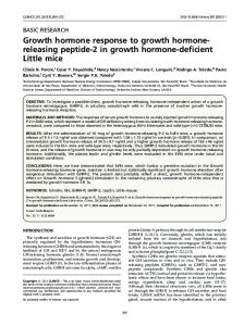
Bone Biomechanical Parameters One leg from each chicken was exarticulated at random at the hip joint and kept frozen until bone measurements were taken. Samples were thawed overnight at 2 C. The tibias were dissected free by removing all muscle tissues, periost, and fibula. Tibias were subjected to three-point bending tests until failure under displacement control (127 mm/min) using a servo Instron Universal Testing Machine. Tests were conducted at room temperature and specimens were kept moist during testing. Span length was 60 mm throughout the experiment and the load was applied at the midpoint of the shaft in antero-posterior direction. A load cell (Model 152 A.50)6 with a maximum capacity of 500 N was used to measure the applied load and the maximum value of the load was recorded as failure load (F). After failure, cortical bone thickness (t) and cross-sectional diameter were measured on two perpendicular axes (a and b; Figure 1) at the fracture site using a caliper. Assuming the bone as a uniform hollow elliptical beam, cross-sectional area (A) and moment of inertia (Ixx) were calculated as structural parameters of resistance (Martin, 1991; 1993) using the following equations (Gere and Timoshenko, 1990): A = p/4[ab – (a – 2t)(b – 2t)] Ixx = p/64 (ab3 – (a – 2t)3) Assuming that material properties are uniformly distributed along the beam, maximum moment (M) and
3Model 450N, Farberware Inc., Bronx, NY 10462. 4Model 205, Beckman Instruments, San Diego, CA 5Model TM, Instron Co., MA 02021. 6Daytronic, Miamisburg, OH 45342. 7Data Translation, Marlboro, MA 01752. 8Waring, New Hartford, CT 06057. 9Savant Instruments, Farmingdale, NY 11735. 10Corning Costar Corp., Cambridge, MA 02140. 11Shimadzu Scientific Inc., Columbia, MD 21046.
92123-1898.
1221
FIGURE 1. Cross-section of the bone model for three-point bending tests. a, b = major and minor diameters of the ellipse, t = wall thickness of the cylindrical tube.
bending strength (ultimate breaking strength) (s) were calculated as (Gere and Timoshenko, 1990) M = FL/4 s = M (b/2)/Ixx where L is the span length.
Ash Weight and Collagen Analysis Following mechanical testing, bones were cut into 0.5- to 1-cm strips using a band saw and subsequently frozen in liquid nitrogen. Frozen samples were powdered in a prechilled stainless steel Waring blender.8 Approximately 4 to 6 mg of each duplicate bone powder sample was transferred to weighed dry crucibles. Dry weight was determined following heating to constant weight at 110 C for 2 h. Ash weight was determined following overnight heating at 600 C in a muffled furnace and expressed as percentage of dry bone weight. Hydroxyproline concentration was measured by the method of Monnier et al., (1986) as an indicator of collagen content. Briefly, 40 mg of duplicate powdered bone sample was delipidated overnight in a chloroform:methanol (2:1) solution. Samples were then rehydrated in 50% methanol and hydrolyzed in 6 N HCl at 110 C for 18 h. Prior to capping for heating, N2 was used to flush all tubes. Following hydrolysis, the samples were evaporated in a Speed Vac centrifuge vacuum drier.9 Subsequently, samples were reconstituted in 250 mL distilled H2O and filtered using a Costar SpinX centrifuge tube filter.10 Samples were analyzed for hydroxyproline concentrations at 564 nm wavelength using a Shimadzu spectrophotometer (Model UV-1201).11
Statistical Analysis Analysis of variance was performed by the GLM procedures of SAS (SAS Institute, 1989) as a three-factor factorial arrangement of treatments in a completely random design. A previous experiment indicated that the variance among chicks within a pen was a valid estimate of experimental error. The Least Significant Difference test was used to compare the means. The CATMOD test was used to compare hatchability. Statements of significance were based on P < 0.05 unless otherwise noted.
1222
KOCAMIS ET AL.
TABLE 1. Effects of in ovo chicken growth hormone (cGH) administration on broiler hatchability (n = 30 for each treatment group, n = 15 for each control group) Injection day
Treatment1
1 4 7 8 9 10 11 12 13 14 15 16 17 18
68 69.9 83.3 92.6 88.9 86.1 85.7 88 86.7 93.1 98 96.5 86.7 89.3
Control2 (%)
± ± ± ± ± ± ± ± ± ± ± ± ± ±
9.3* 9.5* 5.7 5 6 6.5 6.6 7.2 6.3 4.7 2.6 3.4 6.3 5.8
50 66 90.2 84.6 85.7 84.6 92.3 96 86.7 92.8 92.8 86.7 93 93.3
± ± ± ± ± ± ± ± ± ± ± ± ± ±
14.4* 8.1* 8.9 10 9.3 10 7.4 2.1 8.8 6.9 6.9 8.8 2.6 6.4
1Chicken
growth hormone. 2Vehicle-injected control groups. *P < 0.05 compared to other injection days.
RESULTS
Bone Analysis In ovo administration of cGH on Day 11 or Day 16 increased hydroxyproline concentration and crosssectional area of female broiler tibias (P < 0.05, Table 3). This increase was associated with a reduction in the ultimate breaking strength (stress) of female broiler tibias from the same groups (P < 0.05; Table 3); however, there was no significant difference in male broiler tibias for these measurements (Table 3). In ovo administration of cGH did not alter percentage ash, tibia density, weight, length, cortical bone thickness and breaking force of tibias on any given injection day (Table 3).
DISCUSSION A single in ovo administration of cGH increased body weight and tissue development of 42-d-old broilers, although the magnitude of the responses were different between male and female broilers. Feed conversion ratios of all in ovo cGH injected mixed-sex broilers were improved significantly compared to their respective control groups. Although the specific mechanism of GH
There were no significant differences in hatchability between control and cGH treatment groups on any injection day (Table 1). However, injection of either cGH or vehicle on Day 1 or 4 of incubation compared to other injection days reduced hatchability (P < 0.05; Table 1).
Measurements of Growth Performance In ovo administration of cGH on Day 15 or 16 of incubation increased body weight of female broilers (P < 0.01; Figure 2), whereas body weights of male broilers were increased by Day 1 treatment (P < 0.04; Figure 2). Pooled mean cumulative feed conversion ratio of all treatment groups at the end of the treatment period was improved compared to their respective control groups (for cGH treated groups: 1.81 ± 0.02, for control groups: 1.9 ± 0.03; P < 0.05). As there were no significant differences either among the treatment groups or among the control groups in terms of feed conversion ratios, values were represented as pooled means. Breast weights of female broilers from treatment groups Day 15 or 16 were increased (P < 0.01, P < 0.05 respectively; Table 2). On the other hand, breast weights of male broilers were not altered significantly by in ovo administration of cGH on any given injection day (Table 2). Liver weights of female broilers from treatment Day 15 were increased (P < 0.01; Figure 2). In contrast, in ovo administration of cGH on Day 11 of incubation increased liver weights of male broilers (P < 0.03; Figure 2). There were no significant differences between control and treatment groups on any single injection day in terms of heart and leg weights other than sex effects (Table 2). Warner-Bratzler shear force ratios on P. profundus muscle between treatment and control groups were not different on any given injection day.
FIGURE 2. Effects of in ovo chicken growth hormone (cGH) administration on live (panel A) and liver weights (panel B) of 42-d-old broilers. The bars represent the means of males (n = 4 to 6) and females (n = 4 to 6) in each treatment and control (Cont.) group. Means with asterisks are significantly different from their respective control groups (**P < 0.01, *P < 0.05). Vertical lines are SEM.
1223
IN OVO CHICKEN GROWTH HORMONE AND GROWTH IN BROILERS TABLE 2. Effects of in ovo chicken growth hormone (cGH) administration on heart, leg, and breast weights of 42-d-old broilers (x ± SEM, n = 5 to 6 for each treatment and control group) Heart weight Injection day Male
Leg weight
Female
Male
Breast weight
Female
Male
Female
(g) Day 1 cGH Control Day 11 cGH Control Day 15 cGH Control Day 16 cGH Control
11.2 ± 1.2 10.8 ± 1.4
9.1 ± 1.2 8.1 ± 1
143.9 ± 18.9 136.7 ± 26.7
130.3 ± 18.9 114 ± 15.4
502 433
± 34.4 ± 48.7
453 ± 34.4 421.3 ± 28.1
10.1 ± 1.2 11.3 ± 0.8
9.2 ± 0.8 8.9 ± 1.2
166.2 ± 18.9 145.1 ± 13.3
122.1 ± 13.3 133.1 ± 18.9
439.3 ± 34.4 470.8 ± 24.4
403.7 ± 24.3 373.6 ± 34.4
11.4 ± 1 13 ± 1
8.7 ± 1 9.5 ± 1
162.6 ± 15.4 142.3 ± 15.4
128.2 ± 15.4 107.5 ± 15.4
492.8 ± 28.1 468.8 ± 28.1
441.3 ± 28.1** 370.3 ± 28.1
10.3 ± 1 11.6 ± 1
7.7 ± 1 6.8 ± 1
152.4 ± 15.4 117.6 ± 15.4
113.5 ± 15.4 100.2 ± 15.4
466 ± 28.1 451.8 ± 28.1
388.9 ± 28.1* 332.6 ± 18.6
**P < 0.01, *P < 0.05, compared to their respective control groups within the same sex.
Insulin-like growth factor-I is required for normal growth in chickens (Proudman et al., 1994). In a previous study, we demonstrated that in ovo administration of recombinant human IGF-I (100 ng per embryo) altered body weights and tissue development of 42-d-old broilers (Kocamis et al., 1998). Unlike mammalian cell culture, avian growth plate chondrocytes from birds of different ages do not respond to the addition of GH to the culture medium in terms of IGF-I concentrations (Rosselot et al., 1994). Additionally, the effect of GH on IGF-I is not consistent although exogenous studies have demonstrated GH to either increase (Leung et al., 1986; Vasilatos-Younken et al., 1990) or be without effect on IGF-I in the circulation (Cogburn et al., 1989; Rosselot et al., 1995) in avian species. During chicken embryonic development, IGF-I gene expression is GH-independent (Kikuchi et al., 1991; Tanaka et al., 1996). However, IGF-I gene expression in bone tissue is not GH-dependent
action resulting in these various responses in male and female broilers is unknown, partial explanations for the observed findings could be due to sexual-dimorphism, altered thyroid function, and IGF-I and insulin-like growth factor binding proteins (IGFBP) function and expression. Thyroid hormone metabolism has been linked to the regulation of GH action and the growth process of chickens (Singh et al., 1968; Scanes, 1987). The thyroid is functional early in embryonic development and becomes pituitary dependent at Day 10 to 12 of embryogenesis (Thommes and Jameson, 1980). Moreover, GH regulates thyroxine secretion in growing chickens and regulates the circulating concentration of triiodothyronine in adult chickens and embryos by inducing 5′-monodeiodinase activity inside the liver cell (Kuhn et al., 1986; Darras et al., 1990). Thus, it is possible that exogenous GH in this study had the effect of modifying thyroid hormone secretion.
TABLE 3. Effects of in ovo chicken growth hormone (cGH) on biomechanical properties of 42-d-old broiler tibias (n = 5 to 6 for each treatment and control group)1
Injection day
Ash M
F (%)
Day 1 cGH Control Day 11 cGH Control Day 15 cGH Control Day 16 cGH Control 1M
Tibia length
OH-proline M
F
(mg/mg)
M
F (mm)
Density M
F
Weight M
(g/cm3)
F
Cortical thickness M
(g)
F
Breaking force M
(mm)
F (kg)
Stress M
F
Cross-section area M
F (mm2)
(Mpa)
62 59
62 59
9 9
7 10
105 106
106 106
1.3 1.2
1.3 1.2
18 18
17 15
1.4 1.5
1.2 1.2
40 36
35 29
231 189
166 203
22 23
19 17
62 64
62 59
8 8
11* 7
105 107
103 103
1.2 1.2
1.3 1.2
21 20
18 16
1.5 1.4
1.4 1.2
50 37
42 49
172 146
178* 258
29 26
27* 19
61 64
62 63
11 10
9 10
107 102
103 101
1.2 1.2
1.3 1.2
20 20
16 15
1.2 1.7
1.4 1.4
43 40
37 34
157 152
169 176
23 29
24 23
62 62
65 65
8 9
10* 7
106 102
102 97
1.1 1.2
1.2 1.3
21 22
15 12
1.6 1.8
1.4 1.3
37 44
39 30
140 136
145* 296
29 33
25* 16
= male; F = female; OH-proline = hydroxyproline; *P < 0.05 compared to respective control group within same sex.
1224
KOCAMIS ET AL.
either before or after hatching (Tanaka et al., 1996). For this reason, the influence of in ovo administration of IGFI during embryonic development on postnatal long bone growth is under investigation in our laboratory. There are six distinct classes of IGFBP found in extracellular fluid and serum of mammals, termed IGFBP-1 through IGFBP-6 (Hill, 1996). The bioactivity of IGF, particularly in bone tissue, is modulated by these IGFBP. For example, mRNA for IGFBP-5, which is a stimulatory IGFBP for osteoblast proliferation (Bautista et al., 1991), was increased twofold after GH treatment of primary rat osteoblasts (McCarthy et al., 1994). However, only IGFBP-2 (Schoen et al., 1995) and IGFBP-5 (Allander et al., 1995) have been isolated in chicken serum. It is not known whether these IGFBP in chicken are similar in function to those in mammals. It has been suggested that GH may regulate circulating concentrations of chicken IGFBP (Scanes, 1997). Therefore, whether increased concentrations of circulating GH during embryonic development of the chicken has any effect on specific type of IGFBP could be of interest. There is a sexual dimorphism between growth and GH concentrations in chickens. Some evidence implicates testosterone in the physiological control of GH secretion (Harvey et al., 1979). Also, during the early (0 to 3 wk) posthatch period, male broilers grow faster, have larger body size, and display higher circulating GH concentrations than females (Leung et al., 1987; Vasilatos-Younken et al., 1990). However, there is no sex difference in hepatic binding of GH for broiler chickens at Day 1 of age or during the late posthatch period (Leung et al., 1987). These studies could, to some extent, explain the varying response of in ovo injection of GH during different days of incubation between the male and female. In ovo administration of cGH on Day 11 or 16 of incubation resulted in a greater response in the biomechanical properties of the tibias of female broilers. For example, cross-sectional areas and hydroxyproline concentrations of these bones were significantly increased. However, their ultimate breaking strength ratios were reduced, indicating that increased bone mass was not associated with strength. There was no significant effect on percentage ash ratio, tibia length, weight, density, cortical bone thickness, or breaking force ratios found on any given day of injection. These results could be due, in part, to the changes in moisture content, bone cell number, collagen cross-linking, circulating levels of other hormones such as the active vitamin D metabolite, and bone turnover. Chorioallantoic injections of GH (5 mg per embryo) on Day 13 of incubation and continued every other day for 5 d increased hatch weights and tibial growth of chickens (Hsieh et al., 1952). In that study, female chicks were more sensitive to GH than were their male counterparts. In contrast, Hargis et al. (1989) showed that in ovo administration of ovine GH (250 mg per embryo) on Day 11 of incubation resulted in a greater
response in male broilers. In our study, we did not find a significant effect of in ovo cGH on this day for any measures of growth other than increased weight of the male liver. Additionally, there was no significant difference between treated and control groups in terms of hatch weight of broilers. As chicken GH (100 ng per embryo) was administered to embryos from Hubbard × Hubbard broiler strains in this study, these variations in response could be due to the differences in the type of GH, dosage, and genetic line of the broilers. Warner-Bratzler shear force ratios between in ovoinjected treatment and control groups were not different on any given injection day in this study. Likewise, we reported earlier that in ovo administration of recombinant human IGF-I did not alter shear force ratios in either male or female broilers (Kocamis et al., 1998). Therefore, in ovo administration of cGH or recombinant human IGF-I does not detract from product quality. In conclusion, in ovo administration of cGH on Day 1 (males) and Day 15 or 16 (females) of embryonic development increased body weights and altered tissue development of 42-d-old broilers. Increased body weights of female broilers from treatment groups Day 15 or Day 16 were associated with increased breast weights. Moreover, feed conversion ratios of all treatment groups were increased significantly compared to their respective control groups. In ovo administration of cGH on Day 11 or 16 of incubation increased hydroxyproline concentration and cross-sectional area of the tibia from female broilers. There was no significant difference between treatment and control groups in terms of Warner-Bratzler shear force on any given injection day. Therefore, in ovo administration of cGH on specific days may provide a means of increasing broiler growth rate and efficiency without altering quality of the products.
ACKNOWLEDGMENTS The authors wish to thank Edwin Townsend for his assistance in analysis of these data. Pituitary derived cGH was kindly provided by A. F. Parlow, National Hormone and Pituitary Program, CA. This investigation was supported by the Hatch Funds of the West Virginia Agricultural Experiment Station, and is published with the approval of the Director as Scientific Paper No. 2686.
REFERENCES Allander, S. V., E. Ehrenborg, H. Luthman, and D. R. Powell, 1995. Conservation of IGFBP structure during evolution: Cloning of chicken insulin-like growth factor binding protein-5. Prog. Growth Factor. Res. 6:159–165. Andreassen, T. T., and H. Oxlund, 1996. Additive anabolic effects of growth hormone and parathyroid hormone on vertebral body cortical and cancellous bone in old ovariectomized rats. J. Bone Miner. Res. (Suppl. 1)11: S457–S463.
IN OVO CHICKEN GROWTH HORMONE AND GROWTH IN BROILERS Baker, A. R., P. G. Hollingshead, S. Pitts-Meek, S. Hansen, R. Taylor, and T. A. Stewart, 1992. Osteoblast-specific expression growth hormone in transgenic mice. Mol. Cell. Biol. 12:5541–5547. Bautista, C. M., D. J. Baylink, and S. Mohan, 1991. Isolation of a novel insulin-like growth factor binding protein from human bone: a potential candidate for fixing IGF-II in human bone. Biochem. Biophys. Res. Commun. 176: 756–763. Berghman, L. R., V. N. Darras, L. M. Huybrechts, E. Decuypere, S. Vandesande, and E. R. Kuhn, 1989. Evidence for chicken growth hormone as the only hypophysial factor responsible for the stimulation of hepatic 5-monodeionidation activity in the chick embryo. Reprod. Nutr. Dev. 29:197–202. Blumenthal, H. T., K. M. Hsieh, and T. Y. Wang, 1954. The effect of hypophyseal growth hormone on the tibia of the developing chick embryo. Am. J. Pathol. 30:771–787. Bowen, S. J., L. M. Huybrechts, J. A. Marsh, and C. G. Scanes, 1987. Influences of triiodothyronine and growth hormone on growth of dwarf and normal chickens: Interaction of hormone and genotype. Comp. Biochem. Physiol. 86 (A): 137–142. Burke, W. H., and H. L. Marks, 1982. Growth hormone and prolactin levels in nonselected and selected lines of chickens from hatch to eight weeks of age. Growth 46: 283–295. Cogburn, L. A., S. S. Liou, A. L. Rand, and J. P. McMurtry, 1989. Growth, metabolic and endocrine responses of broiler cockerels given a daily subcutaneous injection of natural or biosynthetic chicken growth hormone. J. Nutr. 119:1213–1222. Darras, V. M., L. M. Huybrechts, L. Berghman, E. R. Kuhn, and E. Decuypere, 1990. Ontogeny of the effect of purified chicken growth hormone on the liver 5′monodeiodination activity in the chicken: Reversal of the activity after hatching. Gen. Comp. Endocrinol. 77:212–220. Ernst, M., and E. R. Froesch, 1988. Growth hormone dependent stimulation of osteoblast-like cells in serum-free cultures via local synthesis of IGF-I. Biochem. Biophys. Res. Commun. 151:142–147. Gere, J. M., and S. P. Timoshenko, 1990. Mechanics of Materials, PWS-KENT Publishing Co., Boston, MA. Goddard, C., R. S. Wilkie, and I. C. Dunn, 1988. The relationship between insulin-like growth factor-I, growth hormone, thyroid hormones and insulin in chickens selected for growth. Dom. Anim. Endocrinol. 5:165–176. Goff, J. P., T. J. Caperna, and N. C. Steel, 1990. Effects of growth hormone administration on vitamin D metabolism and vitamin D receptors in the pigs. Dom. Anim. Endocrinol. 7:425–433. Hargis, P. S., S. L. Pardue, A. M. Lee, and G. W. Sandel, 1989. In ovo growth hormone alters growth and adipose tissue development of chickens. Growth Dev. Aging 53:93–99. Hargis, P. S., and S. L. Pardue, 1989. In ovo mammalian somatotropins and growth in chickens. Fed. Proc. 3:3731. (Abstr.) Harvey, S., C. G. Scanes, and P.M.M. Gadden, 1979. Plasma growth hormone levels in normal and testosterone implanted growing chickens. Poultry Sci. 58:745–748. Hedner, E., A. Linde, and A. Nilsson, 1996. Systemically and locally administered growth hormone stimulates bone healing in combination with osteopromotive membranes:
1225
an experimental study in rats. J. Bone Miner. Res. 11: 1952–1960. Hill, D. J., 1996. Relationships of insulin-like growth factors and their binding proteins to embryonic development. J. Anim. Sci. 74 (Suppl. 2):85–93. Hsieh, K. M., T. Y. Wang, and H. T. Blumental, 1952. The diabetogenic and growth promoting activities of growth hormone (somatotropin) in the developing chick embryo. Endocrinology 51:298–301. Jorgensen, P. H., B. Bak, and T. T. Andreassen, 1991. Mechanical properties and biochemical composition of rat cortical femur and tibia after long term treatment with biosynthetic human growth hormone. Bone 12:353–359. Kenney, P. B., S. D. Slider, R. R. Nayak, and J. W. Massey, 1996. Stunning method and time held in a transport coop affect pH decline and muscle quality in broilers. Poultry Sci. 75 (Suppl. 1):220. (Abstr.) Kikuchi, K., F. C. Buonomo, Y. Kajimato, and P. Rotwein, 1991. Expression of insulin-like growth factor-I during chicken development. Endocrinology 128:1323–1328. King, D. B., and C. G. Scanes, 1986. Effect of mammalian growth hormone and prolactin on the growth of hypophysectomized chickens. Proc. Soc. Exp. Biol. Med. 182:201–207. Klindt, J., F. C. Buonomo, T. Wise, and J. T. Yen, 1996. Endocrine and metabolite responses to porcine growth hormone administrated by sustained release important for different lengths of time in male pigs. Endocrinology 137: 3689–3695. Kocamis, H., D. C. Kirkpatrick-Keller, H. Klandorf, and J. Killefer, 1998. In ovo administration of recombinant human insulin-like growth factor-I alters postnatal growth and development of the broiler chicken. Poultry Sci. 77: 1913–1919. Kuhn, E. R., L. M. Huybrechts, E. Decuypere, and P. Merat, 1986. Endocrinological effects of the sex-linked dwarf gene: III prolactin and growth hormone fail to increase the liver T4 5-monodeiodinase activity in the sex-linked dwarf embryo. Pages 965–969 in: Proceeding of the 7th Europian Poultry Conference, Paris, France. Leung, F. C., J. E. Taylor, S. L. Steelman, S. D. Bennett, J. A. Rodkey, R. A. Long, R. Serio, R. M. Weppelman, and G. Olson, 1984. Purification and properties of chicken growth hormone and development of a homologous radioimmunoassay. Gen. Comp. Endocrinol. 56:389–397. Leung, F. C., J. E. Taylor, S. Wien, and A. Van der Iderstein, 1986. Purified chicken growth hormone (GH) and human pancreatic GH-releasing hormone increase body weight gain in chickens. Endocrinology 118:1961–1965. Leung, F. C., W. J. Styles, C. I. Rosenblum, M. S. Lilburn, and J. A. Marsh, 1987. Diminished hepatic growth hormone receptor binding in sex-linked dwarf broiler and leghorn chickens. Proc. Soc. Exp. Biol. Med. 184:234–238. Martin, R. B., and J. Ishida, 1989. The relative effects of collagen fiber orientation, porosity, density, and mineralization on bone strength. J. Biomechanics 22:419–426. Martin, R. B., 1991. Determinants of the mechanical properties of bones. J. Biomechanics 24 (Suppl. 1):79–88. Martin, R. B., 1993. Aging and strength of bones as a structural material. Calcif. Tissue Int. 53 (Suppl. 1):S34–S40. Martinez, D. A., M. W. Orth, K. E. Carr, R. Vanderby, and A. C. Vailas, 1996. Cortical bone growth and maturational changes in dwarf rats induced by human growth hormone. Am. J. Physiol. 270:E51–E59.
1226
KOCAMIS ET AL.
McCarthy, T. L., S. Casinghino, M. Centrella, and E. Canalis, 1994. Complex pattern of insulin-like growth factor binding protein expression in primary rat osteoblast enriched cultures: regulation by prostaglandin E2, growth hormone, and the insulin-like growth factors. J. Cell. Physiol. 160:163–175. McGinness, M. C., and L. A. Cogburn, 1988. Growth and hormonal responses of young broiler cockerels to daily injection of chicken growth hormone. Poultry Sci. 67 (Suppl. 1):117. (Abstr.) Monnier, V. M., V. Vishwanath, K. E. Frank, C. A. Elmets, P. Dauchot, and R. R. Kohn, 1986. Relation between complications of type I diabetes mellitus and collagen-linked fluorescence. N. Eng. J. Med. 314:403–408. Myers, W. R., and R. A. Peterson, 1974. Responses of six and ten week-old broilers to a tryptic digest of bovine growth hormone. Poultry Sci. 53:508–514. Noland, K., P. Orhii, J. Rutstein, and D. Kalu, 1996. Effect of growth hormone therapy on cortical bone in aged ovariectomized rats. J. Bone Miner. Res. 11 (Suppl. 1):S458. Ohlsson, C., J. Tornell, J. Sandstedt, and T. T. Andreassen, 1996. Effect of increased growth hormone on mechanical strength of cortical bone in growth hormone-transgenic mice. J. Bone Miner. Res. 11 (Suppl. 1):S458. Peebles, E. D., W. D. Burke, and H. L. Marks, 1988. Effects of recombinant chicken growth hormone in random bred meat-type chickens. Growth, Dev. Aging. 52:133–158. Porter, T. E., G. S. Couger, and B. Morpurgo, 1995. Evidence that somatotroph differentiation during chicken embryonic development is stimulated by a blood-borne signal. Endocrinology 136:3721–3728. Proudman, J. A., M. C. McGuinness, K. A. Krishman, and L. A. Cogburn, 1994. Endocrine and metabolic responses of intact and hypophysectomized turkey poults given daily injection of chicken growth hormone. Comp. Biochem. Physiol. 109 (C):47–56. Rosselot, G., R. Vasilatos-Younken, and R. M. Leach, 1994. Effect of growth hormone, insulin-like growth factor, basic fibroblast growth factor, and transforming growth factor b on cell proliferation and proteoglycan synthesis by avian postembryonic growth plate chondrocytes. J. Bone Miner. Res. 9:431–439. Rosselot, G., J. P. McMurtry, R. Vasilatos-Younken, and S. Czerwinski, 1995. Effect of exogenous chicken growth hormone administration on insulin-like growth factor-I gene expression in domestic fowl. Mol. Cell. Endocrinol. 114:157–166. SAS Institute, 1989. SAS/STAT User’s Guide. Version 6. 4th Edition. Vol. 2. SAS Institute Inc., Cary, NC. Sass, D. A., C. P. Jerome, A. R. Bowman, A. Bennett-Cain, T. A. Ginn, D. LeRoith, and S. Epstein, 1997. Short-term effect of growth hormone and IGF-I on cancellous bone formation in Rhesus Macaque monkeys. J. Clin. Endocrinol. Metab. 82:1202–1209. Scanes, C. G., S. B. Telfer, A. F. Hackett, R. Nightingale, and B.A.K. Sharifuddin, 1975. Effects of growth hormone on tissue metabolism in broiler chick. Br. Poult. Sci. 16: 405–408. Scanes, C. G., D. R. Duyka, T. J. Lauterio, S. T. Bowen, L. M. Huybrechts, L. M. Bacon, and D. B. King, 1986. Effect of
chicken growth hormone, triiodothyronine and hypophysectomy in growing domestic fowl. Growth 50: 12–31. Scanes, C. G., 1987. The physiology of growth, growth hormone and growth factors in poultry. CRC Critical Rev. Poult. Biol. 1:51–105. Scanes, C. G., 1997. Ontogeny of the hypothalamic-pituitary (growth hormone)-insulin-like growth factor-I axis in birds. Am. Zool. 37:524–535. Schiltz, P. M., T. Ohta, D. Glass, S. Mohan, and D. J. Baylink, 1992. Growth hormone stimulates cortical bone formation in immature hypophysectomized rats. Endocrine Res. 18: 19–30. Schoen, T. J., K. Mazuruk, R. J. Waldbillig, J. Potts, D. C. Beebe, G. J. Chader, and I. R. Rodriguez, 1995. Cloning and characterization of a chick embryo cDNA and gene for IGF-binding protein-2. J. Mol. Endocrinol. 15:49–59. Shah, K. M., C. H. Goh, R. Karunanithy, L. S. Low, S. Das De, and K. Bose, 1995. Effect of decalcification on bone mineral content and bending strength of feline femur. Calcif. Tissue Int. 56:78–82. Singh, A., E. P. Reineke, and R. K. Ringer, 1968. Influence of thyroid status of the chick on growth and metabolism with observations on several parameters of thyroid function. Poultry Sci. 47:212–219. Slootweg M. C., S. C. Van Buul-Offers, M. P. Herrmann-Erle, and S. A. Duursma, 1988. Direct stimulatory effect of growth hormone on DNA synthesis of fetal chicken osteoblasts in culture. Acta. Endocrinol. (Copenh.) 118: 294–300. Stewart, P. A., and K. W. Washburn, 1983. Variation in growth hormone, T3 and lipogenic enzyme activity in broiler strains differing in growth and fatness. Growth 57: 411–425. Tanaka, T, Y. Hayashida, T. Sagaguchi, T. Ohkubo, M. Wakita, S. Hoshino, and K. Nakashima, 1996. Growth hormone independent expression of IGF-I messenger RNA in extrahepatic tissues of chicken. Endocrinology 137:30–34. Thommes, R. C., and K. M. Jameson, 1980. Hypothalamoadenohypophyseal-thyroid interrelationships in the chick embryo. III. Total T4 levels in the plasma of decapitated chick embryos adenohypophyseal transplants. Gen. Comp. Endocrinol. 42:267–269. Tojo, H., H. Noiri, and K. Ogawa, 1978. Fractionation of chicken pituitary for growth hormone. Jpn. J. Zootech. Sci. 49:75–77. Vasilatos-Younken, R., K. S. Gray, W. L. Bacon, K. E. Nestor, D. W. Long, and R. L. Rosenberger, 1990. Ontogeny of growth hormone binding in the domestic turkey: Evidence of sexual dimorphism and developmental changes in relationship to plasma GH. J. Endocrinol. 126:131–139. Vasilatos-Younken, R., and P. G. Zarkower, 1987. Age related changes in plasma immunoreactive growth hormone secretory patterns in broiler pullets. Growth 51:171–180. Wright, N. M., J. Renault, B. Hollis, N. H. Bell, and L. L. Key, 1995. Effect of growth hormone on bone: bone mineral density, trabecular bone volume, and alkaline phosphatase improve or are restored in the dwarf rat treated with growth hormone. J. Bone Miner. Res. 10:127–131.