REVIEW ARTICLE
Platelet-rich Plasma and Plantar Fasciitis Raymond R. Monto, MD
Abstract: Plantar fasciitis is the most common cause of heel pain and can prove difficult to treat in its most chronic and severe forms. Advanced cases of plantar fasciitis are often associated with ankle stiffness, heel spurs, and other conditions and can lead to extensive physical disability and financial loss. Most available traditional treatments, including orthoses, nonsteroidal anti-inflammatory drugs, and steroid injections have a paucity of supportive clinical evidence. More invasive treatments, ranging from corticosteroid and botulinum-A toxin injections to shockwave therapy and plantar fasciotomy, have demonstrated varying clinical success in severe cases but carry the potential for serious complication and permanent disability. Platelet-rich plasma has recently been demonstrated to be helpful in managing chronic severe tendinopathies when other techniques have failed. This review examines the pathophysiology, diagnostic options, nonoperative treatment modalities, and surgical options currently used for plantar fasciitis. It also focuses on the clinical rationale and available evidence for using autologous platelet-rich plasma to treat severe refractory chronic plantar fasciitis. Key Words: platelet-rich plasma, PRP, plantar fasciitis, plantar fasciopathy, heel spur syndrome
(Sports Med Arthrosc Rev 2013;21:220–224)
P
lantar fasciitis is the most common cause of foot pain, affecting 10% of the US population and generating 1 to 2 million office visits per year.1,2 The peak incidence of heel pain occurs between ages 40 and 60 and it is a particularly common problem in older athletes, runners, military recruits, and trades people.2,3 Individual risk factors include obesity, loss of ankle dorsiflexion, and extensive workrelated weight bearing.1 Although 90% of cases resolve with conservative treatment within a few weeks, there is no general consensus regarding treatment paradigms, and severe cases of plantar fasciitis can be disabling. It has been estimated that the annual economic burden of the disease ranges between 192 and 376 million dollars.4 Wood first described the syndrome in 1812, errantly believing it to be a complication of tuberculosis.5 As clinical recognition of the problem improved over time, the condition became associated with other chronic inflammatory syndromes and was given many names including plantar fasciosis, jogger’s heel, plantar fasciitis, heel (calcaneal) spur syndrome, and plantar fasciopathy.6–10 Platelet-rich plasma (PRP) is a bioactive component of whole blood with platelet concentrations elevated above baseline and containing high levels of various growth factors.11 It is postulated that when transplanted into injured tissue, these platelet nests act as rally points for the From the Nantucket Cottage Hospital Nantucket, Nantucket, MA. Disclosure: R.R.M. is on the speaker’s bureau for Exactech Inc. (Gainesville, FL). Reprints: Raymond R. Monto, MD, Nantucket Cottage Hospital, 57 Prospect Street, Nantucket, MA 02554. Copyright r 2013 by Lippincott Williams & Wilkins
220 | www.sportsmedarthro.com
modulation of collagen synthesis and tissue healing by releasing cytokines and chemoattractants.11,12 Early pain relief after PRP transplantation may be due to an antiinflammatory effect resulting from the inhibition of cyclooxygenase-2 enzymes by the cytokines provided by the platelets while later benefits may be due to local cellular proliferation, neoangiogenesis, and increased type 1 collagen production.11–15 PRP has been shown to be helpful in treating chronic severe tendinopathies including Achilles tendinosis and has proven more effective and reliable than traditional cortisone injections in the treatment of lateral epicondylitis.12,16,17 This review will examine the pathophysiology, presentation, diagnosis, and treatment of plantar fasciitis, while also identifying the current and future role of PRP in its management.
ANATOMY The plantar fascia is a durable, longitudinal bundle of thick fibrous bands that originate off the medial tubercle of the calcaneus. These bundles condense to form the arch of the foot before proceeding across the transverse bands of the deep transverse metatarsal ligaments to insert along the proximal phalanges. It functions using a windlass mechanism to support and cushion the foot during gait while efficiently converting potential energy to kinetic energy during toe-off.18,19
PATHOPHYSIOLOGY Once thought to be primarily inflammatory in nature, the source of plantar fasciitis is now believed to be multifactorial and there is a distinct absence of inflammatory tissue in its chronic phase.20–23 Driven by high calcaneal pressures and the combination of repetitive opposing traction by the Achilles tendon and the forefoot windlass effect, microscopic tears occur in the central bundle of the plantar fascia.21,22 Cumulative cellular damage is exacerbated by a failed healing pattern that leads to chaotic vascularity with zones of hyperplasia and hypoplasia.23 Collagen matrix production collapses while cellular protein and enzyme production falter. This results in a disruption of the normal collagen repair cycle and a continuum of cellular damage similar to that seen in Achilles tendinosis and lateral epicondylitis.12,17 Recent research also suggests that there may be a central nervous system role in chronic tendinopathies with programmed cell death (apoptosis) and tissue breakdown.20
HISTORY AND PHYSICAL EXAMINATION A thorough clinical history and physical examination are critical in making a correct diagnosis of plantar fasciitis. The condition typically presents with sharp morning heel pain and “first-step” pain that improves with use during the day and often worsens with heavy use.19 Although bilateral heel pain is seen in 30% of plantar fasciitis cases, other
Sports Med Arthrosc Rev
�
Volume 21, Number 4, December 2013
Sports Med Arthrosc Rev
�
Volume 21, Number 4, December 2013
symptoms such as numbness, tingling, night pain, swelling, and radiating pain are rare.24 The symptoms of plantar fasciitis often begin after exercise or activity modification, shoe change, weight gain, or minor foot injury.25 The physical examination is marked by localized tenderness at the medial tubercle of the calcaneus, loss of ankle dorsiflexion due to gastrocnemius muscle or Achilles tendon tightness, and in more severe cases, increased heel discomfort with passive toe dorsiflexion (the windlass sign).18 Findings of edema, induration, redness, diffuse tenderness, diminished sensation, motor weakness, or vascular compromise, would suggest an alternative diagnosis. A careful and well-documented physical examination is important because the differential diagnosis of heel pain is extensive and includes calcaneal stress fracture, compressive neuropathy, vasculitis, tendon rupture, radiculopathy, tendon failure, and neoplasm.
IMAGING AND TESTS The role and clinical impact of imaging in plantar fasciitis is controversial. Although of limited benefit in acute cases of plantar fasciitis, plain radiographs of the hindfoot may confirm calcifications or spur formation in chronic cases (Fig. 1).26 The clinical importance of calcaneal spurs is uncertain, but it is clear that they are not the cause of plantar fasciitis and an adaptive, physiologic response to chronic traction and irritation in the injury zone. Technetium-99 bone scan imaging typically demonstrates increased uptake along the medial aspect of the calcaneus in plantar fasciitis while displaying more intense and diffuse calcaneal uptake in stress fractures.27 A useful alternative to the low sensitivity of radiographs and equivocal specificity of bone scintigraphy is magnetic resonance imaging (MRI). Highly sensitive and specific MRI of the hindfoot is useful in confirming the diagnosis of plantar fasciitis and excluding other possible causes of heel pain. Typical findings of plantar fasciitis on MRI include fascial thickening, partial fascial tears, and reactive edema (Fig. 2). A more cost-effective and engaging technique for imaging plantar fasciitis is ultrasonography.28 Although operator-dependent, the technology allows the rapid, re-
FIGURE 1. Stippled calcifications in the plantar fascia seen on a lateral radiograph. r
2013 Lippincott Williams & Wilkins
Platelet-rich Plasma and Plantar Fasciitis
FIGURE 2. MRI findings in plantar fasciitis of central fascial thickening, calcaneal spur formation, and reactive osseous edema. MRI indicates magnetic resonance imaging.
producible, and accurate diagnosis of plantar fasciitis. The technique is painless and quick. Typical findings seen on ultrasound are hypoechoic islets and disrupted fascial bundles. One particular advantage of ultrasound over other techniques is that it allows cost-effective interval studies to document the effects of clinical intervention. In bilateral heel pain cases or in the context of systemic illness, blood tests may be warranted. A standard serologic workup would consist of a complete blood count, cellular differential, erythrocyte sedimentation rate, C-reactive protein, uric acid level, antinuclear antibodies, rheumatoid factor, human leukocyte antigen-B27, and Lyme disease antibody testing. Findings of neurologic deficits during the physical examination prompt further evaluation with sensory and motor electromyelographic nerve conduction velocity studies to detect neural disease, peripheral neuropathy, or radiculopathy.
TREATMENT Although there is no clear consensus on the primary medical treatment of plantar fasciitis, it generally accepted that traditional treatment is successful in the majority of cases. These regimens include rest, heel cord stretching, foot orthoses, silicone heel lifts, nonsteroidal anti-inflammatory drugs , eccentric exercise, night splints, and walking boots. Despite the ubiquitous use of these techniques, there have been very few randomized trials assessing their efficacy.29–31 The most common secondary level treatment for recalcitrant plantar fasciitis is the use of corticosteroid injections. Critical reviews of cortisone injection therapy have yielded equivocal short-term findings and disappointing long-term results.32,33 Potentially disabling complications have also been reported, such as rupture of the plantar fascia.34 Botulinum toxin-A injection of the calf muscles and plantar has shown some clinical promise in easing contractures and improving symptoms in short-term studies, but there are no long-term data supporting its use.35,36 Extracorporeal shockwave therapy has become an increasingly available and popular treatment option for refractory plantar fasciitis. Recent clinical studies have shown promising short-term effects but the mechanism of effect remains elusive and long-term results have not surpassed more conservative methods such as plantar specific stretching exercises.10,37–39 The procedure is painful, www.sportsmedarthro.com |
221
Sports Med Arthrosc Rev
Monto
�
Volume 21, Number 4, December 2013
requires local anesthesia, and can be complicated by posttreatment bleeding and osseous pain. Surgical treatments for chronic severe plantar fasciitis, including plantar fasciotomy with and without neurolysis of the calcaneal branches of the tibial nerve, have demonstrated conflicting late clinical results with pain and disability persisting in many patients.40,41 Potential surgical complications include infection, skin slough, nerve injury, and vascular damage. This has led to the adoption of lessinvasive surgical release techniques such as the endoscopic fasciotomy and bipolar radiofrequency microtenotomy.42,43 These procedures have yet to be fully evaluated in extended clinical trials.
ROLE OF PRP Early success in using PRP to treat chronic refractory tendinopathy has led to consideration for its use in the management of recalcitrant cases of plantar fasciitis.12,15–17,44 Lopez-Gavito et al45 treated a small mixed group of patients in an uncontrolled study with a minimum of 12 months of severe chronic plantar fasciitis and/or Achilles tendinosis and noted American Orthopedic Foot and Ankle Society (AOFAS) hindfoot score improvement from 39 to 97 by month 4 after PRP treatment. Visual analogue scale (VAS) scores for pain before treatment dropped from 9 down to 2 after injection. No complications were noted. In a nonblinded, uncontrolled, prospective preliminary study, Martinelli et al46 used PRP to treat a group of 14 patients with chronic plantar fasciitis. Three weekly injections were given and the patients were followed up for 12 months. Average VAS scores decreased from 7.1 to 2.1. They reported excellent final results in 9 patients, good or acceptable results in 4, and poor in 1. No complications were noted in the series. Ragab and Othman47 examined a larger group of 25 patients who were injected with PRP and were then followed up for an average of 10.3 months after treatment. VAS scores improved from 9.1 pretreatment to 1.6 posttreatment. Before treatment, 72% of patients noted severe activity limitations, whereas 28% were moderately limited. After PRP treatment, 60% had no functional limitations, 32% had mild limitations, and 8% noted moderate limitations. Ultrasonography was completed before and after PRP treatment and demonstrated decreased plantar fascial thickening. Akashin et al48 completed a prospective nonrandomized comparison of PRP and corticosteroid injection for plantar fasciitis. Sixty patients who had failed 3 months of conservative care were treated in 2 consecutive groups of 30 each with either 40 mg methylprednisolone or 3 mL of PRP and were followed up for 6 months after treatment. The mean VAS scores dropped from 6.2 to 3.2 in the steroid group and 7.33 to 3.93 in the PRP group at 6-month followup. The authors concluded that while both treatments appeared effective, PRP injection appeared to be the safer of the two (Fig. 3). The author has completed a prospective, blinded, and randomized comparison study of PRP and corticosteroid injection for severe chronic cases of plantar fasciitis (Monto R, “Platelet-rich plasma is more effective than cortisone for chronic severe plantar fasciitis,” podium presentation, 2012 Annual Meeting of the American Academy of Orthopedic Surgeons; manuscript under submission). The study examined 36 patients who had failed 5 months of
222 | www.sportsmedarthro.com
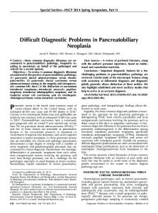
FIGURE 3. PRP injection set-up using ultrasound needle guidance (right foot). PRP indicates platelet-rich plasma.
conservative care including rest, heel lift, physical therapy, nonsteroidal anti-inflammatory drugs, cam walker immobilization, and night splinting. One group received an ultrasound-guided injection of 40 mg depomedrol and the second group received an ultrasound-guided injection of 3 mL PRP (Accelerate System, Exactech Inc.). Both groups initially performed well. The average pretreatment AOFAS score in the steroid group was 52 and improved to 81 at 3 months after treatment. The average pretreatment AOFAS score in the PRP group was 37 and improved to 95 at 3 months after treatment. However, the steroid group scores degraded with a sharp drop in the AOFAS rating to 74 at 6 months and 58 at 12 months after treatment. In stark contrast, the PRP group scores remained high with AOFAS scores of 94 at 6 and 12 months after treatment. No complications were seen in either group. This is the first study to confirm the long-term superiority of PRP over cortisone injection for chronic plantar fasciitis.
DISCUSSION The search for a uniformly successful treatment for plantar fasciitis remains both controversial and elusive. Although the majority of cases are self-limited, a consensus has yet to be reached on a reliable comprehensive treatment strategy. As a result, most clinicians still resort to a speculative collection of traditional conservative treatment regimens that have limited clinical support in the literature. Despite the myriad of available treatments, a 10% failure rate persists. Shockwave treatment, botulinum toxin-A injection, radiofrequency ablation, and surgical procedures have each provided some measure of success but also carry measurable risk for complication and failure. The introduction of PRP into the treatment paradigm as a modulator of angiogenesis and anabolic effects appears to address the pathophysiology of collagen matrix degradation and chaotic vascularity seen in plantar fasciitis.49,50 By combining eccentric exercise and cyclic plantar fascia– specific stretching with PRP injection, enhanced and accelerated healing with excellent long-term results can be achieved in refractory cases.12,51 Future research will focus on optimizing the composition of PRP and compensating for the high individual variability of growth factors among individual patients.52,53 Although the procedure has proven to be safe, a better r
2013 Lippincott Williams & Wilkins
Sports Med Arthrosc Rev
�
Volume 21, Number 4, December 2013
understanding of the systemic effects of PRP will also be needed as recent work by Wasterlain et al54 has documented serologic increases in cytokine levels (VEGF) in patients undergoing PRP treatment. At the present time, given the limited number of studies and heterogenous research methods present in the literature, the use of PRP in the treatment of severe chronic plantar fasciitis may be considered as an alternative to surgical care for use in severe refractory cases of plantar fasciitis where symptoms have persisted longer than 6 months despite prolonged conservative treatment. Patients who are treated with PRP for plantar fasciitis should continue a plantar fascia–specific stretching and eccentric exercise after the injection to optimize their recovery. REFERENCES 1. Riddle DL, Pulisic M, Pidcoe P, et al. Risk factors for plantar fasciitis: a matched case-control study. J Bone Joint Surg Am. 2003;85:872–877. 2. Riddle DL, Schappert SM. Volume of ambulatory care visits and patterns of care for patients diagnosed with plantar fasciitis: a national study of medical doctors. Foot Ankle Int. 2004;25:303–310. 3. Scher DL, Belmont PJ Jr, Bear R, et al. The incidence of plantar fasciitis in the military. J Bone Joint Surg Am. 2009; 91:2867–2872. 4. Tong KB, Furia J. Economic burden of plantar fasciitis in the United States. Am J Orthop. 2010;39:227–231. 5. Leach RE, Seavey MS, Salter DK. Results of surgery in athletes with plantar fasciitis. Foot Ankle. 1986;7:156–161. 6. Aviles E, Arlen M, Miller T. Plantar fibromatosis. Surgery. 1971;69:117–120. 7. Siegel IM. Jogger’s heel. JAMA. 1968;206:2899. 8. Peepelitsa GF, Kuz’min NA, Shmegevskii SA. The treatment of calcaneal spurs with ultrasonics. Klin Khir. 1967;7:39–41. 9. Campbell JW, Inman VT. Treatment of plantar fasciitis and calcaneal spurs with the UCL-BL shoe insert. Clin Orthop Relat Res. 1974;103:57–62. 10. Rompe JD, Cacchio A, Weil L, et al. Plantar fasci-specific stretching versus radial shock-wave therapy as initial treatment of plantar fasciopathy. J Bone Joint Surg Am. 2010;92:2514–2522. 11. Hall MP, Brand PA, Meislin RJ, et al. Platelet-rich plasma: current concepts and application in sports medicine. J Am Acad Orthop Surg. 2009;17:602–609. 12. Monto RR. Platelet rich plasma treatment for chronic Achilles tendinosis. Foot Ankle Int. 2012;33:379–385. 13. Xu K, Kitchen CM, Shu HK, et al. Platelet-derived growth factor-induced stabilization of cyclooxygenase 2 mRNA in rat smooth muscle cells requires the c-Src family of proteintyrosine kinases. J Biol Chem. 2007;282:32699–32709. 14. Bendinelli P, Matteucci E, Dogliotti G, et al. Molecular basis of anti-inflammatory action of platelet-rich plasma on human chondrocytes: mechanisms of NF-(ordM) B inhibition via HGF. J Cell Physiol. 2010;225:757–766. 15. Mischra A, Pavelko T. Treatment of chronic elbow tendinosis with buffered platelet rich plasma. Am J Sports Med. 2006;34: 1774–1778. 16. Kajikawa Y, Morihara T, Sakamoto H, et al. Platelet-rich plasma enhances the initial mobilization of circulation derived cells for tendon healing. J Cell Physiol. 2008;215:837–845. 17. Gosens T, Peerbooms W, van Laar W, et al. Ongoing positive effect of platelet-rich plasma versus corticosteroid injection in lateral epicondylitis: a double blind randomized controlled trial with 2 year follow-up. Am J Sports Med. 2011;39:1200–1208. 18. Neufeld SK, Cerrato R. Plantar fasciitis: diagnosis and treatment. J Am Acad Orthop Surg. 2008;16:338–346. 19. Gill LH. Plantar fasciitis: diagnosis and conservative treatment. J Am Acad Orthop Surg. 1997;5:109–117. 20. Xu Y, Murrell GA. The basic science of tendinopathy. Clin Orthop Relat Res. 2008;466:1528–1538. r
2013 Lippincott Williams & Wilkins
Platelet-rich Plasma and Plantar Fasciitis
21. Lemont H, Ammirati KM, Usen N. Plantar fasciitis: a degenerative process (fasciosis) without inflammation. J Am Podiatr Med Assoc. 2003;93:234–237. 22. Schepsis AA, Leach RE, Gorzyca J. Plantar fasciitis. Etiology, treatment, surgical results, and review of the literature. Clin Orthop Relat Res. 1991;266:185–196. 23. Snider MP, Clancy WG, Mcbeath AA. Plantar fascia release for chronic plantar fasciitis in runners. Am J Sports Med. 1983;11:215–219. 24. Cook JL, Purdam CR. Is tendon pathology a continuum? A pathology model to explain the clinical presentation of loadinduced tendinopathy. Br J Sports Med. 2009;43:409–416. 25. Hicks JH. The mechanics of the foot: II. The plantar aponeurosis and the arch. J Anat. 1954;88:25–30. 26. Levy JC, Mizel MS, Clifford PD, et al. Value of radiographs in the initial evaluation of nontraumatic adult heel pain. Foot Ankle Int. 2006;27:427–430. 27. Graham CE. Painful heel syndrome: rationale of diagnosis and treatment. Foot Ankle. 1983;3:261–267. 28. Akfirat M, Sen C, Gunes T. Ultrasonographic appearance of plantar fasciitis. Clin Imaging. 2003;27:353–357. 29. Gil LH, Kiebzak GM. Outcome of nonsurgical treatment for plantar fasciitis. Foot Ankle Int. 1996;17:527–532. 30. Wapner Kl, Sharkey PF. The use of night splints for treatment of recalcitrant plantar fasciitis. Foot Ankle. 1991;12: 135–137. 31. Donley BG, Moore T, Sferra J, et al. The efficacy of oral nonsteroidal anti-inflammatory medication (NSAID) in the treatment of plantar fasciitis: a randomized, prospective, placebo controlled study. Foot Ankle Int. 2007;28:20–23. 32. Crawford F, Atkins D, Young P, et al. Steroid injections for heel pain: evidence of short-term effectiveness. A randomized controlled trial. Rheumatology. 1999;38:974–977. 33. Tsai WC, Hsu CC, Chen CP, et al. Plantar fasciitis treated with local steroid injection: comparison between sonographic and palpation guidance. J Clin Ultrasound. 2006;34:12–16. 34. Acevedo JI, Beskin JL. Complications of plantar fascia rupture associated with corticosteroid injection. Foot Ankle Int. 1998;19:91–97. 35. Babock MS, Foster L, Pasquina P, et al. Treatment of pain attributed to plantar fasciitis with botulinum toxin A: a shortterm, randomized, placebo controlled, double blind study. Am J Phys Med Rehabil. 2005;84:649–654. 36. Eliondo-Rodriguez J, Araujo-Lopez Y, Moreno-Gonzalez JA, et al. A comparison of Botulinum toxin a and intralesional steroids for the treatment of plantar fasciitis: a randomized, double-blinded study. Foot Ankle Int. 2013;34:8–14. 37. Rompe JD, Decking J, Schoellner C, et al. Shock wave application for chronic plantar fasciitis in running athletes: a prospective, randomized, placebo-controlled trial. Am J Sports Med. 2003;31:268–275. 38. Kudo P, Dainty K, Clarfield M, et al. Randomized, placebocontrolled, double-blind clinical trial evaluating the treatment of plantar fasciitis with an extracorporeal shockwave therapy (ESWT) device: a North American confirmatory study. J Orthop Res. 2006;24:115–123. 39. Sem A, Dimeff R, Iannotti JP. Extracorporeal shock wave therapy in the treatment of chronic tendinopathies. J Am Acad Orthop Surg. 2006;14:95–104. 40. Woelffer KE, Figura MA, Sandberg NS, et al. Five-year follow-up results of instep plantar fasciotomy for chronic heel pain. J Foot Ankle Surg. 2000;39:218–223. 41. Conflitti JM, Tanquinio TA. Operative outcome of partial plantar fasciectomy and neurolysis to the nerve of the abductor digit minimi muscle for recalcitrant plantar fasciitis. Foot Ankle Int. 2004;25:482–487. 42. Urovitz EP, Birk-Urovitz A, Bir-Urovitz E. Endoscopic plantar fasciotomy in the treatment of chronic heel pain. Can J Surg. 2008;51:281–283. 43. Tasto JP. The use of bipolar radiofrequency microtenotomy in the treatment of chronic tendinosis of the foot and ankle. Tech Foot Ankle Surg. 2006;5:110–116.
www.sportsmedarthro.com |
223
Monto
44. Soomekh DJ. Current concepts for the use of platelet-rich plasma in the foot and ankle. Clin Podiatr Med Surg. 2011;28: 155–170. 45. Lopez-Gavito E, Gomez-Carlin LA, Parra-Tellez P, et al. Platelet-rich plasma for managing calcaneus tendon tendinopathy and plantar fasciitis. Acta Ortop Mex. 2011;25:380–385. 46. Martinelli N, Marinozzi A, Carni S, et al. Platelet-rich plasma injections for chronic plantar fasciitis. Int Orthop. 2012;19: S1432–S5195. 47. Ragab EM, Othman AM. Platelet rich plasma for treatment of chronic plantar fasciitis. Arch Orthop Trauma Surg. 2012;132: 1065–1070. 48. Akashin E, Dogruyol D, Yuksel HY, et al. The comparison of the effect of corticosteroids and platelet-rich plasma (PRP) for the treatment of plantar fasciitis. Arch Orthop Trauma Surg. 2012;132:781–785. 49. Schnabel LV, Mohammed HO, Miller BJ, et al. Platelet rich plasma enhances anabolic gene expression patterns in flexor digitorum superficialis tendons. J Orthop Res. 2007;25:230–240.
224 | www.sportsmedarthro.com
Sports Med Arthrosc Rev
�
Volume 21, Number 4, December 2013
50. Gumina S, Campagna V, Ferrazza G, et al. Use of plateletleukocyte membrane in arthroscopic repair of large rotator cuff tears a prospective randomized study. J Bone Joint Surg Am. 2012;94:345–352. 51. Virchenko O, Aspenberg P. How can one platelet injection after tendon injury lead to a stronger tendon after 4 weeks? Interplay between early regeneration and mechanical stimulation. Acta Orthop. 2006;77:806–812. 52. McCarrel T, Minas T, Fortier L. Optimization of leukocyte concentration in platelet-rich plasma for the treatment of tendinopathy. J Bone Joint Surg Am. 2012;94: 1431–1438. 53. Mazzocca A, McCrathy MB, Chowole M, et al. Plateletrich plasma differs according to preparation method and human variability. J Bone Joint Surg Am. 2012;94: 308–316. 54. Wasterlain AS, Braun HJ, Harris AH, et al. The systemic effects of platelet-rich plasma injection. Am J Sports Med. 2013;41:186–193.
r
2013 Lippincott Williams & Wilkins