CLINICAL DENTISTRY AND RESEARCH 2015; 39(2): 74-79
Case Report
PERIODONTITIS AS A MANIFESTATION OF LARGE GRANULAR LYMPHOCYTE (LGL) LEUKEMIA: A CASE REPORT Ali Orkun Topçu DDS, Research Assistant, Department of Periodontology, Faculty of Dentistry, Hacettepe University, Ankara, Turkey
Güliz N. Güncü Associate Professor, Department of Periodontology, Faculty of Dentistry, Hacettepe University, Ankara, Turkey
ABSTRACT The aim of the present paper is to report a case with Large Granular Lymphocyte (LGL) leukemia with severe periodontal destruction. To the best of our knowledge this is the first report of a LGL presented with severe periodontitis. A 20-year-old female presented with neutropenia, hepatosplenomegaly and was taking Mehotrexate 15 mg/day. Intraoral examination revealed apthous ulcerations at labial and buccal mucosa and an inflamed, red, fragile marginal gingiva
Burak Uz
with bleeding upon probing. There were multiple deep pockets
Department of Hematology,
with furcation involments. Initial treatment included extraction
Faculty of Medicine, Hacettepe University,
of hopeless teeth, scaling and root planning and oral hygiene
Ankara, Turkey
motivation. However, the patient refused dental treatment and
Feriha Çağlayan Professor, Department of Periodontology, Faculty of Dentistry, Hacettepe University, Ankara, Turkey
did not show up at recall visits. Hematologists are recommended to have consultations with their patients’ dentists especially when they face with quantitive or qualitative problems associated with neutrophils.
Correspondence Ali Orkun Topcu, DDS Department of Periodontology, Faculty of Dentistry, Hacettepe University,
Keywords:
Large
Granular
Periodontitis, Systemic Interaction
06100 Sıhhiye Ankara, Turkey Phone: +90 312 305 2237 Fax: +90 312 310 4440 E-mail:
[email protected]
74
Submitted for Publication: 01.22.2012 Accepted for Publication : 11.19.2012
Lymphocyte
Leukemia,
CLINICAL DENTISTRY AND RESEARCH 2015; 39(2): 74-79
Olgu Bildirimi
BÜYÜK GRANÜLLÜ LENFOSİTİK LÖSEMİNİN BİR BULGUSU OLARAK PERİODONTİTİS: VAKA RAPORU
Ali Orkun Topçu Araş Gör, Hacettepe Üniversitesi, Diş Hekimliği Fakültesi, Periodontoloji Anabilim Dalı, Ankara, Türkiye
Güliz N. Güncü Doç.Dr., Hacettepe Üniversitesi, Diş Hekimliği Fakültesi,
ÖZ Bu vaka raporunda şiddetli periodontal yıkımları olan büyük granüllü lenfositik lösemi (LGL) hastası sunulmaktadır. Litertür taramasına göre bu vaka raporu şiddetli periodontitise sahip LGL hastasının bildirildiği ilk yayındır. 20 yaşındaki kadın hastada nötropeni ve
Periodontoloji Anabilim Dalı,
hepatosplenomegali bulunmaktadır ve de hasta 15 mg/gün
Ankara, Türkiye
Metotreksat kullanmaktadır. Ağız içi muayene sonucunda dudak
Burak Uz
ve yanak mukozalarında aftöz ülserler ve de sondlamada kanayan
Hacettepe Üniversitesi, Tıp Fakültesi,
iltihaplı, kırmızı, frajil marjinal dişetleri tespit edilmiştir. Hastada
Hematoloji Anabilim Dalı,
furkasyon tutulumlarının da olduğu çoklu derin periodontal cepler
Ankara, Türkiye
bulunmaktadır. Başlangıç tedavisi olarak oral hijyen motivasyonu,
Feriha Çağlayan
diştaşı temizliği ve kök düzeltmesi ile umutsuz dişlerin çekimi
Prof. Dr., Hacettepe Üniversitesi, Diş Hekimliği Fakültesi,
gerçekleştirilmiştir. Ancak hasta diş tedavilerini onaylamayarak idame
Periodontoloji Anabilim Dalı,
randevularına gelmemiştir. Hematologlara hastalarında kalitatif veya
Ankara, Türkiye
kantitatif nötrofil problemleri olduğu durumlarda mutlaka dişhekimi konsültasyonu gerçekleştirmeleri önerilmektedir.
Sorumlu Yazar Ali Orkun Topçu Hacettepe Üniversitesi, Diş Hekimliği Fakültesi, Periodontoloji Anabilim Dalı,
Anahtar Kelimeler: Büyük Granüllü Lenfositik Lösemi, Periodontitis, Sistemik Etkileşim
06100 Sıhhiye Ankara, TURKEY Telefon: +90 312 305 2237 Faks: +90 312 310 4440 E-mail:
[email protected]
Yayın Başvuru Tarihi : 22.01.2012 Yayına Kabul Tarihi : 19.11.2012
75
CLINICAL DENTISTRY AND RESEARCH INTRODUCTION Several hematologic and genetic disorders have been associated with the development of periodontitis in affected individuals and classified as “Periodontitis as a Manifestation of Systemic Diseases”1,2 at the 1999 International Workshop for the Classification of the Periodontal Diseases.3 The major effect of such disorders is through alterations in host defense mechanisms, which have been clearly described for disorders such as neutropenia, leukocyte adhesion deficiencies and leukemia.4 Other non-defense cells such as the red blood cells have crucial roles in maintaining nutrient supply and gas exchange to the periodontium. For efficient hemostasis platelets are needed.1 Leukemia is a serious malignant disease caused by the proliferation of the white blood cell-forming tissues resulting in a marked increase in circulating immature or abnormal white blood cells. In both acute monocytic and chronic lymphocytic leukemia direct leukemic infiltration into the gingival tissues can be seen. Leukemia can result in enlarged gingiva due to infiltration of these tissues by leukocytes in approximately 10% of cases.5,6 Secondary effects from the depression of marrow or lymphoid tissue include hemorrhage, neutropenic ulceration and an increased susceptibility to microbial infections. Clinical periodontal features include anemic pallor of the gingiva, bleeding due to platelet deficiency and reduced resistance to infection due to decreased immune and inflammatory cell numbers. Direct drug toxicity by chemotherapeutic agents may also cause several distinct gingival changes, including erosion and ulceration.5-7 Large granular lymphocyte (LGL) leukemia was first described in 1985 as a clonal disorder involving tissue invasion of marrow, spleen, and liver.8 Clinical presentation is dominated by recurrent infections associated with neutropenia, anemia, splenomegaly, and autoimmune diseases, particularly rheumatoid arthritis (RA).9-11 In 1989, the French-American-British classification identified LGL leukemia as a distinct entity among chronic T-lymphoid leukemias.12 In 1993, the distinction was made between CD3+ T-cell and CD3− NK-cell lineage subtypes of LGL leukemia.9 The real classification in 1994 recommended that LGL leukemia be a distinct clinical entity among peripheral T-cell and NK-cell neoplasms and adopted the suggestion of distinguishing the 2 subtypes of T-cell and NK-cell LGL leukemia.13 The World Health Organization classification, published in 1999, included T- or NK-cell granular lymphocytic leukemia in the subgroup of mature
76
peripheral T-cell neoplasms.14 Furthermore, in 2008, a new provisional entity of chronic lymphoproliferative disorder of NK cells (also known as chronic NK-cell lymphocytosis) was created by World Health Organization to distinguish it from the much more aggressive form of NK-cell leukemia.15 The frequency of T and NK LGL leukemia is not accurately determined and ranges from 2% to 5% of the chronic lymphoproliferative diseases in North America and up to 5% to 6% in Asia. To the best of our knowledge, this is the first report of a LGL presented with severe periodontitis. CASE REPORT
Medical Evaluation A 20-year-old female presented with neutropenia. Her medical history revealed neutropenia ongoing for approximately two years. In December 2010, a bone marrow biopsy was performed which was normocellular and showing an increase in megacaryocytes. Flow cytometry of the bone marrow revealed CD2, CD3, CD4 (25%), CD5, CD7, CD8 (34%), CD38, CD45, HLA-DR positivity. An abdominal ultrasound revealed mild hepatosplenomegaly (vertical lenght of liver 165 mm, spleen 135 mm). Complete blood count was as follows; Hb:12,7 gr/dL (12.00-18.00), WBC: 1.7 x10³/μL (3.60-10.00), PLT: 355 x103/μL (150.00450.00). Hepatitis, viral tests, and autoimmune antibodies were all negative. Sedimentation rate and romatoid factor were in normal limits. A mild hypergammaglobulinemia (IgG: 2120 mg/dL; range: 694-1618) was detected. In peripheral blood smear 4% neutrophil, 56% lymphocyte, 10% large granular lymphocyte, 28% monocyte and 2% eosinophil was reported. A repeat bone marrow biopsy showed an increase in CD3 positive cells, erythroid hyperplasia, and relative incresase of lymphoid cells. Tartrate- resistant acid phosphatase staining was positive in a fine granular manner. Cytogenetic was 46, XX in 20 metaphases. T-cell gene rearrangement was found to be clonal. Flow cytometry of repeat bone marrow was as follows: CD2, CD3, CD4, CD5, CD7, CD8, CD20, CD22, CD45, HLA-DR (+); CD16, CD56, CD57 (-). In conclusion, all these findings led us to a diagnosis of large-granular lymphocyte leukemia (LGL). Mehotrexate 15 mg/day once a week peroral was initiated.
Dental Evaluation The patient was referred from Department of Hematology at Hacettepe University to the Department of Periodontics at the same institution during the spring of 2010. The chief complaint by the patient was her mobile teeth and inability to chew. The patient claimed that she had lost her primary
Periodontitis as a Manifestation of LGL
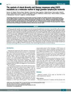
dentition because of the loosening of her teeth. The patient’s medical history revealed that she had been diagnosed for LGL few months ago and was under the control of her physician since then. The patient was taking Mehotrexate 15 mg/day, and that she had no known allergies. There was no family history of any medical problems and she had low social and economic status. The patient reported that she had periodic childhood illnesses i.e. colds and ear infections. Intraoral examination revealed apthous ulcerations at labial and buccal mucosa; (Figure 1) however, hard and soft palates and oropharynx were not affected. The patient displayed poor oral hygiene. Periodontal examination revealed inflamed, red, fragile marginal gingiva and bleeding on probing along with multiple pockets >3 mm most of them being at the posterior site were noticed. Gingival recession was present at all posterior segments. Furcation area was exposed at maxillary and mandibular first molars with class IV involvement (Figures 2, 3). Deepest pockets were of 9 mm, and were located around the maxillary and mandibular premolar-molar sites. Grade 3 mobility for the first and the second molars and grade 2 mobility for the second premolars sites were observed. Panoramic radiograph revealed that there occurred advanced bone loss around these teeth (Figure 4). After clinical and radiographic examination it was noted that the periodontal destruction particularly affected the primary dentition and posterior sites of permanent dentition. Because of the horizontal bone loss noted at incisor sites in both maxilla and mandible, diagnosis of aggressive periodontitis was eliminated. The patient received 2 gr. Amoxicillin one hour before the dental theraphy according to the recommendation of her physician. Initial treatment included extraction of the severly affected first and second molars and second premolars (except lower left second premolar), oral hygiene
Figure 2. Intraoral view left side
Figure 3. Intraoral view right side
Figure 4. Panaromic radiograph.
Figure 1. Frontal intraoral view of the patient
instruction was given to the patient, scaling and root planning was performed and an antimicrobial mouth rinse was prescribed twice a day. Monthly recall/maintenance visits were recommended. However, the patient refused dental treatment and did not show up at recall visits.
77
CLINICAL DENTISTRY AND RESEARCH DISCUSSION LGL leukemia, classified in the indolent non-Hodgkin lymphomas, is a clonal disease, arising most frequently (85%) from a T cell lineage or, less commonly (15%), from a natural killer (NK) cell lineage.9,16 The cause of this rare disorder is not known. Noteworthy, these leukemic cells show all the characteristics of antigen-activated T cells,17 suggesting that an initial step in LGL expansion is an antigen-driven mechanism. The persistence and proliferation of LGLs could be due to the stimulatory effect of various cytokines including interleukin (IL)-12 and IL-15, or to genetic polymorphisms in genes involved in the regulation of immune and inflammatory responses.18-21 IL-15 stimulates LGL, mediating this activity via IL-2 receptor. IL-15 and platelet-derived growth factor are the two key mediators controlling the interactions among the survival pathways.22,23 The median age of onset of T-cell LGL leukemia is 60 years. Only 10% of patients are younger than 40 years of age.24 Symptoms, when present, are usually related to neutropenia, and include fever with recurrent bacterial infections in 20-40% of patients.25 Infections typically involve oropharyngeal, skin and perirectal areas. Fever, fatigue, night sweats, or weight loss are observed in 20-30% of cases.25,26 Nonclonal LGL expansions have been reported in the following clinical situations: Viral infections (eg, EBV, HBV, HCV, HIV, CMV), connective tissue disease, idiopathic thrombocytopenic purpura, non-Hodgkin lymphoma, various skin disorders, and the hemophagocytic syndrome.9,27-29 The myelodysplastic syndrome and solid tumors are sometimes associated with reactive T-cell LGL expansion.28 The immune and inflammatory systems are crucial to our survival. Back-up systems are often present, which means that many functional defects are not fatal, but do result in predisposition to various diseases, such as chronic periodontitis. Systemic diseases affecting the host response as primary immunodeficiencies or secondary defects caused by the lack of nutrients or changes in the local tissues are very often accompanied by severe forms of periodontitis. Thus, blood and systemic disorders of the blood and blood-forming organs may have a profound effect on the periodontium.30 However, inflammatory cell disorders may also have a detrimental effect on the integrity of the periodontium. In addition, specific diseases of the periodontium, i.e. today the early-onset forms of periodontitis, are increasingly linked with leukocyte function abnormalities.31 The clinical manifestation of many of these
78
disorders appears at an early age and may be confused with aggresive forms of periodontitis with rapid attachment loss and the potential for early tooth loss. Although our patient was diagnosed with LGL, her case was unusual as she did not experience recurrent infection although she presented with neutropenia; there were no anti-neutrophil antibodies detected. Periodontal disease was the only medical abnormality that was clinically present. It may be speculated that other signs/symptoms of the neutropenia were subclinical, or the periodontal disease was the first sign of her condition. Persistence and proliferation of LGLs could be due to the stimulatory effect of various immune and inflammatory responses18-21 which could mean that the patient may exhibit overreactive host response. This condition and low white blood cell count could explain the severe alveolar bone loss. Local treatment in combination with systemic antibiotics may in milder forms improve the situation, but in many cases the success is questionable and premature loss of teeth occurs. Even though the patient underestimated her condition, it is important that preventive measures be taken to avoid future periodontal problems. This case also proves that low socioecomic status has important effects on the awareness of the importance of periodontal treatment and establishment of communication. The patient was unwilling to the dental therapy, did not care about losing her teeth and did not return to our calls. Hematologists are recommended to consult their patients to a dentist especially when they have conditions that may cause quantitive or qualitive problems linked with neutrophils. ACKNOWLEDGEMENT All authors report no conflicts of interest related to this study.
Periodontitis as a Manifestation of LGL
REFERENCES 1. Kinane DF. Blood and lymporeticular disorders. Periodontol 2000 1999; 21: 84-93. 2. Kinane DF. Periodontitis modified by systemic factors. Ann Periodontol 1999; 4: 54-64. 3. Armitage GC. Development of a classification system for periodontal diseases and conditions. Ann Periodontol 1999; 4: 1-6. 4. Meyle J, Gonzáles JR. Influences of systemic diseases on periodontitis in children and adolescents. Periodontol 2000 2001; 26: 92-112. 5. Lynch MA, Ship II. Initial oral manifestations of leukaemia. J Am Dent Assoc 1967; 75: 932-940. 6. Stafford R, Sonis S, Lockhart P, Sonis A. Oral pathoses as diagnostic indicators in leukaemia. Oral Surg Oral Med Oral Pathol Oral Radiol Endod 1980; 50: 134-138. 7. Kinane DE, Haematological disorders and the periodontium. In: Newman HN, Rees TD, Kinane DF, editors. Diseases of the periodontium. Northwood: UK Science Reviews; 1993.p.178. 8. Loughran TP Jr., Kadin ME, Starkebaum G, Abkowitz JL, Clark EA, Disteche C et al. Leukemia of large granular lymphocytes: association with clonal chromosomal abnormalities and autoimmune neutropenia, thrombocytopenia, and hemolytic anemia. Ann Intern Med 1985; 102: 169-175. 9. Loughran TP Jr. Clonal diseases of large granular lymphocytes. Blood 1993; 82: 1-14. 10. Lamy T, Loughran TP Jr. Clinical features of large granular lymphocyte leukemia. Semin Hematol 2003; 40: 185-195. 11. Sokol L, Loughran TP Jr. Large granular lymphocyte leukemia. Oncologist 2006; 11: 263-273. 12. Bennett JM, Catovsky D, Daniel MT, Flandrin G, Galton DA, Gralnick HR et al. Proposals for the classification of chronic (mature) B and T lymphoid leukaemias: French-American-British (FAB) Cooperative Group. J Clin Pathol 1989; 42: 567-584. 13. Harris NL, Jaffe ES, Stein H, Banks PM, Chan JK, Cleary ML et al. A revised European-American classification of lymphoid neoplasms: a proposal from the International Lymphoma Study Group. Blood 1994; 84: 1361-1392. 14. Harris NL, Jaffe ES, Diebold J, Flandrin G, Muller-Hermelink HK, Vardiman J et al. World Health Organization classification of neoplastic diseases of the hematopoietic and lymphoid tissues: report of the Clinical Advisory Committee meeting-Airlie House, Virginia, November 1997. J Clin Oncol 1999; 17: 3835-3849. 15. Swerdlow SH, Campo E, Harris NL, Jaffe ES, Pileri SA, Stein H, editors. WHO classification of tumours of haematopoietic and lymphoid tissues. 4th ed. Lyon, France: International Agency for Research on Cancer Press; 2008. 16. Lamy T, Loughran TP Jr. Clinical features of large granular lymphocyte leukemia. Semin Hematol 2003; 40: 185-195. 17. Yang J, Epling-Burnette PK, Painter JS, Zou J, Bai F, Wei S et al. Antigen activation and impaires Fas-induced death-inducing
signaling complex formation in T-large lymphocyte leukemia. Blood 2008; 111: 1610-1616. 18. Nearman ZP, Wlodarski M, Jankowska AM, Howe E, Narvaez Y, Ball E et al. Immunogenetic factors determining the evolution of T-cell large granular lymphocyte leukemia and associated cytopenias. Br J Haematol 2007; 136: 237-248. 19. Gentile TC, Loughran TP Jr. Interleukin-12 is a costimulatory cytokine for leukemic CD3+ large granular lymphocytes. Cell Immunol 1995; 166: 158-161. 20. Zambello R, Trentin L, Cassatella MA, Raimondi R, Cerutti A, Enthammer C et al. IL-12 is involved in the activation of CD3+ granular lymphocytes in patients with lymphoproliferative disease of granular lymphocytes. Br J Haematol 1996; 92: 308-314. 21. Zambello R, Facco M, Trentin L, Sancetta R, Tassinari C, Perin A et al. Interleukin-15 triggers the proliferation and cytotoxicity of granular lymphocytes in patients with lymphoproliferative disease of granular lymphocytes. Blood 1997; 89: 201-211. 22. Zhang R, Shah MV, Yang J, Nyland SB, Liu X, Yun JK, et al. Network model of survival signaling in large granular lymphocyte leukemia. Proc Natl Acad Sci U S A 2008; 105: 16308-16313. 23. Yang J, Liu X, Nyland SB, Zhang R, Ryland LK, Broeg K et al. Platelet-derived growth factor mediates survival of leukemic large granular lymphocytes via an autocrine regulatory pathway. Blood 2010; 115: 51-60. 24. Boeckx N, Uyttebroeck A, Langerak AW, Brusselmans C, Goossens W, Bossuyt X. Clonal proliferation of T-Cell large granular lymphocytes. Pediatr Blood Cancer 2004; 42: 275-277. 25. Lamy T, Loughran TP. Large Granular Lymphocyte Leukemia. Cancer Control 1998; 5: 25-33. 26. Pandolfi F, Loughran TP Jr, Starkebaum G, Chisesi T, Barbui T, Chan WC et al. Clinical course and prognosis of the lymphoproliferative disease of granular lymphocytes. A multicenter study. Cancer 1990; 65: 341-348. 27. Oshimi K. Granular lymphocyte proliferative disorders: report of 12 cases and review of the literature. Leukemia 1988; 2: 617-627. 28. Okuno SH, Tefferi A, Hanson CA, Katzmann JA, Li CY, Witzig TE. Spectrum of diseases associated with increased proportions or absolute numbers of peripheral blood natural killer cells. Br J Haematol 1996; 93: 810-812. 29. Imashuku S, Hibi S, Morigana S, Takagi K, Chen J, Mugishima H et al. Haemophagocytic lymphohistiocytosis in association with granular lymphocyte proliferative disorders in early childhood: characteristic bone marrow morphology. Br J Haematol 1997; 96: 708-714. 30. Consensus Report, Discussion Section I. In: Nevins M, Becker W, Kornman K, editors. Proceedings of the World Workshop in Clinical Periodontics. Chicago: American Academy of Periodontology; 1989: I23-I31. 31. Hart TC, Atkinson JC. Mendelian forms of periodontitis. Periodontol 2000 2007; 45: 95-112.
79