Chirurgia (2011) 106: 535-539 Nr. 4, Iulie - August Copyright© Celsius
Perianal giant condyloma acuminatum (Buschke-Löwenstein tumor). Case report and review of the literature HS. Papiu1, A. Dumnici1, T. Olariu2, M. Onita3, E. Hornung3, D. Goldis1, G. Aiordachioae1, V. Vasca1 1
“Vasile Goldiş” Western University Arad, Department of Surgery, Romania “Vasile Goldiş” Western University Arad, ICU, Romania 3 Department of Surgery, Arad City Hospital, Romania 2
Rezumat Condilomatoza gigantã perianalã (boala BuschkeLöwenstein). Prezentare de caz æi date din literaturã Condilomatoza gigantã perinealã, cunoscutã ca boala Buschke-Löwenstein (BLT), este o afecåiune rarã cu transmitere sexualã, ce afecteazã regiunea anogenitalã. La acest nivel apare o formaåiune conopidiformã cu creştere lentã, care spre deosebire de condilomul obişnuit este agresivã şi distructivã local, degenerând malign în 40-60% din cazuri. Prezentãm un caz cu BLT malignizatã, discuåii pe marginea etiopatogeniei şi tratamentului acestei boli, cu scopul de a sublinia câteva repere în managementul condilomatozei gigante perineale. Concluzii: Pacientul cu BLT trebuie evaluat clinico-imagistic pentru a stabili gradul invaziei viscerale şi astfel amploarea intervenåiei chirurgicale. Excizia largã a leziunilor, cu margini de siguranåã negative histologic este opåiunea de elecåie dacã nu este invadat canalul anal. Chirurgia mai radicalã este rezervatã cazurilor cu invazie visceralã doveditã. Excizia este obligatorie chiar în cazul condiloamelor mici, pentru a preveni evoluåia lor cãtre BLT.
Abstract Giant condyloma acuminatum, also known as BuschkeLöwenstein tumor (BLT) is a very rare sexually transmitted disease that affects the ano-genital region. BLT is a slow growing cauliflower-like tumor, but unlike simple condyloma, it is locally aggressive and destructive, malignant transformation occuring in 40-60% of cases. We present a case of perianal carcinomatous BLT and discuss some controversies about the nature and treatment of this disease. Our aim is to emphasize some basic points in the management of giant perianal condyloma acuminatum. Conclusions: The patient with BLT must be very carefully clinical and imagistic investigated in order to detect the tumor visceral invasion and to establish the extension of the surgical procedure. Wide perineal excision with histopathological margins control is the best surgical choice if the anal canal is not involved. The radical pelvic surgery is indicated only in patients with provable visceral invasion. Excision is mandatory even in very small condilomas to prevent BLT later development. Key words: giant condyloma acuminatum, BuschkeLöwenstein tumor, wide perineal excision
Cuvinte cheie: condilomatozã gigantã, boala BūschkeLöwenstein, excizie perinealã largã
Introduction
Corresponding author:
Horatiu Papiu MD, PhD Municipal Hospital, Clinic of Surgery Pta M.Viteazu 7/8, Arad, Romania E-mail:
[email protected]
Buschke-Löwenstein tumor (BLT) is a very rare sexually transmitted disease that affects the ano-genital region (1,2,3). The incidence of this tumor is probably 0.1% in the general population and it was first described by Buschke (4,5). BLT, also known as giant condyloma acuminatum, is a slow growing
536
cauliflower-like tumor, but unlike simple condyloma, it is locally aggressive and destructive (6). In the pathogenesis of this tumor there are involved HPV types 6 and 11 and, possibly, immune defects (2,3,7). Some authors reported BLT in HIV-infected patients (8). There are controversies regarding the nature of BLT. Some authors have shown that BLT is a verrucous carcinoma and others have described benign forms of this lesion (9,10). Most of the reports have shown that BLT develops on the basis of initially benign condiloma acuminatum and malignant transformation occurs in 40-60% of cases (4). Distant metastases are rare and the risk of developing anal stenosis is low (4,11,12). Common clinical signs that can be found in such patients are bleeding, lymph node enlargement, usually inflammatory, abscess and perianal fistula (4,12). The loco-regional extension must be carrefully assessed to establish the therapeutic strategy. In respect to this, abdominal and pelvic computed tomography and magnetic resonance imaging, pelvic inguinal endoscopy and intraanal ultrasound- endoscopy are useful (13). Besides pathogenesis, there are also controversies on the BLT treatment (5). The majority of authors agree that surgery is the treatment of choice and is efficient in the early stages of the disease (14). Excision must be wide (15,16). Lymph node dissection is indicated only in cases of suspected malignant transformation (4). Some authors prefer abdomino-perineal amputation, but others support the conservative surgery as the best choice, mainly in terms of the patient’s quality of life (13). The high local recurrence rate after excision, of about 50-60%, has suggested the need of pre- and postoperative use of radiation and chemotherapy (12,17). There are reports that locoregional control of recurrent forms was achieved with combined chemoradiotherapy (18,19,20). There are also rare reports of primary radiotherapy producing complete tumor regression (21). Post-treatment monitoring is strongly recommended (4). We present a case with perianal BLT with malignant transformation and discuss some controversies about the nature and treatment of this disease. Our aim is to emphasize some basic points in the management of giant perianal condyloma acuminatum.
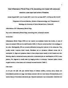
Case report We present a 54 year old man with giant condyloma evolving of about 20 years with no previous surgical or oncologic treatment. The patient, without a history of other pathology, is hospitalized in our clinic presenting a giant cauliflowerlike tumor located at the posterior and anterior perineum, with right parascrotal extension. There are purulent cankers, bleedings, perianal pain and right inguinal lymphadenopathy. The patient has also two scrotal tegument small tumors (Fig. 1). Inpatient bleeding was important from many hemorrhage points. There was an early hemorrhagic shock with pallor, increased pulse frequency and arterial hypotension, malaise and marked fatigue. Laboratory analysis revealed a severe anemia and mild leukocytosis. The patient is admitted to the intensive care unit beeing hydroelectrolitic rebalanced,
Figure 1. Giant perianal and parascrotal condyloma
transfused and receiving antibiotics. A biopsy was performed and histopathology reveals a malignant transformation. The Human Immunodeficiency Virus test result was negative. Surgical intervention was then performed involving, in a first stage, the perianal tumor resection using the electrocision. The right parascrotal condyloma was excised after 12 days, in the second stage. The pathological evaluation showed a solid, exophytic and invasive tumor, hard and white on the surface of section. The microscopic diagnosis was squamous cell carcinoma moderately differentiated (Fig. 2). Tumor cells showed anaplastic features and many mitoses (Fig. 3). Cytokeratin AE1/AE3 was strong but heterogeneously expressed in the tumor area (Fig. 4). Both p53 and Ki67 were expressed by the majority of tumor cells, reflecting the high proliferative potential of the tumor (Fig. 5, 6). No lymphovascular invasion was detected on slides stained with anti-D240/podoplanin, but positive reaction was noticed in lessdifferentiated neoplastic cells, supporting the aggressive and invasive behavior (Fig. 7). The postoperative evolution was favorable with granulation tissue and small necrotic areas that require serial necrectomies (Fig. 8). Controls at 1 and 3 months revealed a slow favorable evolution with epithelization of the wound and remission of the inguinal adenopathy. After 6 months the perineal region is free of tumor recurrence. The patient refused chemoradiotherapy.
Discussions The case presented is a less cooperant, homeless patient with a very long disease evolution, who was hospitalized forced by local massive bleeding. The giant tumor evolved from small cauliflower-like vegetations, as we know that BLT is always preceded by condyloma acuminatum (4,22). The anal canal was carefully examined and it was not involved by the tumoral invasion (5). Malignant transformation was present, beeing suspicioned by massive bleeding,
537
2
3
4
5
6
7
Figures 2-7.
2,3 – low power magnification of the tumor - scuamous cell carcinoma (x100); 4 - heterogeneous expression of cytokeratin AE1/AE3; 5 - p53 expression; 6 - Ki67 expression ; 7 - anti-D2-40/podoplanin stained both lymphatic vessels and tumor cells. Original magnification 7-10 x 400
tumor infiltration, its giant size and lymph node infiltration (4,12). The cancerous nature of the tumor was confirmed by histopathological exam that reveals a squamous cell carcinoma, after a careful sectioning and complete histologic exami-
nation was performed, as it is always required in such cases (9, 23). This tumor shows aggressive local infiltration but does not metastasize (23). There is a controversy in the histopathological diagnosis
538
Figure 8. Aspect 2 weeks after the second excision with granul tion tissue and superficial necrotic areas
of BLT. Most of the authors sustain that BLT is a pseudoépitheliomatous proliferation belonging to verrucous carcinoma group (24), and we have the same opinion, supported by the findings in our case. But there are some reports regarding BLT as an intermediate entity between an ordinary condyloma acuminatum and squamous cell carcinoma (18). Finally, there is a case report of microscopically benign giant condyloma acuminatum of the perianal region (10). Anyway, the histopathological dilemma is not so important, because wide excision is always mandatory when is possible. So far, no definitive therapeutic strategy has been established (5). The management of BLT must be implemented knowing the pelvic tumor extension which needs clinical and imaging exams. Unfortunately, we did not have quick access to computed tomography or magnetic resonance facilities and the developing haemorrhage made the operation mandatory. The majority of authors agree that surgery is the treatment of choice and is effective especially in the early stages of the disease (15). Wide local excision remains the mainstay of therapy, that can be followed, if it is necessary, by delayed split thickness skin grafts (1,11,25,9). There are some controversies between radical abdomino-perineal amputation and conservative surgery supporters (13). We think that wide perineal excision with histopathological margins control is the best surgical choice in the treatment of BLT if the anal canal is not involved. The radical pelvic surgery is reserved only for patients with provable visceral invasion. Wide resection is indicated even in rare cases of benign nature BLT because of the high risk of carcinomatous transformation and the great discomfort produced by the giant perineal tumor. We also emphasize that surgical excision is mandatory in all cases of BLT when it is technically possible and also in all condylomas, even in very small ones, in order to prevent their BLT transformation. There is no consensus about the use of pre- and postoperative oncologic treatment in BLT. Some authors recommend that reduction of the tumoral mass through
radiotherapy or chemotherapy to precede surgical excision (4,15). Others assert that radiotherapy is rarely used, usually after an incomplete excision, in recurrences and when excision is not recommended. There are rare reports of primary radiotherapy producing complete tumor regression (21). Radiotherapy has been suspected of being responsible for the alteration of BLT into anaplastic carcinoma (15,16,26). We can not express a clear opinion about the oncological treatment in our case because our patient refused radiochemotherapy. Some authors reports the use of imiquimod and others present a case of BLT successfully treated using carbon dioxide laser vaporization and systemic interferon therapy after failure of 3 surgical excisions (2,27,28,29,30). The patient must be long time closely followed-up (4, 31). We think prospective studies are necessary to further define the nature and treatment of this very rare disease (32).
Conclusions BLT is a very rare sexually transmitted disease characterized by giant slow growing condyloma acuminatum that is, unlike simple condyloma, locally aggressive and destructive. The histopathological nature of BLT is not well defined, but it is clear that malignant transformation rate is high. The patient with BLT must be very carefully clinical and imagistic investigated in order to detect the tumor visceral invasion and to establish the extension of the surgical procedure. Wide perineal excision with histopathological margins control is the best surgical choice if the anal canal is not involved. The radical pelvic surgery is indicated only in patients with provable visceral invasion. Excision is mandatory even in very small condilomas to prevent BLT later development.
References 1.
2.
3.
4.
5.
6.
Balik E, Eren T, Bugra D. A surgical approach to anogenital Buschke Loewenstein tumours (giant condyloma acuminata). Acta Chir Belg. 2009;109(5):612-6. Erkek E, Basar H, Bozdogan O, Emeksiz MC. Giant condyloma acuminata of Buschke-Löwenstein: successful treatment with a combination of surgical excision, oral acitretin and topical imiquimod. Clin Exp Dermatol. 2009;34(3):366-8. Miranda Aranzubía O, García Rodríguez J, González Alvarez RC, Alvarez Mújica M, Rodríguez Robles L, Regadera Sejas J. Giant condyloma acuminatum (Buschke-Löwenstein tumor). Actas Urol Esp. 2008;32(9):951. Hicheri J, Jaber K, Dhaoui MR, Youssef S, Bouziani A, Doss N. Giant condyloma (Buschke-Löwenstein tumor). A case report. Acta Dermatovenerol Alp Panonica Adriat. 2006; 15(4):181-3. Paraskevas KI, Kyriakos E, Poulios EE, Stathopoulos V, Tzovaras AA, Briana DD. Surgical management of giant condyloma acuminatum (Buschke-Loewenstein tumor) of the perianal region. Dermatol Surg. 2007;33(5):638-44. Talwar A, Puri N, Singh M. Giant condyloma acuminatum of Buschke and Lowenstein: successful surgical treatment. Int J
539
7.
8.
9. 10.
11.
12.
13.
14.
15.
16.
17.
18.
19.
STD AIDS. 2010;21(6):446-8. De Toma G, Cavallaro G, Bitonti A, Polistena A, Onesti M G, Scuderi N. Surgical management of perianal giant condyloma acuminatum (Buschke-Löwenstein tumor). Report of three cases. Eur Surg Res. 2006;38(4):418–422. Kreuter A, Potthoff A, Brockmeyer NH, Gambichler T, Swoboda J, Stñcker M, et al. Anal carcinoma in human immunodeficiency virus-positive men: results of a prospective study from Germany. Br J Dermatol 2010; 162(6):1269-77. Longacre TA, Kong CS, Welton ML. Diagnostic problems in anal pathology. Adv Anat Pathol. 2008;15(5):263-78. Ben Brahim E, Chadli-Debbiche A, Fraoua-Abdelmoula F, Lahmar-Boufaroua A, Bouchoucha S, Khalfallah MT, et al. Buschke-Loewenstein giant condyloma in the perianal region with inguinal invasion: a case report. Tunis Med. 2000;78(3): 205-9. Klaristenfeld D, Israelit S, Beart RW, Ault G, Kaiser AM. Surgical excision of extensive anal condylomata not associated with risk of anal stenosis.Int J Colorectal Dis. 2008;23(9):853-6. Chu Q D, Vezeridis M P, Libbey N P, Wanebo H J. Giant condyloma acuminatum (Buschke-Lowenstein tumor) of the anorectal and perianal regions. Analysis of 42 cases. Dis Colon Rectum. 1994;37(9):950–957. Parise P, Sarzo G, Finco C, Marino F, Savastano S, Merigliano S. Giant condyloma acuminatum of the anorectum (BuschkeLowenstein tumour): a case report of conservative surgery. Chir Ital. 2004;56(1):157-61. Sarzo G, Mistro A, Finco C, Frayle-Salamanca H, Marino F, Franzetti M, et al. Extensive anal condylomatosis: prognosis in relation to viral and host factors.Colorectal Dis. 2010;12(7 Online):e128-34. El Mejjad A, et al. Le condylome acuminé géant-tumeur de Buschke Loewenstein (à propos de 3 cas).Prog Urol 2003; 13(3): 513–7. Qarro A, et al. Tumeur de Buschke-Loewenstein à localisation anorectale (à propos de trois cas). Ann Chir 2005; 130: 96–100. Tytherleigh M G, Birtle A J, Cohen C E, Glynne-Jones R, Livingstone J, Gilbert J. Combined surgery and chemoradiation as a treatment for the Buschke-Löwenstein tumour. Surgeon. 2006;4(6):378–383. Chao MW, Gibbs P. Squamous cell carcinoma arising in a giant condyloma acuminatum (Buschke-Lowenstein tumour). Asian J Surg. 2005;28(3):238-40. Haque W, Kelly E, Dhingra S, Carpenter LS. Successful treatment of recurrent Buschke-Lowenstein tumor by radiation
20.
21. 22.
23.
24.
25. 26.
27.
28.
29. 30.
31.
32.
therapy and chemotherapy. Int J Colorectal Dis. 2010;25(4): 539-40. Armstrong N, Foley G, Wilson J, Finan P, Sebag-Montefiore D. Successful treatment of a large Buschke-Lowenstein tumour with chemo-radiotherapy.Int J STD AIDS. 2009;20(10):732-4. Wietfeldt ED, Thiele J. Malignancies of the anal margin and perianal skin. Clin Colon Rectal Surg. 2009;22(2):127-35. Cusini M, Gaiani F, Girgenti V, Cantoni G, Ramoni S. Perianal Buschke-Löwenstein tumour: progressive growth despite immune restoration in a man positive for human immunodeficiency virus. Clin Exp Dermatol. 2010;35(4):e163-4. Gholam P, Enk A, Hartschuh W. Successful surgical management of giant condyloma acuminatum (Buschke-Löwenstein tumor) in the genitoanal region: a case report and evaluation of current therapies. Dermatology. 2009;218(1):56-9. Epub 2008 Oct 21. Ali Sbai M, Balti W, Dhahak S, Ben Romdhane S, Tabib M, Balti H. Buschke Lowenstein tumor: unusual bilateral localization. Tunis Med. 2009;87(9):627-9. Cintron J R. Buschke-Lowenstein tumor of the perianal and anorectal region. Semin Colon Rectal Surg. 1995;6:135–139. Martin F, Dalac S, Lambert D. Verrucous carcinoma. Nosologic aspects, apropos of 4 cases. Ann Dermatol Venereol. 1995;122(67):399-403. [Article in French] Perniola G, d’Itri F, Di Donato V, Achilli C, Lo Prete E, Panici PB. Recurrent Buschke-Löwenstein tumor treated using CO(2) laser vaporization.J Minim Invasive Gynecol. 2010;17(5): 662-4. Frega A, Stentella P, Tinari A, Vecchione A, Marchionni M. Giant condyloma acuminatum or buschke-Lowenstein tumor: review of the literature and report of three cases treated by CO2 laser surgery. A long-term follow-up. Anticancer Res. 2002; 22(2B):1201-4. Lévy A, Lebbe C. Buschke-Löwenstein tumour: diagnosis and treatment. Ann Urol (Paris). 2006;40(3):175-8. Perisic Z, Lazic JP, Terzic B, Perisic S, Rasic R. Condylomata gigantea in anal and perianal region: surgical and CO2 laser treatment.Arch Gynecol Obstet. 2003;267(4):263-5. Oniåa M, Şupialã CD, Olariu Teodora, Stoica AL. Condilomatozã gigantã perinealã – prezentare de caz şi date din literaturã. Jurnal Medical Arãdean 2004;2(VII):53-7. Trombetta LJ, Place RJ. Giant condyloma acuminatum of the anorectum: trends in epidemiology and management: report of a case and review of the literature. Dis Colon Rectum. 2001; 44(12):1878-86.