ORIGINAL STUDY
Acta Orthop. Belg., 2010, 76, 237-242
Percutaneous suture of acute Achilles tendon rupture A study of 60 cases Jean-Louis ROUVILLAIN, Thomas NAVARRE, Octavio LABRADA-BLANCO, Emmanuel GARRON, Wael DAOUD
From the University Hospital Pierre Zobda Quitman, La Meynard, Fort De France, Martinique, France
Conservative treatment of Achilles tendon ruptures may lead to re-rupture. Open surgical repair entails a risk of skin necrosis or infection. Several percutaneous techniques have been used, such as Tenolig® or Achillon®, but these techniques are costly and may be marred by wound healing problems. Ma and Griffith described a technique for percutaneous repair which left the suture and the knot under the skin, thus reducing the risk for infection. From January 2001 to September 2006, we used this percutaneous treatment for 60 acute ruptures of tendo Achillis. The repair was made under local anaesthesia, using a single or double absorbable suture. Postoperative care was 3 weeks immobilistion in a non-weight bearing cast in equinus position, followed by another 3 weeks in a cast with the ankle at 90° and progressive weight bearing. Mean followup was 19 months. Complications were 2 re-ruptures at 2 and 5 months respectively, 1 infection in a patient who presented with re-rupture after a previous surgical treatment, and 1 Achilles tendonitis. There was no sural nerve lesion. Mean time to return to working activities was 85 days ; mean time to return to sports activities was 5 months. Clinical results were good with no loss in range of motion. This low-cost technique appeared as an interesting alternative to surgical or conservative treatment, providing strong repair with a low complication rate. Keywords : Achilles tendon ; acute rupture ; percutaneous suture.
No benefits or funds were received in support of this study
INTRODUCTION Achilles tendon ruptures are encountered in middle-aged adults. The two main methods of treatment are conservative treatment and open surgical repair. Conservative treatment, which consists in immobilisation in an equinus cast for six to twelve weeks, carries a significant risk of re-rupture. Surgical treatment carries a limited risk of rerupture but a greater risk of infection and wound healing problems. In order to overcome these complications, techniques of percutaneous tenorraphy of the Achilles tendon have been developed (3,5) but they do not entirely eliminate cutaneous risks. In 1977, Ma and Griffith (11) suggested a fully percutaneous technique which left the suture and knot
Jean-Louis Rouvillain, MD, Orthopaedic Surgeon. Thomas Navarre, MD, Orthopaedic Surgeon. Octavio Labrada-Blanco, MD, Orthopaedic Surgeon. Emmanuel Garron, MD, Orthopaedic Surgeon. Wael Daoud, MD, Orthopaedic Surgeon. Orthopaedic and Traumatologic Surgery Department, University Hospital Pierre Zobda Quitman, Martinique, France. Correspondence : Jean-Louis Rouvillain, Orthopaedic and Traumatologic Surgery Department, University Hospital Pierre Zobda Quitman, La Meynard, 97261 Fort de France, Martinique, France. E-mail :
[email protected] © 2010, Acta Orthopædica Belgica. ■ ■ ■ ■ ■
Acta Orthopædica Belgica, Vol. 76 - 2 - 2010
238
J.-L. ROUVILLAIN, T. NAVARRE, O. LABRADA-BLANCO, E. GARRON, W. DAOUD
under the skin, thus reducing the risk of infection. This technique has been systematically used for all acute Achilles tendon ruptures in our unit since January 2001. MATERIALS AND METHOD Percutaneous suture of the Achilles tendon was prospectively performed in a continuous series of patients from January 2001 to September 2006. It involved 59 patients, 11 women and 48 men, with a mean age of 44 years (range : 16-79). The injury was caused by a sports accident in 40 cases (football : 15, basketball : 4 ; tennis : 3 ; handball : 2 ; volleyball : 1 ; not specified : 15), an indirect trauma with excessive strain on the tendon (fall, stumbling) in 16 cases ; there were 2 cases with an open injury to the tendon and 2 cases of direct blunt trauma. A total of 28 patients (47%) had a regular sporting practice at the time they presented their injury : three of them on a competitive level and 25 on a recreational level. The rupture occurred following chronic tendinopathy in four patients, of whom three had been treated by infiltration and one by surgical combing of the tendon. Re-rupture occurred in two patients, eight and ten years respectively after conservative treatment of the index tendon rupture. The mean time interval between the rupture and the surgical repair was 2.1 days (range : 0-16). Forty-seven patients (78%) underwent surgical repair within less than 24 hours, six patients between 9 and 16 days after the injury. One patient underwent surgery 16 days after the injury, i.e. the maximum time interval considered acceptable for this technique. The rupture was located in the main body of the tendon. In 49 patients (81%) and at the musculotendinous junction in 11 patients (19%). All the patients consented to be treated by percutaneous suture following a method derived from Ma and Griffith’s original technique (11). Operative technique The patient was placed in prone position with both legs on a gel cushion, leaving the talocrural joint and the foot free to be mobilised. The rupture zone was easily detected by palpation (fig 1). The first needle penetration point was at the lateral edge of the proximal part of the ruptured tendon. All the anticipated needle penetration points and suture paths through the tendon were drawn
Acta Orthopædica Belgica, Vol. 76 - 2 - 2010
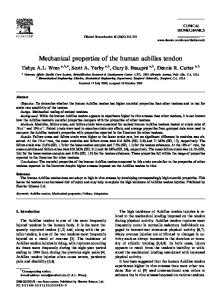
Fig. 1. — Detecting the rupture zone
Fig. 2. — Pre-operative marking and local anaesthesia
on the skin (fig 2), usually using an “X” in the proximal tendon and a “U” or an “X” in the distal part, depending on the patient’s morphology. The final suture pathway in the distal part of the tendon was directed back to the first point of penetration. Local anaesthesia with xylocaine 1% was administered to infiltrate all the suture points and pathways anticipated. A special needle was used, sufficiently long and rigid to enter the skin and easily penetrate at least 5 cm of the tendon, then to exit the skin. All operations were performed using a custom needle with an eyelet at its proximal end. This needle is now avaible as Suturach® (FH Orthopedics, Heimsbrunn, France). The suture selected was inserted through the needle’s eyelet. Its proximal end was held by a forceps. After passing through the tendon for the first time, the needle and suture were brought out on the medial border of the proximal tendon, then reinserted transversely through the same skin point. The lateral exit should be very close to the tendon to avoid damaging the sural nerve. All the exit points of the needle were re-used for
PERCUTANEOUS SUTURE OF ACUTE ACHILLES TENDON RUPTURE
the next suture path. After retrieving the suture on the medial side of the rupture zone, the two strands were used to tighten the suture in the proximal tendon and ensure a good hold. The same was done in the distal part of the tendon. At the end, the suture was brought back to its first point of penetration. The ankle was then placed in equinus position, which brought the two ends of the tendon together and the suture could be properly tightened. After ensuring that good approximation of the tendon ends was achieved and the suture was holding securely, the knot was made and then buried subcutaneously. No cutaneous suture was usually necessary. A plaster cast was applied with the ankle in the equinus position obtained while suturing. This post-operative cast was removed after three weeks and replaced by a second cast with the ankle in slight equinus position, almost at a right angle, for another period of three weeks. Weight-bearing was permitted from the third week. The procedure was performed under local anaesthesia in 48 cases (80%), under plexus block in six cases (10%), under spinal anaesthesia in three cases (5%) and under general anaesthesia in three cases (5%). It was carried out by a senior surgeon in 18 cases (30%), by a graduate resident in 30 cases (50%) and by a resident in 12 cases (20%). The technique was strictly percutaneous. The suture used at the start of the series was a non-resorbable Ethibond® n° 2 suture in 18 cases (30%), then a resorbable Vicryl® n° 2 suture in 42 cases (70%). The patients were followed up and a proforma was used to grade the aspects for evaluation at the consultation. It included the following :
• Appreciation of the local cutaneous condition • Time interval to resuming weight-bearing, returning
239
Complications There was one early infection necessitating further surgery for debridement and immobilisation. This patient had undergone surgical repair of a previous tendon rupture ten years previously. In two cases re-rupture of the tendon occurred after two months and five months respectively. One re-rupture was secondary to an accidental fall and was treated by a Bosworth plasty (1). The other patient had previously undergone repeated steroid infiltrations and open surgical repair of a previous tendon rupture. There was one case of tendinopathy requiring surgical combing of the tendon six months after the repair. There were no neurological complications, particularly involving the sural nerve. Other complications included deep vein thrombosis in two patients and complex regional pain syndrome in one ; one patient developed a painful subcutaneous nodule due to the knot of the nonresorbable suture which was used initially. Return to professional activities Patients returned to work on average 85 days (range : 15-270) after the initial trauma. Two patients did not go back to work as one had retired in the meantime and the other was given invalidity status ; his injury was the result of an occupational accident, and he presented the only infection in the series.
to work and playing sports
• Objective measurement of amyotrophy of the calf • Measurement of the width of the repaired tendon compared with the healthy tendon
• Ability to stand on one foot, hop and walk on tiptoe • Comparison of the range of ankle dorsiflexion and plantar flexion
• Patient’s subjective evaluation of the result RESULTS The mean duration of follow-up was 26 months (range : 18-58). Three patients were lost to followup.
Return to sporting activities Patients resumed their sporting activities on average 5.2 months (range : 3-12) after the initial injury. The three competitive sportsmen returned to sports at six months, at the same level. Twenty two (88%) of the recreational sportsmen and women were able to return to sports. Of these, 15 (68%) resumed sports at the same level of performance, and 7 (32%) at a lower level : 6 of them for fear of re-rupture and one because of a painful tendon. Three recreational sportsmen and women (12%) did not play sports again. Among these three cases were Acta Orthopædica Belgica, Vol. 76 - 2 - 2010
240
J.-L. ROUVILLAIN, T. NAVARRE, O. LABRADA-BLANCO, E. GARRON, W. DAOUD
Two patients had virtually permanent tendon pain and two others had mild occasional pain. The patients’subjective evaluation was as follows : 18 rated the outcome as very satisfactory, 40 as satisfactory and two as poor. DISCUSSION
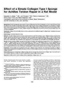
Fig. 3. — Increase in width of the operated right tendon, with the patient standing on tiptoe with the same degree of plantar flexion.
the two patients who had suffered re-rupture and feared a further rupture. Clinical report The local condition of the skin was good in 44 cases (77%). Eleven patients (19%) presented an increase in the circumference of the ankle. In one patient, there was a keloid scar at the point of entry of the needle, and the patient who was re-operated for infection has a permanent adherent scar. Amyotrophy of the calf was observed in six patients. An increase in the width of the sutured tendon compared with the healthy contralateral tendon was observed in 23 patients (fig 3). Monopodal weight-bearing was possible for all the patients except one. Hopping was not possible in eight cases. Walking on tiptoe was not possible in four cases. A 5° limitation of dorsiflexion of the ankle was observed in four patients. Acta Orthopædica Belgica, Vol. 76 - 2 - 2010
There is no consensus on the management of acute Achilles tendon ruptures. Treatment is either surgical (open surgery or tenorrhaphy) or nonsurgical (strict immobilisation in a cast). Although surgical methods have a lower rate of re-rupture than non-surgical methods, they entail a greater risk of complications such as cutaneous necrosis and sural nerve damage (10). The percutaneous suture technique used in this series differs from other methods of surgical repair in being inexpensive. The only specific equipment required is a long needle with an eyelet, sufficiently rigid to transfix the tendon. In this study we had used a custom needle as the prototype of the “Suturach®” needle which we now use. The main concern with percutaneous techniques is the possibility of damaging the sural nerve, which has reportedly occurred in 0 to 10% of cases (7,8). Some authors recommend direct visualisation of the nerve to reduce the risk of such lesions (13). When using our technique, we did not attempt to visualize the nerve but made sure that when passing the needle, it was very much in the “posterior” part of the tendon. The painful subcutaneous nodules described in certain reports (6,9), and which we encountered at the beginning of our study, are probably due to using a non-resorbable suture. Using a slowly resorbable suture avoids this pitfall and does not seem to affect the quality of the repair. The technique used produced satisfactory results with respect to re-ruptures, as we only noted two reruptures (3%). This is better than using non-surgical treatment, with which the re-rupture rate may be as high as 6 to 10% (17,18). Our results are very close to those obtained with open repair, with which the re-rupture rate reportedly ranges from 0% to 6% (4,8).
PERCUTANEOUS SUTURE OF ACUTE ACHILLES TENDON RUPTURE
The technique does not require expensive surgical material and above all, does not leave any foreign body externally in contact with the skin (5) which could be a source of local inflammation, or even of cutaneous necrosis (12). This is particularly important for countries with a hot climate where it is not customary to wear closely fitting shoes. The technique used is reliable, reproducible and easily taught. In this series, it was performed by a number of operators with various levels of training. The immobilisation in a plaster cast that we recommend is for three weeks in the equinus position and three further weeks with the ankle close to 90° flexion. Some physiotherapy protocols recommend early passive mobilisation of the talocrural joint after surgery (1). This is meant to improve vascularisation of the tendon (6) and thus produce more rapid recovery of mobility while limiting the risk of tendon shortening (15). In our series we only observed four cases of significant reduction in dorsiflexion of the ankle. We therefore consider that the strict immobilisation protocol of six weeks that we recommend does not have any major influence on the long-term functional result and allows patients to limit the painful constraints of immediate mobilisation. Weight-bearing in our series was resumed at three weeks. Some authors recommend immediate weight-bearing, on the grounds of obtaining a better functional result (2), but they restrict immediate weight-bearing to selected patients. We considered it difficult to know in advance which patients might benefit from early weight-bearing. Delayed resumption of weight-bearing did not seem to affect the functional result in our experience. Contraindications to this technique are ruptures too close to the calcaneal insertion of the tendon, which do not permit a good distal hold on the tendon, re-ruptures, and ruptures which occurred some considerable time prior to consultation. We consider that two weeks is the maximum time interval that should be accepted after tendon rupture if this technique is to be used. Overall, percutaneous suture of the Achilles tendon appears as a simple, rapid, effective, reproducible and inexpensive technique. It combines the advantages of open surgery with a low risk of
241
re-rupture and those of functional treatment with a lowt risk of infection. The risk of damage to the sural nerve can be reduced by adhering to a rigorous technique when passing a specific needle through the tendon. The very long-term results of this method remain to be evaluated, in particular as regards high-level sportsmen and women. A simple physiotherapy protocol is enough to achieve good functional results, even with non-compliant patients.
REFERENCES 1. Bosworth DM. Repair of defects in the tendo Achillis. J Bone Joint Surg 1956 ; 38-A : 111-114. 2. Costa ML, MacMillan K, Halliday D et al. Randomised controlled trials of immediate weight-bearing mobilisation for rupture of the tendo Achillis. J Bone Joint Surg 2006 ; 88-B : 69-77. 3. Cretnik A, Kosanovic M, Smrkolj V. Percutaneous suturing of the ruptured Achilles tendon under local anesthesia. J Foot Ankle Surg 2004 ; 43 : 72-81. 4. Cretnik A, Kosanovic M, Smrkolj V. Percutaneous versus open repair of the ruptured Achilles tendon : a comparative study. Am J Sports Med 2005 ; 33 : 1369-1379. 5. Delponte P, Potirer L, De Poulpiquet P, Buisson P. Treatment of subcutaneous ruptures of the Achilles tendon by percutaneous tenorraphy. Rev Chir Orthop Réparatr Appar Mot 1992 ; 78 : 404-407. 6. Gelberman R, Menon J, Gonsalves M, Akeson WH. The effects of mobilization on the vascularistion of healing flexor tendons in dogs. Clin Orthop Relat Res 1980 ; 153 : 283-289. 7. Gorschewsky O, Vogel U, Schweizer A, van Laar B. Percutaneous repair of acute Achilles tendon rupture. Foot Ankle Int 2004 ; 25 : 219-224. 8. Haji A, Sahai A, Symes A, Vyas JK. Percutaneous versus open tendo Achillis repair Foot Ankle Int 2004 ; 25 : 215218. 9. Ingvar J, Tagil M, Eneroth M. Nonoperative treatment of Achilles rupture : 196 consecutive patients with a 7% rerupture rate. Acta Orthop 2005 ; 76 : 597-601. 10. Khan RJ, Fick D, Keogh A et al. Treatment of acute Achilles tendon ruptures. A meta-analysis of randomized controlled trials. J Bone Joint Surg 2005 ; 87-A : 22022210. 11. Ma GW, Griffith TG. Percutaneous repair of acute closed ruptured Achilles tendon : a new technique. Clin Orthop Relat Res 1977 ; 128 : 247-255. 12. Maes R, Copin G, Averous C. Is percutaneous repair of the Achilles tendon a safe technique ? A study of 124 cases. Acta Orthop Belg 2006 ; 72 : 179-183.
Acta Orthopædica Belgica, Vol. 76 - 2 - 2010
242
J.-L. ROUVILLAIN, T. NAVARRE, O. LABRADA-BLANCO, E. GARRON, W. DAOUD
13. Majewski M, Rohrbach M, Czaja S, Ochsner P. Avoiding sural nerve injuries during percutaneous Achilles tendon repair. Am J Sports Med 2006 ; 34 : 793-798. 14. Mortensen HM, Skov O, Jensen PE. Early motion of the ankle after operative treatment of a rupture of the Achilles tendon. A prospective, randomized clinical and radiographic study. J Bone Joint Surg 1999 ; 81-A : 983-990. 15. Roberts CP, Palmer S, Vince A, Deliss LJ. Dynamised cast management of Achilles tendon ruptures. Injury 2001 ; 32 : 423-426. 16. Strauss EJ, Ishak C, Jazrawi L, Sherman O, Rosen J. Operative treatment of acute Achilles tendon ruptures : An
Acta Orthopædica Belgica, Vol. 76 - 2 - 2010
institutional review of clinical outcome. Injury 2007 ; 38 : 832-838. 17. Wallace RG, Traynor IE, Kernohan WG, Eames MH. Combined conservative and orthotic management of acute ruptures of the Achilles tendon. J Bone Joint Surg 2004 ; 86-A : 1198-1202. 18. Wong J, Barrass V, Maffulli N. Quantitative review of operative and nonoperative management of Achilles tendon ruptures. Am J Sports Med 2002 ; 30 : 565-575.