ORIGINAL ARTICLE
PATTERN OF PATHOLOGIES ON HYSTEROSALPINGOGRAPHY IN PRIMARY INFERTILITY AND REVIEW OF LITERATURE Saima Naqi, Syed Shafqat-Ul-Islam, Sibtain Raza, Haji Haroon Karachi X-rays & CT scan/Ultrasound centre, Karachi-Paksitan. PJR July - September 2008; 18(3): 82-86
ABSTRACT Hysterosalpingography (HSG) is the radiographic evaluation of the uterus and fallopian tubes and is used predominantly in the evaluation of infertility. It plays an important role in the evaluation of abnormalities related to the uterus and fallopian tubes. Tubal abnormalities seen at HSG can be either congenital or due to spasm, occlusion, or infection. Uterine anomalies can also be due to congenital abnormalities of uterine shape, luminal filling defects, abnormalities of uterine contour. A prospective cross sectional study for a period of 9 months in which 1124 patients who were referred for Hysterosalpingography with infertility were included for the evaluation of pattern of pathologies encountered on hysterosalpingography in primary infertility. K EY W O RDS: Hysterosalpingography, Primary infertility, tubal patency
Introduction Hysterosalpingography (HSG) is used predominantly in the evaluation of infertility. Other indications include postoperative evaluation of women who have undergone tubal ligation or reversal of tubal ligation. It plays an important role in the evaluation of abnormalities related to the uterus and fallopian tubes including congenital anomalies, polyps, leiomyomas, surgical changes, synechiae, adenomyosis, tubal occlusion, hydrosalpinx, and peritubal adhesions etc. But its primary role is in the evaluation of fallopian tubes.1 The aim of this study was to describe the pattern of pathologies encountered on hysterosalpingography in females presenting with primary infertility.
M ate rialand M e th ods A prospective cross sectional study was carried out for a period of 9 months, from 1st April 2008 to 31st December 2008. 1124 patients who were referred from different infertility clinics to radiology department at Karachi X-rays & CT scan/Ultrasound centre were included in the study. Corre s ponde nce : Dr. Shafqat ul Islam Karachi Xrays and CT scan/ Ultrasound Center, Karachi, Paksitan. Tel. No.: 2229001 E-mail:
[email protected]
PA K I S TA N J O U RN A L O F R A D I O L OG Y
Patients with primary infertility irrespective of age and duration of infertility were included in study. Patients with secondary infertility, history of prior pelvic surgery, or known allergy to contrast media were excluded. 474 patients were excluded, who had prior history of pelvic surgery (n=76) and patients with secondary infertility (n=398). A total of 650 patients with primary infertility were finally included in the study. All patients had hysterosalpingogram examination under fluoroscopic guidance. Procedure performed during 7th-12th day of menstrual cycle. All patients were premedicated with 3mg bromazepam, the night before procedure. An informed consent was obtained from all patients. A detailed performa for each patient was filled up prior to procedure. Intravenous Buscopan was also administered to all patients just before starting the examination. All examinations were performed under fluoroscopic guidance. Water-soluble contrast material is then slowly instilled, with intermittent fluoroscopy and images obtained to evaluate the uterus and fallopian tubes. Image interpretation was made by a consultant radiologist.
Re s ul ts A total 650 patients out of initially recruited 1124 patients were finally evaluated for this study. All examinations were technically successful. No significant PJR July - September 2008; 18(3)
82
post procedure complication noted except mild pain and per-vaginal bleeding in a minority of patients. Normal uterine cavity with free bilateral peritoneal spillage of contrast noted in 446(68.6%) patients. While different pathologies encountered in 204(31.3%). Hysterosalpingographic results of 650 patients are presented in table below; NORMAL EXAMINATION
446
68.6%
ABNORMAL FINDINGS
204
31.3%
TUBAL BLOCKAGE
114
17.5%
HYDROSALPINX
24
3.6%
LOCULATED SPILL
20
3.0%
FIBROIDS
32
4.9%
ADENOMYOSIS
2
0.3%
CONGENITAL ABNORMALITIES
12
1.8%
Most commonly observed pathology was tubal blockage and out of 114 (17.5%) patients with tubal blockage 74(11.3%) had unilateral while 40(6.1%) patients had bilateral tubal blockage. Second most commonly observed pathology was uterine fibroids noted in 32(4.9%) patients. Loculated spillage of contrast noted in 20(3.0%) patients, which was unilateral in 12(1.8%) and bilateral in 8(1.2%) patients. Hydrosalpinx noted in 24(3.6%), unilateral in 14 (2.1%) and bilateral in 10(1.5%) patients. Findings of adenomyosis were seen in 2(0.3%) patients. Congenital abnormalities noted in 12(1.8%) patients, Arcuate shape uterine cavity noted in 6 (0.9%) patients, unicornuate unicollis in 3 (0.4%), bicornuate unicollis in 2 (0.3%) patients, bicornuate bicollis in 1(0.1%) patient.
Dis cus s ion Hysterosalpingography is one of the most commonly used techniques for the initial evaluation of cause of infertility. Primarily used for the evaluation of fallopian tubes, it’s a reliable procedure, quick and easy to perform, safe and cost effective in comparison with other modalities. Hysterosalpingography is contraindicated during menstruation and pregnancy. PA K I S TA N J O U RN A L O F R A D I O L OG Y
Other contra-indications include pelvic sepsis, a recent dilatation and curettage, severe cardiac or renal disease, uterine malignancy i.e. endometrial carcinoma and sensitivity to contrast media.2 Some of the known complications are bleeding and infection which happen to be the most common. Other potential complications are extremely rare like reactions due to the contrast material or perforation of uterus.3 Pelvic exposure to radiation is another side effect of hysterosalpingography. Hysterosalpingography has both therapeutic and diagnostic value. Following hysterosalpingography, certain minor uterine adhesions and partial tubal occlusion are lysed. An increase in pregnancy rate has been observed in the months after Hysterosalpingography without any other gynaecological intervention.4 HSG is considered to have a high sensitivity (60-98%) but a low specificity (15-80%).5 The lack of accuracy could be influenced by the faulty technique. Accuracy could be improved by optimizing the technique for performing the procedure and training the personnel. On the other hand diagnostic accuracy of HSG can also be influenced due to variability among observer.6 A study by Mol et al analyzed interobserver as well as intraobsever reproducibility on four HSG items- proximal tubal obstruction, distal tubal obstruction, hydrosalpinx and peritubal adhesion. The authors found that reproducibility within and between observers for proximal tubal occlusion was almost perfect, for distal obstruction and hydrosalpinx only substantial and for adhesion from slight to fair.7 The results by Renbaum and coworkers were similar.8 The possible variability among observers was not taken into account in our study. The most commonly observed pathology in our study is tubal blockage. It may be difficult to differentiate tubal obstruction from cornual spasm and those due to technical reasons.9 Horwitz et al described cornual spasm radiologically, as spasm characterized by rounded smooth corneal margin whereas cornual occlusion was characterized by pointed or irregular cornual margin. 10 These radiological features are difficult to evaluate objectively. The high incidence of tubal blockage in primary infertility in our study may be a reflection of the high prevalence rate of pelvic inflammatory diseases & especially tuberculosis in our environment. Second most common abnormality noted in our study is uterine fibroid. Uterine cavity abnormalities can be the cause of infertility in 10-15% of women. Abnormal uterine findings occur in approximately PJR July - September 2008; 18(3)
83
50% of infertile women. 11 Because of the high prevalence of uterine abnormalities, inspection of the uterine cavity is routinely performed in the evaluation of infertile women. In a study conducted by T.O.Bello et al, normal examinations were noted in 60% cases. Most commonly observed pathology in their study was hydrosalpinx.9 Another study conducted by Poonam et al showed among tubal pathologies, tubal blockage is most commonly observed.12 Another study conducted by Anna Roma Dalfó et al to detect intrauterine abnormalities on hystersalpingography combined with hysteroscopy, showed normal uterine cavity in 47% and some abnormalities in 53% cases.13 Another study conducted by Preeti Kanal et al to evaluate causes of infertility by diagnostic laparoscopy combined with Hysterosalpingography, also found tubal blockage without adhesions as a most common cause of infertility.14
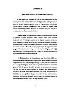
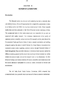
Figure 3: Adenomyosis
Figure 4: Bicornuate unicollis uterine cavity
Figure 1: Bilaterally blocked fallopian tubes giving typical appearance of salpingitis isthmica nodosa.
Figure 2: Multiple filling defects in uterine cavity (fibroids)
PA K I S TA N J O U RN A L O F R A D I O L OG Y
Other modalities used for evaluation of cause of infertility include Ultrasound examination, Hysterosonography, Selective Salpingography and Fallopian Tube Catheterization, MRI, Hysteroscopy, Laproscopy. Per abdominal/ transvaginal ultrasound both play an important role in evaluation of cause of primary infertility. Main indications include evaluation of uterine cavity and ovaries like polycystic ovaries, uterine fibroids, adenomyosis etc. Contrast material–enhanced sonohysterography, a relatively new modality has been suggested as a means of assessing tubal patency.15,16 It does not however allow visualization of the entire tube. Sonohysterography is similar to HSG for evaluating uterine abnormalities but it is inferior to HSG for evaluating tubal anomalies.17 The procedure is less painful and is also radiation free. Selective salpingography and fallopian tube catheterization combination has improved diagnosis PJR July - September 2008; 18(3)
84
& management of infertility caused by tubal obstruction. The technique is useful in differentiating spasm from true obstruction. In fallopian tube recanalization a catheter and guide wire is used to clear proximal tubal obstructions. Results reported to date indicate that procedure is successful in 62%–90% of patients and provides important diagnostic information in all these women.18 MRI, a useful modality as an adjunct for routine infertility work-ups especially for evaluation of uterus and ovaries, visualization of pituitary adenomas, differentiation of müllerian duct anomalies, leiomyoma, adenomyosis and endometriosis etc. Its radiation free, non invasive and less observer dependent. Its major limitations include cost, phobias and inavailability. MR hysterography with a heavily T2-weighted sequence (a procedure that is similar to MR cholangiopancreatography) has also been suggested for visualization of the fallopian tubes.19 However, the size of the normal fallopian tube is at or below the resolution of most MR imagers.20 Moreover, even if the tube is visualized, MR hysterography does not help to assess tubal patency. Instillation of saline solution or water through an HSG catheter in conjunction with MR hysterography is possible, but this procedure has not gained wide acceptance.19 Hysteroscopy is a minimally invasive approach done to visualise the interior of the uterus and to detect the abnormalities & simultaneous treatment can be done by hysteroscopic surgery. A comparative study by Anna Roma Dalfó et al compared the results of hysterosalpingography with hysteroscopy, estimated sensitivity of HSG 81.2% and specificity 80.4% & overall agreement correlation between HSG and hysteroscopy was 73%.13 The accuracy of HSG and hysteroscopy for detecting uterine abnormalities in infertile patients has been discussed by various authors. 21,22 Hysteroscopy has an increased accuracy over HSG, although the magnitude of this discrepancy is controversial. Although some authors have suggested that HSG should be completely replaced by hysteroscopy22,23,24, others have found that hysteroscopy added little information when HSG results were negative.25 Diagnostic laparoscopy provides a direct visual access to inner pelvic anatomy without major abdominal surgery so that ovaries, fallopian tubes & uterus can be studied in more detail. It is used as a reference standard in most clinical studies. However data of
PA K I S TA N J O U RN A L O F R A D I O L OG Y
some studies found the choice of Laproscopy as a gold standard, questionable. Findings in a meta-analysis comparing results of HSG and laproscopy for diagnosis of tubal pathology indicated that 35% of the tubes found to be occluded at Laproscopy showed patency at HSG.25 Another study conducted by Egle Tvarijonaviciene and co-workers for evaluating tubal pathologies on Hysterosalpingography, used laproscopy as reference standard, concluded that HSG is more accurate in diagnosing tubal occlusion while for peritubal adhesion its accuracy is poor.6
Concl us ion Despite new innovations and recent advances in imaging field, hysterosalpingography remains a valuable imaging tool for assessment of pathologies including patency of fallopian tubes and morphology of uterine cavity etc, in the evaluation of cause of infertility.
Re fe re nce s 1. S i m p s o n . W. L , B e i t i a . L . G , M e s t e r . J . Hysterosalpingography: A Reemerging Study. RadioGraphics 2006; 26:419–31. 2. Kathryn Grumbach. Hysterosalpingography. A concise Textbook of Clinical Imaging. 2nd edition by David Sutton; Jeremy W.R. Young: 702-3. 3. Ott DJ, Fayez JA, Chen MYM. Techniques of hysterosalpingography. In: Ott DJ, Fayez JA, Zagoria RJ, eds. Hysterosalpingography: a text and atlas. 2nd ed. Baltimore, Md: Williams & Wilkins, 1998; 11–27. 4. De Muyldex X. Role of hysterosalpingography in the evaluation of infertility in Black Africa. Med. Trop 1995; 55(2): 160-4. 5. Golan A, Eilat E, Ron-El R, Herman A, Soffer Y, Bukovsky I. Hysteroscopy is superior to hysterosalpingography in infertility investigation. Acta Obstet Gynecol Scand1996; 75:654-6. 6. Tvarijonaviciene.E, Nadišauskiene.R.J. The value of hysterosalpingography in the diagnosis of tubal p a th o l o g y a mo n g i n fe r ti l e p a ti e n ts. Medicina(Kaunas) 2008; 44(6):439-48. PJR July - September 2008; 18(3)
85
7. Mol BW, Swart P, Bossuyt PM, van Beurden M, van der Veen F. Reproducibility of the interpretation of hysterosalpingography in the diagnosis of tubal pathology. Hum Reprod 1996 Jun;11(6):1204-8. 8. Renbaum L, Ufberg D, Sammel M, Zhou L, Jabara S, Barnhart K. Reliability of clinicians versus radiologists for detecting abnormalities on hyste rosalpi ngogram fi lms. Fer t il St er il 2002;78(3):614-8. 9. Bello.T.O. Pattern of tubal pathology in infertile women on Hysterosalpingography in Ilorin, Nigeria. Annals of African Medicine 2004;3(2):77-9. 10. Horwitz RC, Morton PC, Shaft MI, Hugo PA. R ad i ol o gi ca l a p pr oa ch t o in fer t i l it y –hysterosalpingography. Br J Radiol 1979; 52: 255–62. 11. Brown SE, Coddington CC, Schnorr J, Toner J, Gibbons W, Oehninger S. Evaluation of outpatient hysteroscopy, saline infusion hysterosonography, and hysterosalpingography in infertile women: a prospective, randomized study. Fertil Steril 2000;74:1029–34. 12. Poonam. The role of hysterosalpingography in cases of subfertility. Kathmandu University Medical Journal (2007), Vol. 5, No. 4, Issue 20, 456-60. 13. Dalfó.A.R, Úbeda.B, Úbeda.A. Diagnostic Value of Hysterosalpingography in the Detection of Intrauterine Abnormalities: A Comparison with Hysteroscopy. AJR 2004;183:1405–9. 14. Kanal.P, Sharma.S. Study of Primary Infertility in females by Diagnostic Laparoscopy. Internet Journal of Medical Update, Vol. 1, No. 2, Jul-Dec 2006. 15. Reis MM, Soares SR, Cancado ML, Camargos AF. Hysterosalpingo contrast sonography (Hy-CoSy) with SH U 454 (Echovist) for the assessment of tubal patency. Hum Reprod 1998;13(11): 3049–52.
17. D a r w i s h . A . M , Yo u s s e f. A . A . S c r e e n i n g Sonohysterography in Infertility. Gynecol Obstet Invest1999;48:43-7. 18. Thurmond.A.S, Machan.S.L, Maubon.A.J, et al. A Review of Selective Salpingography and Fallopian Tube Catheterization. RadioGraphics 2000;20: 1759-68. 19. Rouanet De Lavit JP, Maubon AJ, Thurmond AS. MR hysterography performed with saline injection and fluid attenuation inversion recovery sequences: initial experience. AJR Am J Roentgenol 2000; 175(3):774–6. 20. Hagspiel KD, Altes TA, Mugler JP, Brookeman JR. Magnetic resonance hysterography and hysterosalpingography using hyperpolarized (3)He: demonstration of feasibility in an animal model. J Magn Reson Imaging 2000;12(6):1009–13. 21. Gaglione R, Valentini A, Pistilli E, Nuzzi NP. A comparison of hysteroscopy and hysterosalpingography. Int J Gynaecol Obstet 1996;52:151-3. 22. Golan A, Eilat E, Ron-El R, Herman A, Soffer Y, Bukovsky I. Hysteroscopy is superior to hysterosalpingography in infertility investigation. Acta Obstet Gynecol Scand1996; 75:654-6. 23. Cicinelli E, Matteo M, Causio F, Schonauer LM, Pinto V, Galantino P. Tolerability of the mini-panendoscopic approach (transvaginal hydrolaparoscopy and minihysteroscopy) versus hysterosalpingography in an outpatient infertility investigation. Fertil Steril2001; 76:1048-51. 24. Gordts S, Campo R, Puttemans P, et al. Investigation of the infertile couple: a one-stop outpatient endoscopy-based approach. Hum Reprod2002; 17:1684-7. 25. Fayez JA, Mutie G, Schneider PJ. The diagnostic value of hysterosalpingography and hysteroscopy in infertility investigation. Am J Obstet Gynecol1987; 156:558-60.
16. Killick SR. Hysterosalpingo contrast sonography as a screening test for tubal patency in infertile women. J R Soc Med 1999;9 2(12):628–31.
PA K I S TAN J O U R N A L O F R A D I O LO G Y
PJR July - September 2008; 18(3)
86