Global Veterinaria 12 (6): 891-896, 2014 ISSN 1992-6197 © IDOSI Publications, 2014 DOI: 10.5829/idosi.gv.2014.12.06.102143
Pathogenicity, Plasmid Profile and Effectiveness of Some Herbal Extracts on Pseudomonas fluorescens Isolated from Oreochromis niloticus Fish A.M. Younes and A.Y. Gaafar Department of Hydrobiology, National Research Centre, Giza, Egypt Abstract: Pseudomonas fluorescens is one of serious fish diseases in Egypt responsible for sever economic losses. Four P. fluorescens isolates were previously isolated from outbreaks in Al-Abbassa and Al-Fayoum farms were used in this study. The plasmid profile of all isolates showed the pattern of two bands. Disc diffusion method was used to test the potency of some natural plant extracts in controlling P. fluorescens infections. The most effective methanolic extracts of plants and substances were Propolis, Thyme (Thymus vulgaris) whole plant, Juniper (Juniperus communis) fruits, Myrrh (Commiphora molmol) oleoresin, Aloe (Aloe vera L.,) dried gel, Tamarind (Tamarindus indica) paste and Tea (Camellia sinensis) leaves. Other extracts didn’t show any inhibition zone and presumptively not effective. Key words: Pseudomonas fluorescens
Oreochromis niloticus
INTRODUCTION
Natural Plant Extracts
grow at 37°C [4]. Plasmids are common in many species of Pseudomonas. Conjugative plasmids are an essential factor in the evolution of these bacteria. Plasmids of Pseudomonas spp. are carrying genes for virulence, antibiotics resistance, phytotoxin and phytohormone biosynthesis, UV tolerance and bioremediation of polluted environments [5-7]. Natural plant products present a good alternative to chemotherapeutics and antibiotics and other banned drugs being safer for the reared organism and humans. Moreover, these substances also possess other valuable properties; they are nontoxic, biodegradable and biocompatible, inexpensive, easy to get and to produce locally in our country.
Increasing the demand for protein, Nile Tilapia (Oreochromis niloticus) has attained a great economic importance in Egypt. Infectious diseases of cultured fish are the most impediments on the expansion and development of aquaculture. Bacterial fish pathogens under these stress factors of intensive culture would prevail causing severe economic losses with 80% of mortalities and intricate health hazard both in Egypt and worldwide [1, 2]. P. fluorescens is gram-negative, motile, rod shape and highly heterogeneous species of -Proteobacteria known for their versatile metabolism and for their ability to colonize different ecological niches that inhabit soil, plants and water surfaces. The optimum growth temperature is between 25 to 30°C. It has been reported to cause disease in a wide range of fishes. Pseudomonas infection in fish leads to development of the so called Red skin disease, which occurs throughout the year especially at low temperatures. The disease is characterized by petechial hemorrhage, darkness of the skin, detached scales, abdominal ascites and exophthalmia [2, 3]. Some strains of P. fluorescens found to be opportunistic human pathogens. MFN1032 is a clinical strain, belonging to P. fluorescens biovar I which was isolated from a patient with a lung infection and is able to Corresponding Author:
Plasmids
MATERIALS AND METHODS Samples and Biochemical Identification: A total of four strains of P. fluorescens were isolated from moribund O. niloticus from Al-Abbassa governmental fish farms and Al-Fayoum private fish farms, Egypt. The strains were isolated from the kidney of individual diseased fish and cultivated on Pseudomonas selective agar base (LabM, UK) supplemented with CFC supplement. The suspected Pseudomonas isolates were identified by oxidase, catalase. The isolates were then biochemically identified using API20NE strips (BioMerieux, France) according to manufacturer’s instructions.
Alkhateib Y. Gaafar, Veterinary Research Division, National Research Centre, Postal code: 12622, Dokki, Giza, Egypt. E-mail:
[email protected].
891
Global Veterinaria, 12 (6): 891-896, 2014
Plasmid DNA Isolation: Plasmid miniprep kit (Thermo Scientific, USA) was used, as recommended by the manufacturer, to extract 10kb (Fig 1). Histopathology: Circulatory, degenerative, proliferative and necrotic changes were evident. Moreover, some bacterial agents were detected in histological sections. Haemorrhages were noticed extensively in haemopoeitic tissues. Moreover, Vascular and sinusoidal congestions diffuse vacuolar degeneration and discrete necrotic changes were commonly evident in hepatocytes as well as pancreatic acinar cells (Fig. 2a). Congestion of renal and splenic blood vessels also detected. Moreover, diffuse vacuolation of tubular epithelial cells and necrotic areas were frequently noticed in glomerulo-tubular and interstitial tissues. These necrotic areas were replaced by empty spaces and tissue debris. Furthermore, multifocal depletion of renal and splenic heamopoeitic elements was eminent concurrently with hyper-activation of melanomacrophage centers and multifocal infiltration of mononuclear cells (Fig. 2b, c). 892
Global Veterinaria, 12 (6): 891-896, 2014
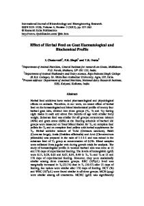
Fig. 1: Agarose gel electrophoresis of plasmid profile of some P. fluorescens isolates. Lane 1: kb molecular DNA marker. Lane 2 and 3: two P. fluorescens strains from Al-Fayoum governorate bearing two plasmids. Lane 4 and 5: two P. fluorescens strains from Al-Abbasa governmental farms bearing the same plasmids.
Fig. 2: (a) Hepatopancreas of O. niloticus experimentally infected with P. fluorescens after 48 hours showing congestion of main blood vessels and sinusoidal spaces with diffuse vacuolar degeneration and hepatocytic necrosis. (b) Posterior kidney of O. niloticus experimentally infected with P. fluorescens after 48 hours showing multifocal necrosis of the interstitial tissue with discrete tubular necrosis leaving cyst like spaces. (c) Spleen of O. niloticus experimentally infected with P. fluorescens after 48 hours showing congestion and multifocal necrosis with discrete depletion of white pulp with activation of melanomacrophage centers. (d) Gills of O. niloticus experimentally infected with P. fluorescens after 48 hours showing separation in-between the epithelial cell lining of the secondary gill lamellae and the underlying capillary bed with hyperplasia of the epithelial lining at the base of the secondary gill lamellae. (Bar = 50µm). 893
Global Veterinaria, 12 (6): 891-896, 2014 Table 1: Effects of some herbal extracts against three Pseudomonas fluorescens strains by using disc diffusion method. P. fluorescens strains ----------------------------------
P. fluorescens strains -----------------------------------
Plant extract or substance
Concentration
FP2
FP6
BP1
Plant extract or substance
Concentration
FP2
FP6
BP1
Linden (Tilia plalyphyllys) leaves Chinese Dar (Cinnamomum cassia) park
25% 10%
-
-
-
Myrrh (Commiphora molmol) oleoresin Cinnamon (Cinnamomum verum) park
50% 25%
+ -
+ -
+ -
Salvia (Salvia miltiorrhiza) leaves Meswak (Salvadora persica) roots
7% 10%
-
-
-
Cinchona (Cinchona officinalis) park Basil (Ocimum basilicum) whole plant
20% 25%
+
+
+
Propolis Thyme (Thymus vulgaris) whole plant
20% 10%
+ -
+ -
+ -
Bees wax (Cera alba) Aloe (Aloe vera) dried gel
10% 10%
+
+
+
Oregano (Origanum vulgare) whole plant Juniper (Juniperus communis) fruits
10% 50%
+
+
-
Tamarind (Tamarindus indica) paste Tea (Camellia sinensis) leaves
10% 10%
+ +
+ +
+ +
The gills showed congestion of lamellar blood vessels, diffuse and multifocal hyperplasia of epithelial lining of the secondary lamellae (Fig. 2d).
which inactivate certain antibiotics and S-factor plasmids capable of initiating chromosomal gene transfer. Plasmids in Pseudomonas species are also known to code for heavy metal resistance and degradation of organic compounds [12]. Analysis of 10 strains, only one strain of P. fluorescens A506, harbored a conjugative plasmid of 57-kb [7]. Also, plasmid DNA was isolated from P. aeruginosa and designated as pBC15. The size of the plasmid DNA was approximately 23 kb. The Pseudomonas aeruginosa isolate exhibited resistance to heavy metals such as cadmium, chromium, nickel and lead [13]. Members of the Enterobacteriaceae family and the genus Pseudomonas carry plasmids belonging to more than 30 Incompatibility (Inc) groups especially (IncP, W, N and Q) which can transfer between and maintain themselves in both enteric bacteria and strains of Pseudomonas [14]. Regarding histopathological lesions noticed in infected fishes, circulatory, degenerative, proliferative and necrotic changes were evident. Destruction of the critical components of the circulatory and immune system by toxic bacterial extracellular products is thought to be the corner stone behind these pathological alterations [15, 16]. Among these ECPs, proteases and haemolysins are at the forefront, contributing significantly to the ruinous nature of these diseases [17, 18]. Plant essential oils have been used for hundreds of years as natural medicines to combat bacteria, fungi and viruses. Several essential oils confer antimicrobial activity by damaging the cell wall and membrane, leading to cell lysis, leakage of cell contents and inhibition of proton motive force [19, 20]. Herbs or herbal products also have a role in aquaculture at present time, because it is natural and biodegradable and has antimicrobial activity. Several medicinal plants have shown activity against many important pathogenic bacteria of fish. Similar to our results Kavanaugh and Ribbeck [20] found that cassia, clove (Syzygium aromaticum), Peru balsam (Myroxylon balsamum), red thyme (Thymus vulgaris) and tea tree (Melaleuca alternifolia) oils were effective in killing P. aeruginosa. The essential oil
Potency of Herbal Extracts Against P. fluorescens: Disc diffusion method was followed to evaluate the antibacterial activity against three P. fluorescens isolates. No zone of inhibition was seen in the control disc without antibiotics. As shown in Table 1, some herbal extracts had variable zone of inhibition and were found to be effective in controlling the Pseudomonas infection in Tilapia fish. These extracts and substances were; Propolis, Thyme (Thymus vulgaris) whole plant, Juniper (Juniperus communis) fruits, Myrrh (Commiphora molmol) oleoresin, Aloe (Aloe vera L.,) dried gel, Tamarind (Tamarindus indica) paste and Tea (Camellia sinensis) leaves. Other extracts didn’t show any inhibition zone and presumptively not effective. These findings indicated that these extracts can be developed as preferred natural agents for the treatment of P. fluorescens isolates. DISCUSSION P. fluorescens is an aquaculture pathogen that has been reported to cause disease in a wide range of fish species, including Indian major carps, black carp, common carp and Japanese flounder. P. fluorescens is considered to be one of the primary causes of pseudomonas septicemia, tail and fin rot in fish, with all over the year with maximum prevalence in summer [3, 10]. P. fluorescens is one of the most pathogenic bacteria affecting fish farms in Egypt. P. fluorescens Biovar I (15 isolates), P. fluorescens Biovar II (17 isolates), P. fluorescens Biovar III (13 isolates) were isolated from 480 fish in Egypt [11]. The result of plasmid isolation showed that all isolates either from Al-Abbassa or from Al-Fayoum have the same plasmid profile bearing two plasmid of about ~3.5kb and other plasmid band was >10kb. In accordance with this result, a variety of plasmids have been isolated from Pseudomonas species such as R-factor plasmids, 894
Global Veterinaria, 12 (6): 891-896, 2014
composition of Thymus vulgaris include mainly thymol and carvacrol [21]. In addition, five herbs Allium sativum, Calotropis gigantea, Momordica charantia, Polygonum hydropiper, Psidium guajava found to be highly effective against three high virulent aquaculture bacteria Aeromonas hydrophila, Pseudomonas fluorescens and Edwardsiella tarda [22]. Similarly, aqueous extracts of common fruits, herbs and spices (raspberry, blueberry, blackberry, cranberry, grape, oregano, thyme, basil, kale, ginger, turmeric) significantly inhibited quorum sensing by the inhibition of swarming motility in the case of pathogenic P. aeruginosa and E. coli [23]. Also, propolis may be a useful adjunctive agent for traditional antibiotic therapy for P. aeruginosa keratitis in rabbits [24].
5.
6.
7.
8. 9.
CONCLUSION The result of plasmid isolation of P. fluorescens showed that all isolates either from Al-Abbassa or from Al-Fayoum have the same plasmid profile bearing two plasmids and could play a role in pathogenesis and resistance of this pathogen. Due to the increased prevalence of antibiotic resistance, using of antibiotics in aquaculture needs to be reduced and replaced with alternative effective medicinal plants against the bacterial diseases. These preliminary findings provided that some extracts were found to be effective in treatment of P. fluorescens and can be used for treating bacterial infection in fish farms.
10.
11.
12.
REFERENCES 13. 1. 2.
3.
4.
Plumb, J.A., 1999. Overview of warmwater fish diseases. Journal of Applied Aquaculture, 9(2): 1-10. Austin, B. and D.A. Austin, 2007. Bacterial fish pathogens: disease of farmed and wild fish, Springer Science & Business Media, pp: 552. Swain, P., A. Behura, S. Dash and S. Nayak, 2007. Serum antibody response of Indian major carp, Labeo rohita to three species of pathogenic bacteria; Aeromonas hydrophila, Edwardsiella tarda and Pseudomonas fluorescens. Veterinary immunology and Immunopathology, 117(1): 137-141. Chapalain, A., G. Rossignol, O. Lesouhaitier, A. Merieau, C. Gruffaz, J. Guerillon, J.M. Meyer, N. Orange and M. Feuilloley, 2007. Comparative study of 7 fluorescent pseudomonad clinical isolates. Canadian Journal of Microbiology, 54(1): 19-27.
14.
15.
16. 17.
895
Frost, L.S., R. Leplae, A.O. Summers and A. Toussaint, 2005. Mobile genetic elements: the agents of open source evolution. Nature Reviews Microbiology, 3(9): 722-732. Strateva, T. and D. Yordanov, 2009. Pseudomonas aeruginosa–a phenomenon of bacterial resistance. Journal of Medical Microbiology, 58(9): 1133-1148. Stockwell, V.O., E.W. Davis, A. Carey, B.T. Shaffer, D.V. Mavrodi, K.A. Hassan, K. Hockett, L.S. Thomashow, I.T. Paulsen and J.E. Loper, 2013. pA506, a conjugative plasmid of the plant epiphyte Pseudomonas fluorescens A506. Applied and Environmental Microbiology, 79(17): 5272-5282. Roberts, R.J., 2012. Fish pathology, John Wiley & Sons., pp; 581. Wikler, M.A., 2006. Performance standards for antimicrobial susceptibility testing: Sixteenth informational supplement, Clinical and Laboratory Standards Institute. Zhang, W.W., Hu, Y.H., Wang, H.L. and L. Sun, 2009. Identification and characterization of a virulenceassociated protease from a pathogenic Pseudomonas fluorescens strain. Veterinary Microbiology, 139(1): 183-188. Eissa, N., E.A. El-Ghiet, A. Shaheen and A. Abbass, 2010. Characterization of pseudomonas species isolated from Tilapia Oreochromis niloticus in Qaroun and Wadi-El-Rayan lakes, Egypt. Global Veterinaria Journal, 5: 116-121. Palchaudhuri, S. and A. Chakrabarty, 1976. Isolation of plasmid deoxyribonucleic acid from Pseudomonas putida. Journal of Bacteriology, 126(1): 410-416. Raja, C.E. and G. Selvam, 2009. Plasmid profile and curing analysis of Pseudomonas aeruginosa as metal resistant. International Journal of Environmental Science & Technology, 6(2): 259-266. Popowska, M. and A. Krawczyk-Balska, 2013. Broadhost-range IncP-1 plasmids and their resistance potential. Front. Microbiol, 4(44): 1-8. Kimura, H. and R. Kusuda, 1979. Studies on the Pathogenesis of Streptococcal Infection in Cultured Yellowtails Seriola spp. - Effect of the Cell Free Culture on Experimental Streptococcal Infection. Journal of Fish Diseases, 2(6): 501-510. Woo, P.T., 2011. Fish Diseases and Disorders: Volume 3: Viral, Bacterial and Fungal Infections, Cabi. Pazos, F., 1997. Flexibacter maritimus: estudio fenotípico, inmunológico y molecular. Universidad Santiago de Compostela Thesis. Doctoral.
Global Veterinaria, 12 (6): 891-896, 2014
18. Li, J., L.R. Zhou and N.Y.S. Woo, 2003. Invasion route and pathogenic mechanisms of Vibrio alginolyticus to silver sea bream Sparus sarba. Journal of Aquatic Animal Health, 15(4): 302-313. 19. Burt, S., 2004. Essential oils: their antibacterial properties and potential applications in foods—a review. International Journal of Food Microbiology, 94(3): 223-253. 20. Kavanaugh, N.L. and K. Ribbeck, 2012. Selected antimicrobial essential oils eradicate Pseudomonas spp. and Staphylococcus aureus biofilms. Applied and Environmental Microbiology, 78(11): 4057-4061. 21. Thompson, J.D., J.C. Chalchat, A. Michet, Y.B. Linhart and B. Ehlers, 2003. Qualitative and quantitative variation in monoterpene co-occurrence and composition in the essential oil of Thymus vulgaris chemotypes. Journal of Chemical Ecology, 29(4): 859-880.
896
22. Muniruzzaman, M. and M. Chowdhury, 2013. Evaluation of medicinal plants through fish feed against bacterial fish disease. Progressive Agriculture, 19(2): 151-159. 23. Vattem, D., K. Mihalik, S. Crixell and R. McLean, 2007. Dietary phytochemicals as quorum sensing inhibitors. Fitoterapia, 78(4): 302-310. 24. Onlen, Y., C. Tamer, H. Oksuz, N. Duran, M.E. Altug and S. Yakan, 2007. Comparative trial of different antibacterial combinations with propolis and ciprofloxacin on Pseudomonas keratitis in rabbits. Microbiological Research, 162(1): 62-68.