University of Wollongong
Research Online University of Wollongong Thesis Collection
University of Wollongong Thesis Collections
2010
Patellar Tendon Loading During Dynamic Landings: How this is Moderated by Fatigue and the Presence of a Patellar Tendon Abnormality Suzi Edwards University of Wollongong
Recommended Citation Edwards, Suzi, Patellar Tendon Loading During Dynamic Landings: How this is Moderated by Fatigue and the Presence of a Patellar Tendon Abnormality, Doctorate of Philosophy thesis, School of Health Sciences, University of Wollongong, 2010. http://ro.uow.edu.au/theses/3205
Research Online is the open access institutional repository for the University of Wollongong. For further information contact Manager Repository Services:
[email protected].
Patellar Tendon Loading During Dynamic Landings: How this is Moderated by Fatigue and the Presence of a Patellar Tendon Abnormality
A thesis submitted in fulfilment of the requirements for the award of the degree
Doctorate of Philosophy from University of Wollongong by Suzi Edwards BAppSc (Ex Sport Sc), MSc (Hons)
School of Health Sciences 2010
Dedication
I dedicate this thesis to my parents, Bev and Bill Edwards. Through all the hard times that I have endured, you have been by my side providing me with unconditional love and support in helping me achieve my dreams. I love you so much and I thank you from my heart for everything that you have done for Zale and I.
i
Declaration
I, Suzi Edwards, declare that this thesis “Patellar Tendon Loading During Dynamic Landings: How this is Moderated by Fatigue and the Presence of a Patellar Tendon Abnormality”, submitted in fulfilment of the requirements for the award of Doctor of Philosophy, in the School of Health Sciences, University of Wollongong, is wholly my own work unless otherwise referenced or acknowledged in this thesis. This thesis has not been submitted for a degree at any other university or institution.
Suzi Edwards 17 February 2010
ii
Publications This thesis is composed of chapters that have been written as journal articles and includes:
Chapter 2:
Edwards S, Steele JR, Cook JL, Purdam CR, McGhee DE. Lower limb symmetry cannot be assumed when investigating the biomechanics of a stop-jump landing: Implications for experimental design.
Journal of
Science and Medicine in Sport. Submitted for publication February, 2010. Chapter 3:
Edwards S, Steele JR, McGhee DE, Cook JL, Purdam CR. Reliability and validity of a lower limb stretch-shortening cycle fatigue protocol. Medicine & Science in Sports & Exercise.
Submitted for publication
February, 2010. Chapter 4:
Edwards S, Steele JR, Cook JL, Purdam CR, McGhee DE, Munro BJ. Characterising patellar tendon loading during the landing phases of a stopjump task. Scandinavian Journal of Medicine & Science in Sports. 2010: Early View: doi: 10.1111/j.1600-0838.2010.01119.x.
Chapter 5:
Edwards S, Steele JR, Purdam CR, Cook JL, McGhee DE. Alterations to landing technique and patellar tendon loading in response to fatigue. Medicine & Science in Sports & Exercise.
Submitted for publication
February, 2010. Chapter 6:
Edwards S, Steele JR, McGhee DE, Beattie S, Purdam CR, Cook JL. Landing strategies of athletes with an asymptomatic patellar tendon abnormality. Medicine & Science in Sports & Exercise. 2010: 42(11): doi: 10.1249/MSS.0b013e3181e0550b.
Chapter 7:
Edwards S, Steele JR, Purdam CR, McGhee DE, Beattie S, Cook JL. Asymptomatic athletes with a patellar tendon abnormality do not adapt when fatigued.
British Journal of Sports Medicine.
Submitted for
publication February, 2010.
As the primary, supervisor, I, Professor Julie Steele, declare that the greater part of the work in each article listed above is attributed to the candidate, Suzi Edwards. In each of the above manuscripts, Suzi has contributed to study design, was solely responsible for data collection and data analysis, and was largely responsible for data interpretation. iii
The first draft of each manuscript was written by the candidate and Suzi was then responsible for responding to the editorial suggestions of her co-authors. The coauthors, Julie Steele (Chapters 2-7), Deirdre McGhee (Chapters 2-7), Jill Cook (Chapters 2-7) and Craig Purdam (Chapters 2-7) were responsible for assisting in study design, data interpretation and editing in the manuscripts. Bridget Munro (Chapter 2) assisted in study design and editing in the manuscript. Sue Beattie was responsible for data collection of the ultrasonographic images of the patellar tendons. Suzi has been solely responsible for submitting each manuscript for publication to the relevant journals, and she has been primarily in charge of responding to reviewer‟s comments, with assistance from her co-authors.
Suzi Edwards
Professor Julie R Steele
Candidate
Primary Supervisor
17 February 2010
17 February 2010
iv
Acknowledgements I would like to express my sincere thanks to all the following people without whose assistance this thesis would not have been possible. My biggest thanks of all are to my beautiful little chatterbox son Zale who has been by my side throughout this journey. He has given me all the cuddles, hugs, laughs and love that I needed to brighten up my day, especially when he sings his song about how he loves his mummy and his dog Zebra. No longer will he hear his Mum say that she is still at Uni or, most importantly, that we can‟t afford to buy things! In completing this thesis, I wish to thank my primary supervisor Professor Julie Steele for her foresight, support and enthusiasm, which has been instrumental in shaping my future direction. Despite my colourful path that I have encountered in completing my PhD thesis, Julie has offered her guidance and persistence to get me through it and, most importantly, she has developed my skills and knowledge to become an independent and lateral thinking researcher and biomechanist. For everything, thank you. I am extremely indebted for my co-supervisor Deirdre McGhee, not only her guidance and assistance through her diverse knowledge and experience, but who taught me how to write manuscripts and kept my thesis moving along. Thank you also to my co-supervisors Associate Professor Jill Cook and Craig Purdam whose expansive wealth of knowledge has given added depth to my PhD thesis. Without your input, this thesis would have not reached the potential that it has. I would also like to thank the members of the Biomechanics Research Laboratory who assisted with testing, John Whitting, Laura Buckley, Catherine Wild, and Karen Mickle. I am very grateful to all of the participants who volunteered their time and bodies for testing, and came back for retesting despite the severe muscle soreness that they sustained each time. Special thanks to the New South Wales Sporting Injury Committee (Australia) for their foresight, belief in the project and financial support.
v
Abstract BACKGROUND Patellar tendinopathy is a complex overuse knee injury common in sports involving repetitive jumping and landing, in which repetitive loading is the most frequently reported causative factor associated with patellar tendinopathy. In order to provide evidence for the development of prevention and rehabilitation programs for patellar tendinopathy, research is required to broaden our understanding of the risk factors associated with patellar tendinopathy. THESIS AIM The primary purpose of this thesis was to systematically investigate and characterise the patellar tendon loading generated during dynamic landings, and the influence of fatigue and the presence of a patellar tendon abnormality on diagnostic imaging (PTA) on these loads. METHODS To achieve the thesis aim, the thesis was completed in three parts. Part I (Chapter 2) assessed whether lower limb symmetry during a stop-jump landing task in 16 male athletes with normal patellar tendons could be assumed. Then the validity and reliability of an experimental protocol designed to induce lower limb fatigue was assessed (Chapter 3), in which 13 healthy male athletes performed the fatigue protocol on three separate occasions. The participants performed sets of 30 submaximal stretchshortening cycle efforts immediately followed by 30 seconds rest during which the participants‟ kinetics and kinematics were quantified and blood lactate samples recorded. The experimental protocol that was developed in Part I (Chapter 2), was then used to investigate the landing technique and patellar tendon loads generated during the landing phases of a stop-jump task by 16 male athletes with healthy patellar tendons (Chapter 4), and between seven male athletes with a PTA but with no previous history or clinical signs of patellar tendon injury who were then matched to seven male athletes with normal patellar tendons (Chapter 6). Part II and III used the experimental protocol that was developed in Part I (Chapter 3) to investigate the effect of fatigue on landing technique and patellar tendon loads generated during a stop-jump movement by 16 male athletes with normal patellar tendons (Chapter 5) and by seven asymptomatic athletes with a PTA (Chapter 7). During each stop-jump trial (Chapters 2, 4-7), the participants‟ ground reaction forces (GRF) were recorded, three-dimensional kinematics estimated, and FPT calculated by dividing the net knee joint moment by the patellar tendon moment arm. MAJOR CONCLUSIONS In characterising the patellar tendon loads generated during the landing phases of a stop-jump task, it was evident that athletes with normal patellar tendons were able to reduce their patellar tendon loads when fatigued. This was achieved by altering their landing technique in a way which may have a protective effect and potentially decrease the likelihood of patellar tendon pathologies in vulnerable athletes. In contrast, asymptomatic athletes with a PTA utilised a different lower limb landing technique than their healthy counterparts with normal patellar tendons, by landing with greater knee flexion and utilising a hip extension rather than a hip flexion strategy. These asymptomatic athletes with a PTA, however, were unable to modify either their patellar vi
tendon load or their landing technique in response to fatigue. It was speculated that these asymptomatic athletes with a PTA, may be less able to adapt to changes evoked by fatigue and are therefore at risk of developing patellar tendinopathy due to higher patellar tendon loading.
vii
Table of Contents Page Dedication ...................................................................................................................... i Declaration ..................................................................................................................... ii Publications .................................................................................................................... iii Acknowledgements ............................................................................................................. v Abstract .................................................................................................................... vi Table of Contents ............................................................................................................. viii List of Tables .................................................................................................................... xi List of Figures ................................................................................................................... xii Chapter 1
The Problem ................................................................................................ 1 INTRODUCTION....................................................................................... 1 STATEMENT OF THE PROBLEM .......................................................... 5 REFERENCES ............................................................................................ 8
PART I
Establishing a Valid and Reliable Experimental Protocol ........................ 18
Chapter 2
Lower Limb Symmetry Cannot be Assumed when Investigating the Biomechanics of a Stop-Jump Landing: Implications for Experimental Design ....................................................................................................... 19 ABSTRACT .............................................................................................. 19 INTRODUCTION..................................................................................... 20 METHODS ............................................................................................... 20 Participants ................................................................................. 22 Experiment Task .......................................................................... 22 Experimental Procedure ............................................................. 23 Data Reduction ............................................................................ 25 Data Analysis .............................................................................. 25 Statistical Analysis ...................................................................... 26 RESULTS ................................................................................................. 27 Patellar Tendon Loading and Ground Reaction Forces ............. 27 Joint Kinematics .......................................................................... 27 Muscle Activation Patterns ......................................................... 30 DISCUSSION ........................................................................................... 32 CONCLUSION ......................................................................................... 34 REFERENCES .......................................................................................... 35
Chapter 3
Reliability and Validity of a Lower Limb Stretch-Shortening Cycle Fatigue Protocol ........................................................................................ 41 ABSTRACT .............................................................................................. 41 INTRODUCTION..................................................................................... 42 METHODS ............................................................................................... 44 Participants ................................................................................. 44 Experimental Protocol ................................................................ 45 Stretch-Shortening Cycle Exercises ............................................ 45 Fatigue Protocol ......................................................................... 47 Data Collection ........................................................................... 48 Data Analysis .............................................................................. 48 viii
Statistics ...................................................................................... 49 RESULTS ................................................................................................. 49 Fatigue Criteria Variables .......................................................... 49 Fatigue Protocol Reliability Variables ....................................... 50 DISCUSSION ........................................................................................... 52 CONCLUSION ......................................................................................... 56 REFERENCES .......................................................................................... 56 PART II
Patellar Tendon Loading of Athletes with Normal Patellar Tendons and How this is Moderated by Fatigue ..................................................... 62
Chapter 4
Characterising Patellar Tendon Loads During the Landing Phases of a Stop-Jump Movement ............................................................................... 63 ABSTRACT .............................................................................................. 63 INTRODUCTION..................................................................................... 64 METHODS ............................................................................................... 65 Participants ................................................................................. 65 Experimental Task ....................................................................... 66 Experimental Procedure ............................................................. 67 Data Reduction ............................................................................ 67 Data Analysis .............................................................................. 68 Statistical Analysis ...................................................................... 69 RESULTS ................................................................................................. 70 Patellar Tendon Loading and Ground Reaction Forces ............. 70 Joint Kinematic Data .................................................................. 71 DISCUSSION ........................................................................................... 75 CONCLUSIONS ....................................................................................... 79 REFERENCES .......................................................................................... 80
Chapter 5
Alterations to Landing Technique and Patellar Tendon Loading in Response to Fatigue .................................................................................. 86 ABSTRACT .............................................................................................. 86 INTRODUCTION..................................................................................... 87 METHODS ............................................................................................... 89 Participants ................................................................................. 89 Experimental Protocol ................................................................ 90 Experimental Task ....................................................................... 90 Fatigue Protocol ......................................................................... 92 Experimental Procedures ............................................................ 92 Data Reduction ............................................................................ 93 Data Analysis .............................................................................. 94 Statistical Analysis ...................................................................... 95 RESULTS ................................................................................................. 95 Fatigue Variables ........................................................................ 95 Patellar Tendon Loading ............................................................ 97 Ground Reaction Forces ............................................................. 97 Joint Kinematic Data .................................................................. 97 Muscle Activation Patterns ....................................................... 100 DISCUSSION ......................................................................................... 102 CONCLUSION ....................................................................................... 106 REFERENCES ........................................................................................ 106 ix
PART II
Patellar Tendon Loading of Athletes with a Patellar Tendon Abnormality and How this is Moderated by Fatigue .............................. 112
Chapter 6
Landing Strategies of Athletes with an Asymptomatic Patellar Tendon Abnormality ............................................................................................ 113 ABSTRACT ............................................................................................ 113 INTRODUCTION................................................................................... 114 METHODS ............................................................................................. 116 Participants ............................................................................... 116 Experimental Task ..................................................................... 117 Experimental Procedure ........................................................... 118 Data Reduction .......................................................................... 119 Data Analysis ............................................................................ 120 Statistics Analysis ...................................................................... 121 RESULTS ............................................................................................... 121 Patellar Tendon Loading .......................................................... 121 Ground Reaction Forces ........................................................... 123 Joint Kinematic Data ................................................................ 123 Muscle Activation Patterns ....................................................... 124 DISCUSSION ......................................................................................... 127 CONCLUSION ....................................................................................... 132 REFERENCES ........................................................................................ 133
Chapter 7
Asymptomatic athletes with a patellar tendon abnormality do not adapt when fatigued ................................................................................ 139 ABSTRACT ............................................................................................ 139 INTRODUCTION................................................................................... 140 METHODS ............................................................................................. 142 Participants ............................................................................... 142 Experimental Protocol .............................................................. 142 Experimental Task ..................................................................... 143 Fatigue Protocol ....................................................................... 143 Experimental Procedures .......................................................... 144 Data Reduction .......................................................................... 145 Data Analysis ............................................................................ 146 Statistical Analysis .................................................................... 147 RESULTS ............................................................................................... 147 Fatigue Variables ...................................................................... 147 Patellar Tendon Loading & Ground Reaction Forces .............. 148 Joint Kinematic Data ................................................................ 149 Muscle Activation Patterns ....................................................... 150 DISCUSSION ......................................................................................... 152 CONCLUSION ....................................................................................... 154 REFERENCES ........................................................................................ 155
Chapter 8
Summary, Conclusions and Recommendations for Future Research ..... 160 SUMMARY ............................................................................................ 160 CONCLUSIONS ..................................................................................... 164 RECOMMENDATIONS FOR FUTURE RESEARCH ......................... 164
x
List of Tables Page Table 1.
Fatigue protocol variables displayed by the 13 participants for each of the three test sessions. ............................................................................... 50
Table 2.
Fatigue protocol variables derived from the 100 log transformed data for the 13 participants during Test Sessions 1 v 2 and Test Sessions 2 v 3. ............................................................................................................. 51
Table 3.
PTA measurements in PTA Participants ................................................. 117
Table 4.
Lower limb muscle recruitment order relative to the time of the peak patellar tendon force during the horizontal and vertical landing phases of a stop-jump task. ................................................................................. 127
xi
List of Figures Page Figure 1.
Schematic representation of structure of this thesis to achieve thesis aim. .............................................................................................................. 7
Figure 2.
Phases of the stop-jump task (A) horizontal landing, (B) two-foot jump vertically upwards to strike a ball, and (C) vertical landing phase. ......................................................................................................... 23
Figure 3.
Mean (± SD) of the forces (normalised to body weight) generated during the horizontal and vertical landing phases of a stop-jump task by the dominant and non-dominant lower limbs of 16 male participants with healthy patellar tendons. ................................................ 28
Figure 4.
Mean (± SD) joint angles ( ) displayed during the horizontal and vertical landing phases of a stop-jump task for the dominant and nondominant lower limbs of 16 male participants with healthy patellar tendons. ..................................................................................................... 29
Figure 5.
Mean (± SD) joint velocities ( .s-1) generated during the horizontal and vertical landing phases of a stop-jump task by the dominant and non-dominant lower limbs of 16 male participants with healthy patellar tendons. ........................................................................................ 30
Figure 6.
Mean (± SD) for the times of the (A) onset of muscle activation and (B) the peak muscle activity relative to the time of the peak patellar tendon force (FPT) generated during the horizontal and vertical landing phases of a stop-jump movement by the dominant and non-dominant lower limbs of 16 male participants with healthy patellar tendons. .......... 31
Figure 7.
Flow chart illustrating the experimental protocol. .................................... 46
Figure 8.
The five phases of the stop-jump movement identified from the vertical ground reaction force-time curve include: (1) preparation for the horizontal landing; (2) the first horizontal landing phase; (3) preparation for the take-off of the vertical jump; (4) preparation for the vertical landing; and (5) the vertical landing phase. ........................... 69
Figure 9.
Mean (± SD) of the forces generated during the horizontal and vertical landing phases of a stop-jump movement (normalised to body weight) of 16 male participants with healthy patellar tendons. .............................. 70 xii
Figure 10.
Representative data for the ankle, knee and hip joint angles (º), and the patellar tendon and ground reaction forces (BW) for a single participant with healthy patellar tendons during the horizontal and vertical landing phases of a stop-jump task. ............................................. 71
Figure 11.
Mean (± SD) joint angles ( ) displayed at initial foot-ground contact (IC), at the time of the peak vertical ground reaction force (FV) and at the time of the peak patellar tendon force (FPT) during the two landing phases of the stop-jump task of 16 male participants with healthy patellar tendons. ........................................................................................ 72
Figure 12.
Mean (± SD) joint velocities ( .s-1) displayed at initial foot-ground contact (IC), at the time of the peak vertical ground reaction force (FV) and at the time of the peak patellar tendon force (FPT) during the two landing phases of the stop-jump task of 16 male participants with healthy patellar tendons............................................................................. 73
Figure 13.
Lower limb alignment during the horizontal and vertical landing phases of a stop-jump movement at initial foot-ground contact (IC) and at the time of the peak patellar tendon force (FPT). ............................ 74
Figure 14.
Flow chart illustrating the experimental protocol. .................................... 91
Figure 15.
Mean (± SD) of the forces generated during the horizontal and vertical landing phases of a stop-jump task (normalised to body weight) of athletes of 16 male participants with healthy patellar tendons.. ............... 96
Figure 16.
Means (
SD) of joint angles ( ) displayed at initial foot-ground
contact (IC), at the time of the peak vertical ground reaction force (FV) and at the time of the peak patellar tendon force (FPT) during the two landing phases of the stop-jump task during the non-fatigued (NF) and fatigued (F) conditions of 16 male participants with healthy patellar tendons. ........................................................................................ 98 Figure 17.
Means ( SD) of joint velocities ( .s-1) displayed at initial foot-ground contact (IC), at the time of the peak vertical ground reaction force (FV) and at the time of the peak patellar tendon force (FPT) during the two landing phases of the stop-jump task of 16 male participants with healthy patellar tendons............................................................................. 99
xiii
Figure 18.
Means (± SD) for the times of the (A) onset of muscle activity relative to the time of the FPT, represented as Time 0, and (B) Peak muscle activity relative to the time of the FPT, represented as Time 0, during the two landing phases of the stop-jump task. ........................................ 101
Figure 19.
Mean (SD) values of the peak patellar tendon forces and the peak vertical ground reaction forces (normalised to body weight) generated by the PTA group and the controls during (1) the horizontal and (2) the vertical landing phases of a stop-jump task. ..................................... 122
Figure 20.
Means (SD) values of joint angles ( ) displayed at initial foot-ground contact (IC), at the time of the peak vertical ground reaction force (FV) and at the time of the peak patellar tendon force (FPT) during the two landing phases of the stop-jump task for the PTA and control groups. ..................................................................................................... 124
Figure 21.
Means (SD) values of joint velocities ( .s-1) displayed at initial footground contact (IC), at the time of the peak vertical ground reaction force (FV) and at the time of the peak patellar tendon force (FPT) during the two landing phases of the stop-jump task for the PTA and control groups. ........................................................................................ 125
Figure 22.
Means (SD) values for the times of (1) the onset of muscle activation and (2) the peak muscle activity relative to the time of the peak patellar tendon force (FPT) generated during the horizontal (H) and vertical (V) landing phases of a stop-jump task for the PTA and control groups. ........................................................................................ 126
Figure 23.
Mean (± SD) of the forces generated during the horizontal and vertical landing phases of a stop-jump task (normalised to body weight) of 7 asymptomatic male athletes with a PTA. ................................................ 148
Figure 24.
Means (
SD) of joint angles ( ) displayed at initial foot-ground
contact (IC), at the time of the peak vertical ground reaction force (FV) and at the time of the peak patellar tendon force (FPT) during the two landing phases of the stop-jump task of 7 asymptomatic male participants with a PTA.. ......................................................................... 149 Figure 25.
Means ( SD) of joint velocities ( .s-1) displayed at initial foot-ground contact (IC), at the time of the peak vertical ground reaction force xiv
(FV) and at the time of the peak patellar tendon force (FPT) during the two landing phases of the stop-jump task of 7 asymptomatic male participants with a PTA.. ......................................................................... 150 Figure 26.
Mean (± SD) for the times of the (A) onset of muscle activation and (B) the peak muscle activity relative to the time of the peak patellar tendon force (FPT) generated during the horizontal and vertical landing phases of a stop-jump movement during the non-fatigued and fatigued condition of 7 asymptomatic male participants with a PTA. .................. 151
xv
Chapter 1: The Problem
Chapter 1 The Problem INTRODUCTION Patellar tendinopathy is a insidious overuse knee injury with a reported prevalence ranging from 10% to 45% (1-3) in sports involving repetitive jumping and landing, such as volleyball (1-3), basketball (2-4) and soccer (2,3). This is a complex condition that is difficult to treat (5), rest does not alleviate pain, and it can severely limit or potentially end an athletic career (6).
Within the literature patellar
tendinopathy has been described using a myriad of different terminology including jumper‟s knee and patellar tendinitis. Advances in our understanding of this injury, however, indicate that the condition is not caused by an inflammatory process but rather from degeneration of the patellar tendon (7-12). The correct label is therefore patellar tendinopathy (9-16) and this label will be used to describe this overuse injury of the patellar tendon from here onwards. In an attempt to provide a basis for prevention and treatment of patellar tendinopathy, research has investigated the numerous intrinsic and extrinsic risk factors thought to be associated with this overuse knee injury. An overview of these risk factors and their implications for rehabilitation programs has been presented by Crossley et al. (13) and Kountouris & Cook et al. (17). Although a wide range of intrinsic risk factors has been investigated to provide a more scientific basis for rehabilitation of patellar tendinopathy (13), many of these intrinsic risk factors are not readily moderated. Therefore, emphasis within this thesis is focused on extrinsic factors, which arise from outside the body and can be more easily modified.
1
Chapter 1: The Problem One of the major extrinsic risk factors in the development of patellar tendinopathy is thought to be repetitive landing (18-22).
As the structure,
composition, and mechanical properties of tendons can be altered in response to mechanical loading (23), the patellar tendon will display histological adaptation in reaction to loading (24). For example, as patellar tendinopathy is an insertional tendinopathy in which the pathologic lesion is located at or near the insertion site of the tendon, known as the enthesis, adaptations will occur at the enthesis. An overview of the structure of the enthesis has been presented by Benjamin et al. (25,26) and the patellar tendon enthesis by Toumi et al. (27). However, as the patellar tendon sustains regional strain pattern variations, the stress-shielding side of the enthesis adapts to the compression loads, which leads to cartilage-like changes in the tendon (28). Nevertheless, such adaptations may reduce the ability of the tendon to withstand higher loads (28,29). Therefore, repetitive loading is the most frequently reported causative factor associated with patellar tendinopathy (13) and rehabilitation strategies have frequently focused on altering patellar tendon loading (6,30-35). As loading the patellar tendon at higher knee flexion angles increases stress-shielding and also potentially increases the compressive strain (36), lower limb landing techniques displayed by athletes may influence the development of patellar tendinopathy. In order to identify factors that might contribute to high loading of the knee during repetitive landing tasks, research has investigated the biomechanics of lower limb landing techniques displayed by athletes with patellar tendinopathy while they perform vertical landing movements (19,37-39). This research has found that athletes with patellar tendinopathy alter their lower limb landing strategies relative to athletes without patellar tendinopathy (19,37-40), such as landing with greater knee flexion (19,38,39). These landing studies, however, have not quantified the patellar tendon
2
Chapter 1: The Problem forces or knee joint loads generated by athletes with patellar tendinopathy during a dynamic landing task. Furthermore, no research has established the magnitude and/or loading patterns of the patellar tendon during a dynamic landing task of asymptomatic athletes with a normal patellar tendon against which comparisons can be made to athletes with patellar tendinopathy. Another limitation of these previous studies is that they have used a drop landing movement as the experimental task.
The
ecological validity of drop landing movements, however, is questionable, particularly in terrns of whether drop landings simulate the landing phase of a whole jump-landing movement (41).
Furthermore, drop landings are rarely performed in a sporting
context. Ideally, a whole jump-landing task, such as the stop-jump movement, should be used to investigate patellar tendon loading during landing in an attempt to identify factors that might contribute to high and/or altered loading patterns of the patellar tendon and, in turn, patellar tendinopathy. Fatigue is also thought to be a major risk factor in the development of knee joint injuries (42-45), particularly overuse injuries such as patellar tendinopathy, as a higher incidence of injuries occur towards the end of both halves (43) or in the later part of competitive team games (42,43). Lower limb fatigue may also contribute to patellar tendinopathy by altering the way an individual lands and, in turn, the magnitude and/or pattern of patellar tendon loading. Altered lower limb landing strategies have been observed both in healthy athletes as a consequence of being fatigued (44-46) and in athletes with patellar tendinopathy (19,37-40). It remains unknown, however, whether fatigue-induced alterations to an athlete‟s lower limb landing technique lead to an increased risk of developing patellar tendinopathy in sports such as soccer, basketball or volleyball, which involve both repetitive landings and incur a high prevalence of patellar tendinopathy (2).
3
Chapter 1: The Problem Although there has been extensive research into the effects of lower limb fatigue during landing tasks, conflicting results have been found regarding the effect of lower limb fatigue on landing technique (44,45). Fatigue during a landing task has been noted to increase (47,48), decrease (49-55) or not alter (48,56-60) the vertical ground reaction force generated during landing, as well as decrease (61) or not alter (44,53,62,63) the magnitude of knee flexion. Interpretation of between-study results are confounded by differences in experimental design, such as movement task (63,64) and/or fatigue protocol (63,65), as the mechanism of fatigue may vary according to the details of the task used to elicit fatigue (66-69).
In order to systematically
investigate the relationships among lower limb fatigue, landing technique, and knee joint injuries, a valid and reliable method of inducing lower limb fatigue needs to be developed. One risk factor that could provide insight into the development of patellar tendinopathy, in terms of potentially altering an athlete‟s lower limb landing technique and loading pattern of the patellar tendon, is the presence of a patellar tendon ultrasound abnormality on diagnostic imaging (PTA). A PTA is a hypoechoic region within the patellar tendon, as defined by Cook et al. (70), which indicates structural changes within the patellar tendon. Although a PTA and tendon pain are diagnostic criteria for patellar tendinopathy, a PTA can also be evident in athletes without tendon pain, with a prevalence of 22% to 32% (1,71-75). The likelihood of an asymptomatic athlete with a PTA developing patellar tendinopathy increases four times in basketball players (72) and 17% in elite soccer players compared to asymptomatic athletes with no evidence of a PTA (75). Despite being confirmed as a risk factor (70,72,75), a PTA can resolve, remain unchanged or worsen in athletes (73,75)
without
predicting
symptoms
4
of
patellar
tendinopathy
(1,71,74).
Chapter 1: The Problem Nevertheless, although altered lower limb landing strategies have been associated with patellar tendinopathy (19,37-40), it remains unknown whether the development of a PTA is caused by a specific lower limb landing strategy that might contribute to the development of patellar tendinopathy via a high and/or altered loading patterns of the patellar tendon. Furthermore, it also remains unknown how lower limb fatigue affects the lower limb landing strategies in asymptomatic athletes with a PTA, and whether fatigue increases the risk of these athletes developing patellar tendinopathy.
STATEMENT OF THE PROBLEM The primary purpose of this thesis was to systematically investigate and characterise the patellar tendon loading generated during dynamic landings, and the influence of fatigue and the presence of a patellar tendon abnormality on diagnostic imaging on these loads. To achieve this purpose, the thesis was completed in three parts. Part I aimed to establish valid and reliable experimental methods that could be used in Part II and III of this thesis to investigate the relationship among patellar tendon loading during dynamic landings, fatigue and the presence of a PTA. This involved firstly determining whether the experimental design could assume lower limb symmetry during a stop-jump landing (Chapter 2). The validity and reliability of an experimental protocol to induce lower limb fatigue was then established (Chapter 3). The primary purpose of Part II of the thesis was to characterise the patellar tendon loads generated during a dynamic landing task by asymptomatic athletes with normal patellar tendon (Chapter 4), and to indicate how this was moderated by fatigue (Chapter 5). Part III then aimed to characterise the patellar tendon loads generated during a dynamic landing task by asymptomatic athletes with a PTA (Chapter 6), and to indicate how this was moderated by fatigue (Chapter 7). The summary of the 5
Chapter 1: The Problem findings of the thesis, with recommendations for future research and clinical practice, are then summarised in Chapter 8.
The way in which each of these chapters
contributed to the overall aim of the thesis is depicted in Figure 1.
6
Chapter 1: The Problem
Thesis Aim The primary purpose of this thesis was to systematically investigate and characterise the patellar tendon loading generated during dynamic landings, and the influence of fatigue and the presence of a patellar tendon abnormality on these loads.
Part I Establishing the Experimental Protocol Chapter 2
Chapter 3
Lower limb symmetry cannot be assumed when investigating the biomechanics of a stop-jump landing: Implications for experimental design
Reliability and validity of a lower limb stretch shortening cycle fatigue protocol
+
Part II
Part III
Characterising Patellar Tendon Loading of Athletes with Normal Patellar Tendons and How this is Moderated by Fatigue
Characterising Patellar Tendon Loading of Asymptomatic Athletes with a Patellar Tendon Abnormality and How this is Moderated by Fatigue
Chapter 4
Chapter 6
Characterising patellar tendon loads during the landing phases of a stop-jump movement
Landing strategies of athletes with an asymptomatic patellar tendon abnormality
Chapter 5
Chapter 7
Alterations to landing technique and patellar tendon loading in response to fatigue
Asymptomatic athletes with a patellar tendon abnormality do not adapt when fatigued
Thesis Recommendations Summary on how patellar tendon loading is altered during dynamic landings and how this is moderated by fatigue and the presence of a patellar tendon abnormality. Provide important landing assessment criteria against which clinicians can identify athletes at possible risk of developing a patellar tendon abnormality and, in turn, patellar tendinopathy.
Figure 1. Schematic representation of structure of this thesis to achieve thesis aim.
7
Chapter 1: The Problem
REFERENCES 1.
Gisslen K, Gyulai C, Soderman K, & Alfredson H. High prevalence of jumper's knee and sonographic changes in Swedish elite junior volleyball players compared to matched controls. British Journal of Sports Medicine. 2005: 39(5): 298-301.
2.
Lian OB, Engebretsen L, & Bahr R. Prevalence of jumper's knee among elite athletes from different sports: a cross-sectional study. American Journal of Sports Medicine. 2005: 33(4): 561-567.
3.
Witvrouw E, Bellemans J, Lysens R, Danneels L, & Cambier D. Intrinsic risk factors for the development of patellar tendinitis in an athletic population. A two-year prospective study. American Journal of Sports Medicine. 2001: 29(2): 190-195.
4.
Hickey GJ. Injuries of young elite female basketball players over a six-year period. Clinical Journal of Sport Medicine. 1997: 7(4): 252-256.
5.
Cook JL. A cross sectional study of 100 athletes with jumper's knee managed conservatively and surgically. The Victorian Institute of Sport Tendon Study Group. Clinical Journal of Sport Medicine. 1997: 7(3): 199-206.
6.
Young MA, Cook JL, Purdam CR, Kiss ZS, & Alfredson H. Eccentric decline squat protocol offers superior results at 12 months compared with traditional eccentric protocol for patellar tendinopathy in volleyball players. British Journal of Sports Medicine. 2005: 39(2): 102-105.
7.
Alfredson H, Forsgren S, Thorsen K, & Lorentzon R. In vivo microdialysis and immunohistochemical analyses of tendon tissue demonstrated high amounts of free glutamate and glutamate NMDAR1 receptors, but no signs of inflammation, in Jumper's knee. Journal of Orthopaedic Research. 2001: 19(5): 881-886. 8
Chapter 1: The Problem
8.
Cook JL, & Khan KM. What is the most appropriate treatment for patellar tendinopathy? British Journal of Sports Medicine. 2001: 35(5): 291-294.
9.
Khan KM, Cook JL, Tauton JE, & Bonar F. Overuse tendinosis, not tendinitis: Part 1: A new paradigm for a difficult clinical problem. The Physician and Sportsmedicine. 2000: 28(5): 38-48.
10.
Khan KM, Cook JL, Maffulli N, & Kannus P. Where is the pain coming from in tendinopathy? It may be biochemical, not only structural, in origin. British Journal of Sports Medicine. 2000: 34(2): 81-83.
11.
Peers KHE, & Lysens RJJ. Patellar Tendinopathy in Athletes: Current Diagnostic and Therapeutic Recommendations. Sports Medicine. 2005: 35: 7187.
12.
King JB, Cook JL, Khan KM, & Maffulli N. Patellar tendinopathy. Sports Medicine & Arthroscopy Review. 2000: 8(1): 86-95.
13.
Crossley KM, Thancanamootoo K, Metcalf BR, Cook JL, Purdam CR, & Warden SJ. Clinical features of patellar tendinopathy and their implications for rehabilitation. Journal of Orthopaedic Research. 2007: 25(9): 1164-1175.
14.
Khan KM, Cook JL, Kannus P, Maffulli N, & Bonar SF. Time to abandon the "tendinitis" myth. British Medical Journal. 2002: 324(7338): 626-627.
15.
Rees JD, Wilson AM, & Wolman RL. Current concepts in the management of tendon disorders. Rheumatology. 2006: 45(5): 508-521.
16.
Maffulli N, Wong J, & Almekinders LC. Types and epidemiology of tendinopathy. Clinics in Sports Medicine. 2003: 22(4): 675-692.
17.
Kountouris A, & Cook JL. Rehabilitation of Achilles and patellar tendinopathies. Best Practice & Research Clinical Rheumatology. 2007: 21(2): 295-316.
9
Chapter 1: The Problem
18.
Cook JL, Khan KM, Kiss ZS, & Griffiths L. Patellar tendinopathy in junior basketball players: a controlled clinical and ultrasonographic study of 268 patellar tendons in players aged 14-18 years. Scandinavian Journal of Medicine & Science in Sports. 2000: 10(4): 216-220.
19.
Richards DP, Ajemian SV, Wiley JP, & Zernicke RF. Knee joint dynamics predict patellar tendinitis in elite volleyball players. American Journal of Sports Medicine. 1996: 24(5): 676-683.
20.
Ferretti A, Ippolito E, Mariani P, & Puddu G. Jumper's knee. American Journal of Sports Medicine. 1983: 11(2): 58-62.
21.
Lian O, Engebretsen L, Ovrebø RV, & Bahr R. Characteristics of the leg extensors in male volleyball players with jumper's knee. American Journal of Sports Medicine. 1996: 24(3): 380-385.
22.
Sanchis-Alfonso V, Rosello-Sastre E, & Subias-Lopez A. Neuroanatomic basis for pain in patellar tendinosis ("jumper's knee"): a neuroimmunohistochemical study. American Journal of Knee Surgery. 2001: 14(3): 174-177.
23.
Wang JHC. Mechanobiology of tendon. Journal of Biomechanics. 2006: 39(9): 1563-1582.
24.
Hamilton B, & Purdam C. Patellar tendinosis as an adaptive process: a new hypothesis. British Journal of Sports Medicine. 2004: 38(6): 758-761.
25.
Benjamin M, & McGonagle D. Entheses: tendon and ligament attachment sites. Scandinavian Journal of Medicine & Science in Sports. 2009: 9999(9999).
26.
Benjamin M, Toumi H, Ralphs JR, Bydder G, Best TM, & Milz S. Where tendons and ligaments meet bone: attachment sites ('entheses') in relation to exercise and/or mechanical load. Journal of Anatomy. 2006: 208(4): 471-490.
10
Chapter 1: The Problem
27.
Toumi H, Higashiyama I, Suzuki D, Kumai T, Bydder G, McGonagle D, et al. Regional variations in human patellar trabecular architecture and the structure of the proximal patellar tendon enthesis. Journal of Anatomy. 2006: 208(1): 47-57.
28.
Almekinders LC, Weinhold PS, & Maffulli N. Compression etiology in tendinopathy. Clinics in Sports Medicine. 2003: 22(4): 703-710.
29.
Maganaris CN, Narici MV, Almekinders LC, & Maffulli N. Biomechanics and pathophysiology of overuse tendon injuries: Ideas on insertional tendinopathy. Sports Medicine. 2004: 34(14): 1005-1017.
30.
Cook JL, Khan KM, & Purdam CR. Conservative treatment of patellar tendinopathy. Physical Therapy in Sport. 2001: 2: 54-65.
31.
Jensen K, & Di Fabio RP. Evaluation of eccentric exercise in treatment of patellar tendinitis. Physical Therapy. 1989: 69(3): 211-216.
32.
Purdam CR, Jonsson P, Alfredson H, Lorentzon R, Cook JL, & Khan KM. A pilot study of the eccentric decline squat in the management of painful chronic patellar tendinopathy. British Journal of Sports Medicine. 2004: 38(4): 395-397.
33.
Visnes H, Hoksrud A, Cook J, & Bahr R. No effect of eccentric training on jumper's knee in volleyball players during the competitive season: a randomized clinical trial. Clinical Journal of Sport Medicine. 2005: 15(4): 227-234.
34.
Jonsson P, & Alfredson H. Superior results with eccentric compared to concentric quadriceps training in patients with jumper's knee: a prospective randomised study. British Journal of Sports Medicine. 2005: 39(11): 847-850.
35.
Fredberg U, Bolvig L, & Andersen NT. Prophylactic training in asymptomatic soccer players with ultrasonographic abnormalities in Achilles and patellar tendons: the Danish Super League Study. American Journal of Sports Medicine. 2008: 36(3): 451-460.
11
Chapter 1: The Problem
36.
Almekinders LC, Vellema JH, & Weinhold PS. Strain patterns in the patellar tendon and the implications for patellar tendinopathy. Knee Surgery, Sports Traumatology, Arthroscopy. 2002: 10(1): 2-5.
37.
Stacoff A, Kaelin X, & Stuessi E (1988). The impact of landing after a volleyball block. In G de Groot, AP Hollander, PA Huijing & G van Ingen Schenau (Eds.), Biomechanics XI-B (pp. 694-700). Amsterdam: Free University Press.
38.
Bisseling RW, Hof AL, Bredeweg SW, Zwerver J, & Mulder T. Relationship between landing strategy and patellar tendinopathy in volleyball. British Journal of Sports Medicine. 2007: 41(7): e8.
39.
Bisseling RW, Hof AL, Bredeweg SW, Zwerver J, & Mulder T. Are the take-off and landing phase dynamics of the volleyball spike jump related to patellar tendinopathy? British Journal of Sports Medicine. 2008: 42(6): 483-489.
40.
Grau S, Maiwald C, Krauss I, Axmann D, Janssen P, & Horstmann T. What are causes and treatment strategies for patellar-tendinopathy in female runners? Journal of Biomechanics. 2008: 41(9): 2042-2046.
41.
Edwards S, & Steele JR (2002, 2002 Nov 28-30 ). Effects of fatigue on landing in Beach Volleyball. Paper presented at the 4th Australiasian Biomechanics Conference, Melbourne (Australia).
42.
Ostenberg A, & Roos H. Injury risk factors in female European football. A prospective study of 123 players during one season. Scandinavian Journal of Medicine & Science in Sports. 2000: 10(5): 279-285.
43.
Hawkins RD, Hulse MA, Wilkinson C, Hodson A, & Gibson M. The association football medical research programme: an audit of injuries in professional football. British Journal of Sports Medicine. 2001: 35(1): 43-47.
12
Chapter 1: The Problem
44.
McLean SG, Felin RE, Suedekum N, Calabrese G, Passerallo A, & Joy S. Impact of fatigue on gender-based high-risk landing strategies. Medicine & Science in Sports & Exercise. 2007: 39(3): 502-514.
45.
Chappell JD, Herman DC, Knight BS, Kirkendall DT, Garrett WE, & Yu B. Effect of fatigue on knee kinetics and kinematics in stop-jump tasks. American Journal of Sports Medicine. 2005: 33(7): 1022-1029.
46.
Edwards S, Steele JR, & McGhee DE. Does a drop landing represent a whole skill landing and is this moderated by fatigue? Scandinavian Journal of Medicine & Science in Sports. 2010: 20(3): 516-523.
47.
Grant MJ, Wrigley TV, & Purdam GR (1996, 1996 1-2 Feb). Ground reaction forces and kinematic patterns during landings by male basketball players. Paper presented at the First Australasian Biomechanics Conference, Sydney (Australia).
48.
Nummela A, Rusko H, & Mero A. EMG activities and ground reaction forces during fatigued and nonfatigued sprinting. Medicine & Science in Sports & Exercise. 1994: 26(5): 605-609.
49.
Moritani T, Oddson L, & Thorstensson A. Electromyographic evidence of selective fatigue during the eccentric phase of stretch/shortening cycles in man. European Journal of Applied Physiology. 1990: 60: 425-429.
50.
Augustsson J, Thomee R, Linden C, Folkesson M, Tranberg R, & Karlsson J. Single-leg
hop
testing
following
fatiguing
exercise:
reliability
and
biomechanical analysis. Scandinavian Journal of Medicine & Science in Sports. 2006: 16(2): 111-120. 51.
Komi PV. Stretch-shortening cycle: a powerful model to study normal and fatigued muscle. Journal of Biomechanics. 2000: 33(10): 1197-1206.
13
Chapter 1: The Problem
52.
Coventry E, O'Connor KM, Hart BA, Earl JE, & Ebersole KT. The effect of lower extremity fatigue on shock attenuation during single-leg landing. Clinical Biomechanics. 2006: 21(10): 1090-1097.
53.
Madigan ML, & Pidcoe PE. Changes in landing biomechanics during a fatiguing landing activity. Journal of Electromyography & Kinesiology. 2003: 13(5): 491498.
54.
Nicol C, Komi PV, & Marconnet P. Fatigue effects of marathon running on neuromuscular performance I. Changes in muscle force and stiffness characteristics. Scandinavian Journal of Medicine & Science in Sports. 1991: 1(1): 10-17.
55.
Christina KA, White SC, & Gilchrist LA. Effect of localized muscle fatigue on vertical ground reaction forces and ankle joint motion during running. Human Movement Science. 2001: 20(3): 257-276.
56.
Viitasalo JT, Hämäläinen K, Mononen HV, Salo A, & Lahtinen J. Biomechanical effects of fatigue during continuous hurdle jumping. Journal of Sports Sciences. 1993: 11(6): 503-509.
57.
Edwards S (2002). The effects of fatigue on landing in Beach Volleyball: Implications for patellar tendinosis. Unpublished Master of Science (Hon), University of Wollongong, Wollongong.
58.
Edwards S, Peikenkamp K, Steele JR, & Bandholm T (2001, 2001 July 8-13). Does fatigue induced by repetitive weighted standing vertical jumps affect muscle activation patterns during landing in beach volleyball? Paper presented at the 18th International Society of Biomechanics Congress, Zurich (Switzerland).
14
Chapter 1: The Problem
59.
Rodacki AL, Fowler NE, & Bennett SJ. Vertical jump coordination: fatigue effects. Medicine & Science in Sports & Exercise. 2002: 34(1): 105-116.
60.
Kellis E, Katis A, & Vrabas IS. Effects of an intermittent exercise fatigue protocol on biomechanics of soccer kick performance. Scandinavian Journal of Medicine & Science in Sports. 2006: 16(5): 334-344.
61.
Benjaminse A, Habu A, Sell TC, Abt JP, Fu FH, Myers JB, et al. Fatigue alters lower extremity kinematics during a single-leg stop-jump task Knee Surgery, Sports Traumatology, Arthroscopy. 2008: 34(1): 43-54.
62.
Fagenbaum R, & Darling WG. Jump landing strategies in male and female college athletes and the implications of such strategies for anterior cruciate ligament injury. American Journal of Sports Medicine. 2003: 31(2): 233-240.
63.
Sanna G, & O'Connor KM. Fatigue-related changes in stance leg mechanics during sidestep cutting maneuvers. Clinical Biomechanics. 2008: 23(7): 946954.
64.
Horita T, Komi V, Hamalainen I, & Avela J. Exhausting stretch-shortening cycle (SSC) exercise causes greater impairment in SSC performance than in pure concentric performance. European Journal of Applied Physiology & Occupational Physiology. 2003: 88(6): 527-534.
65.
Falvo MJ, & Bloomer RJ. Review of exercise-induced muscle injury: relevance for athletic populations. Research in Sports Medicine. 2006: 14(1): 65-82.
66.
Enoka RM, & Stuart DG. Neurobiology of muscle fatigue. Journal of Applied Physiology. 1992: 72(5): 1631-1648.
67.
Enoka RM, & Duchateau J. Muscle fatigue: what, why and how it influences muscle function. Journal of Physiology. 2008: 586(1): 11-23.
15
Chapter 1: The Problem
68.
Enoka RM. Mechanisms of muscle fatigue: Central factors and task dependency. Journal of Electromyography and Kinesiology. 1995: 5(3): 141-149.
69.
Bigland-Ritchie B, Rice CL, Garland SJ, & Walsh ML (1995). Task-dependent factors in fatigue of human voluntary contractions. In SC Gandevia, RM Enoka, AJ McComas, DG Stuart & CK Thomas (Eds.), Fatigue: Neural and Muscular Mechanisms (Vol. 384, pp. 361-380). New York, USA: Plenum Press.
70.
Cook JL, Khan KM, Kiss ZS, Coleman BD, & Griffiths L. Asymptomatic hypoechoic regions on patellar tendon ultrasound: A 4-yr clinical and ultrasound followup of 46 tendons. Scandinavian Journal of Medicine & Science in Sports. 2001: 11(6): 321-327.
71.
Cook JL, Khan KM, Harcourt PR, Kiss ZS, Fehrmann MW, Griffiths L, et al. Patellar tendon ultrasonography in asymptomatic active athletes reveals hypoechoic regions: a study of 320 tendons. Victorian Institute of Sport Tendon Study Group. Clinical Journal of Sport Medicine. 1998: 8(2): 73-77.
72.
Cook JL, Khan KM, Kiss ZS, Purdam CR, & Griffiths L. Prospective imaging study of asymptomatic patellar tendinopathy in elite junior basketball players. Journal of Ultrasound in Medicine. 2000: 19(7): 473-479.
73.
Malliaras P, Cook JL, Ptasznik R, & Thomas S. Prospective study of change in patellar tendon abnormality on imaging and pain over a volleyball season. British Journal of Sports Medicine. 2006: 40(3): 272-274.
74.
Lian O, Holen KJ, Engebretsen L, & Bahr R. Relationship between symptoms of jumper's knee and the ultrasound characteristics of the patellar tendon among high level male volleyball players. Scandinavian Journal of Medicine & Science in Sports. 1996: 6(5): 291-296.
16
Chapter 1: The Problem
75.
Fredberg U, & Bolvig L. Significance of ultrasonographically detected asymptomatic tendinosis in the patellar and achilles tendons of elite soccer players: a longitudinal study. American Journal of Sports Medicine. 2002: 30(4): 488-491.
17
Part I
Establishing a Valid and Reliable Experimental Protocol
18
Part I: Chapter 2
Chapter 2 Lower Limb Symmetry Cannot be Assumed When Investigating the Biomechanics of a Stop-Jump Landing: Implications for Experimental Design This chapter is an amended version of the manuscript: Edwards S, Steele JR, Cook JL, Purdam CR, McGhee DE.
Lower limb symmetry cannot be assumed when
investigating the biomechanics of a stop-jump landing: Implications for experimental design. Journal of Science and Medicine in Sport. Submitted for publication February, 2010. ABSTRACT When investigating lower limb landing biomechanics, researchers often assume movement symmetry for the simplicity of data collection and analysis, despite the fact that landing tasks often involve dual-limb motion. As it is unknown whether lower limb symmetry can be assumed when investigating dynamic, sport-specific movements such as the stop-jump, this study aimed to investigate whether there were any significant between-limb differences in selected kinetic, kinematic and muscle activation patterns characterising lower limb biomechanics of the landing phases of a stop-jump task. Sixteen male athletes with normal patellar tendons on diagnostic imaging performed five successful stop-jump trials. Patellar tendon forces, ground reaction forces, threedimensional kinematics, and electromyographic activity of seven lower limb muscles were recorded for the dominant and non-dominant lower limb during each trial. Most biomechanical variables did not significantly vary as a function of lower limb dominance, implying relative lower limb symmetry throughout the landing phases of the stop-jump task. However, during the horizontal landing phase, the dominant lower limb 19
Part I: Chapter 2
sustained a significantly higher peak patellar tendon force (FPT), and a higher peak knee joint moment compared to the non-dominant lower limb. Furthermore, during the vertical landing phase, the dominant lower limb sustained significantly lower vertical but higher posterior ground reaction forces compared to non-dominant lower limb. It is recommended that researchers clearly identify their primary outcome variables and ensure their experimental design, particularly in terms of lower limb dominance, provides an appropriate framework to investigate possible mechanics underlying unilateral and bilateral knee joint injuries during dual-limb movements such as the stopjump task.
INTRODUCTION Researchers investigating the biomechanics of dual-limb landing tasks often assume movement symmetry (1,2) for the simplicity of data collection and analysis (1,3). However, whether this assumption is valid when analysing dynamic dual-limb landing tasks has not been established. Lower limb asymmetry is an important factor to consider as it can lead to preferential overloading of one lower limb (2), and, in turn, can contribute to the development of unilateral lower limb injuries, such patellar tendinopathy (4-6). Lower limb asymmetry can also place both limbs at an increased risk of a knee joint injury (7-9), as the dominant lower limb may sustain higher forces from its increased dependence and loading, whereas the weaker lower limb‟s ability to tolerate typical forces may also be compromised (7).
Furthermore, unilateral and
bilateral patellar tendinopathy are thought to have different aetiologies (4,5), potentially indicating that these are distinct entities necessitating separate treatment (6). Therefore, lower limb symmetry in dual-limb landing tasks is an important experimental design consideration.
20
Part I: Chapter 2
The biomechanics of dual-limb dynamic sporting movements, such as the stopjump, have been extensively investigated in an attempt to identify potential risk factors associated with common knee joint injuries, such as patellar tendinopathy (10-16). The rationale for selecting tasks such as the stop-jump as the experimental movement is that these movements are repetitively performed in sports that have a high prevalence of patellar tendinopathy (17-20). Furthermore, stop-jumps are a common sporting skill performed in a variety of sports such as basketball, volleyball and soccer (12). Previous researchers investigating lower limb landing mechanics of a stop-jump task have frequently collected and/or analysed data for only one lower limb, relying on the assumption of movement symmetry when interpreting the data. The limb tested during these dual-limbed stop-jump tasks has varied from the dominant lower limb, (12,15,16,21), the right lower limb (11,13,22) or has not been reported (10,14,23). Although movement symmetry has been investigated in drop landings (2,7,24), conflicting results have been reported due to different research designs and statistical analyses (2). The application of the results of these studies to knee injury mechanisms is also limited as drop landing tasks do not replicate game-like situations (25,26), and it is questionable whether they simulate the landing phase of a whole jump-landing movement (25). It remains unknown, therefore, whether lower limb symmetry can be assumed when investigating a dynamic, dual-limb sport-specific movement such as the stop-jump. Given the paucity of research investigating lower limb symmetry during dynamic movements, this study aimed to investigate whether there was any significant between-limb differences in selected kinetic, kinematic and muscle activation patterns characterising lower limb biomechanics of the landing phases of a stop-jump task. We hypothesised that there would be significant differences in the biomechanical variables
21
Part I: Chapter 2
characterising the dominant and non-dominant lower limb during the horizontal but not vertical landing phase of a stop-jump task. It was hypothesised that the dominant lower limb would sustain higher patellar tendon loading and a different lower limb landing strategy during the horizontal landing phase, but not the vertical landing phase of the stop-jump task compared to the non-dominant lower limb.
METHODS Participants Sixteen male basketball, soccer and volleyball athletes (mean age = 22.4 years; height = 182.1
8.7 cm; mass = 75.7
2.9
10.1 kg), who reported no history of
traumatic lower limb injuries were recruited. The patellar tendon morphology of all participants was documented as normal on diagnostic imaging by an experienced musculoskeletal radiologist using a 13 MHz linear array ultrasound transducer (Siemens Antares, Siemens AG, Germany). Written informed consent was obtained from each participant prior to data collection and all methods were conducted in accordance with the Institution‟s Human Research Ethics Committee (HE06/205) requirements. Experiment Task The stop-jump task performed in this study involved two landing phases, a horizontal landing phase, immediately followed by a vertical landing phase (Figure 2). The horizontal landing phase required the participants to accelerate forwards for four steps towards two force platforms (mean (SD) approach speed 4.5 (0.4) m.s-1 measured using infrared timing lights; OnSpot, University of Wollongong), to then jump off one lower limb and, to stop abruptly, performing a simultaneous two-foot landing with each foot contacting a separate force platform. Participants then immediately performed a two-foot jump vertically upwards to strike a ball, suspended from the ceiling, with both hands (mean (SD) vertical jump height 57 (5) cm). They then landed on both feet a 22
Part I: Chapter 2
second time, with each foot again contacting a separate force platform simultaneously (vertical landing phase). During the stop-jump task familiarisation, the effort among the participants at which they performed the task was standardised by using a set starting position away from the force platform. Jump height effort was standardised among the participants by positioning the ball at the maximum height each participant could touch the ball with both hands after performing a stop-jump movement during task familiarisation.
(A)
(B)
Figure 2.
(C)
Phases of the stop-jump task (A) horizontal landing, (B) two-foot jump vertically upwards to strike a ball, and (C) vertical landing phase.
Experimental Procedure Each participant‟s height, body mass, lower limb dimensions and ankle joint range of motion (27) were evaluated before determining their dominant lower limb, which was based on their preferred kicking leg (7). All participants were right leg dominant.
After completing a 5-10 minute warm-up, each participant was then
familiarised with the stop-jump task before performing approximately five successful stop-jump trials, whereby a successful trial was defined as a participant placing each foot wholly on a separate force platform during both landing phases and contacting the suspended ball with both hands. During each trial the ground reaction forces generated at landing were recorded (1000 Hz) using two multichannel force platforms (Type 23
Part I: Chapter 2
9281B; Type 9253B; Kistler, Winterthur, Switzerland) embedded in the floor, with each platform connected to a multichannel charge amplifier (Type 9865A; Type, 9865B; Kistler, Winterthur, Switzerland). The participant‟s three-dimensional lower limb motion was recorded (100 Hz) using an OPTOTRAK® 3020 motion analysis system (Northern Digital, Waterloo, Canada). Infrared light-emitting diodes were placed on each participant‟s dominant lower limb and pelvis, on the shoe at the first and fifth metatarsal head and mid anterior foot, lateral and medial malleolus, lateral leg, anterior distal and anterior proximal leg, lateral and medial femoral epicondyle, lateral femur, anterior distal femur, anterior proximal femur, greater trochanter, anterior superior iliac spine and iliac crest. To avoid losing view of the infrared light-emitting diodes, the participants wore minimal clothing (a t-shirt and shorts). Socks and sports shoes were worn by the participants during the stop-jump. Electromyographic activity was recorded bilaterally for vastus lateralis (VL), rectus femoris (RF), vastus medialis (VM), biceps femoris (BF), semitendinosus (ST), tibialis anterior (TA), and medial gastrocnemius (MG) using two Telemyo systems (Noraxon, Arizona, USA). Following standard preparation (28), bipolar silver-silverchloride surface electrodes (Ambu® Blue Sensor M, electrode size = 40.8 x 32 mm, detection area = 13.2 mm) were placed longitudinally on each muscle belly (interelectrode distance of 20 mm). A common reference electrode was located on the tibial tuberosity of each lower limb. The electromyographic signals for each lower limb were sampled (1000 Hz, bandwidth 16-500 Hz) and relayed from two TeleMyo 900 battery powered transmitters (Noraxon, Arizona, USA), firmly fixed around the participant‟s waist, to two TeleMyo 900 receivers. The kinetic, kinematic and electromyographic data were time synchronised and collected using First Principles software (Version 1.00.2, Northern Digital, Waterloo, Canada).
24
Part I: Chapter 2
Data Reduction Analysis of the kinematic and kinetic data was performed using Visual 3D software (Version 3, C-Motion, Maryland, USA). The raw ground reaction force data were initially filtered using a fourth-order zero-phase-shift Butterworth digital low pass filter (fc = 50 Hz) before calculating the ground reaction force variables. The raw kinematic coordinates, ground reaction forces, free moments and center of pressure data were then filtered using a fourth-order zero-phase-shift Butterworth digital low pass filter (fc = 18 Hz) before calculating individual joint kinematics, internal knee joint moments and patellar tendon forces (24,29). The patellar tendon forces were calculated by dividing the net knee joint moment by the patellar tendon moment arm (30). Patellar tendon moment arms were calculated as a function of knee joint angle using the method of Herzog and Read (30). The raw electromyographic signals were filtered using a fourth-order zero-phase shift Butterworth (high-pass ƒc = 15 Hz) to eliminate any movement artefact. To quantify temporal characteristics of the muscle bursts, the filtered electromyographic data were full-wave rectified, filtered with a 20 Hz low pass filter and then full-wave rectified to create linear envelopes that were then screened using a threshold detector (8% of the maximum amplitude) (31) via custom software (LabVIEW 8, National Instruments, Austin, Texas, USA).
Each individual muscle‟s filtered signal was
visually inspected to confirm the validity of the calculated results of the temporal characteristics of the muscle bursts to minimise the probability of error. Data Analysis The jump height attained by each participant during the stop-jump movement was defined as the difference in the maximum vertical displacement of the greater trochanter marker minus the vertical displacement of the same marker measured while
25
Part I: Chapter 2
each participant stood motionless.
The two landing phases within the stop-jump
movement were then identified from the vertical ground reaction force-time curve as (i) the “horizontal” landing phase and (ii) the “vertical” landing phase.
The primary
outcome variable analysed during these two landing phases was the peak patellar tendon force (FPT). Secondary variables analysed during the same two landing phases included the peak vertical ground reaction force (FV); peak anterior-posterior ground reaction force (FAP); ankle, knee and hip joint kinematics; and the time of the onset and peak muscle activity of each of the seven lower limb muscles relative to the time of the FPT in each landing phase. Loading rate of the FV (LR FV, BW.s-1) was calculated by dividing the FV by the time interval between IC to the time of the FV. Loading rate of the FPT (LR FPT, BW.s-1) was calculated by dividing the FPT by the time interval between IC to the time of the FPT. The temporal events (IC, and at the time of the peak FV and FAP) were defined using the 18 Hz filtered kinetic data, with initial contact defined when the ground reaction force exceeded 30 N. Statistical Analysis Means and standard deviations were calculated for each kinetic, kinematic and muscle activity outcome variable for the participants‟ dominant and non-dominant lower limbs during the horizontal and vertical landing phases of the stop-jump task. After confirming normality and equal variance, the data were analysed using a series of paired t-tests to determine whether there were any significant differences (p < 0.05) in the variables when comparing the dominant and non-dominant lower limb data. It was assumed that limb symmetry was evident when no statistically significant between-limb difference was identified. Although multiple statistical tests were conducted, increasing the chance of incurring an error, no adjustment to the alpha level was deemed necessary given the exploratory nature of the study and the low cost associated with incurring a
26
Part I: Chapter 2
Type I error. All statistical procedures were conducted using SPSS Statistical Software (Version 15, SPSS Inc., Chicago, USA).
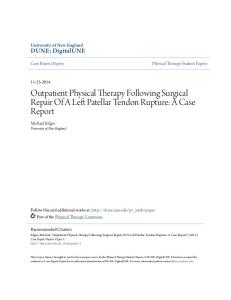
RESULTS Patellar Tendon Loading and Ground Reaction Forces During the horizontal landing phase, the participants‟ dominant lower limb generated a significantly higher FPT (p = 0.016) and peak net knee joint extension moment (p = 0.02) relative to their non-dominant lower limb, although the FV and FAP were symmetrical. In contrast, during the vertical landing phase, the patellar tendon loading and the ground reaction forces were symmetrical (Figure 3). During both landing phases, participants displayed symmetry in timing of their foot placement in that both lower limbs contacted the ground at the same time (negative value indicates the right lower limb contacted the ground first; horizontal landing phase = -10 vertical landing phase = -11
18 ms;
21 ms).
Joint Kinematics Most of the lower limb kinematic variables analysed during the horizontal and vertical landing phases of the stop-jump task did not differ significantly between the participants‟ dominant and non-dominant lower limbs (Figure 4 and Figure 5). However, knee joint asymmetries were evident during both landing phases, whereby the participants displayed significantly less knee flexion at IC (p = 0.039), and greater knee external rotation during the entire landing phase from IC (p = 0.047) to the times of the FV (p < 0.01) and the FPT (p = 0.027) when landing on their dominant lower limb compared to their non-dominant lower limb (Figure 4). Furthermore, the dominant lower limb displayed significantly increased forefoot abduction velocity at the time of the FV (p = 0.042), and increased ankle dorsiflexion velocity (p = 0.035) and tibial
27
Part I: Chapter 2
Horizontal Landing
Vertical Landing
Figure 3.
Mean (± SD) of the forces (normalised to body weight) generated during the horizontal and vertical landing phases of a stop-jump task by the dominant and non-dominant lower limbs of 16 male participants with healthy patellar tendons. (A) Peak vertical ground reaction force (FV), peak anterior-posterior ground reaction force (FAP), peak patellar tendon forces (FPT) and peak net knee joint extension moment. (B) The loading rate of the FV (LR FV) and the loading rate of the FPT (LR FPT). (C) The time from initial foot-ground contact (IC) to the time of the FV (IC to FV), the time from IC to the time of the FAP (IC to FAP) and the time from IC to the time of the FPT (IC to FPT). *Indicates a significant between-limb condition difference (p < 0.05).
segment velocity (dominant = -335 ± 82; non-dominant = -283 ± 64; p = 0.042) at the time of the FPT during the horizontal landing phase when compared to the non-dominant lower limb.
The dominant lower limb displayed a significantly greater forefoot
abduction angle at the time of the FV (p = 0.033) during the vertical landing phase when
28
Part I: Chapter 2
Figure 4.
Mean (± SD) joint angles ( ) displayed during the horizontal and vertical landing phases of a stop-jump task for the dominant and non-dominant lower limbs of 16 male participants with healthy patellar tendons. *Indicates a significant between-limb difference (p < 0.05). IC, initial foot-ground contact; FV, peak vertical ground reaction force; FPT, peak patellar tendon force.
compared to the non-dominant lower limb. Hip kinematics did not significantly differ between limbs throughout the entire horizontal landing phase. However, during the vertical landing phase, the participants displayed significantly less hip flexion at IC (p = 0.013) and at the time of the FV (p