Patella tendonitis Dr Keith Holt
Patella tendonitis (or tendinosis as is it sometimes referred to) is a relatively common problem in which part of the patella tendon, usually the deep part just underneath the kneecap, becomes damaged and causes localised pain. It is usually seen in sports that involve jumping, and indeed, it is sometimes known as jumpers knee.
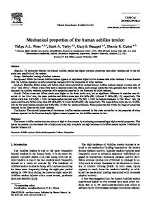
The patella tendon is the rope like structure that connects the patella to the tibia or shin bone. It is an extension of the quadriceps muscle of the thigh and, as such, is a continuation of the tendon of that muscle. The patella (knee cap) is a small bone that develops in the middle of that tendon, dividing it into the quadriceps tendon above, and the patella tendon below. This arrangement makes for better leverage of the quadriceps muscle, with the bone holding the tendon further away from the knee joint. The patella also has a smooth under-surface like the rest of the knee joint and, as such, it acts like a modified pulley, running in a groove in the femur (the thigh bone) during knee flexion.
What does the tendon do? By connecting the quadriceps muscle to the tibia (shin bone), the tendon acts as the rope which, when pulled on, straightens the knee. It is the only tendon that acts to straighten the knee, and therefore, it is necessary for everyday standing and walking. In the jumping athlete however, it is the essential tendon for transmitting the huge force that enables the quadriceps to snap the knee straight and create a jump.
How is the tendon damaged? In the process of jumping, very large forces are generated which are then transmitted to the tibia via the patella tendon. To tolerate those forces this tendon has to be very strong and indeed, it turns out that a healthy tendon is strong enough to suspend a small car without rupture. Being this strong, the normal tendon rarely ruptures due to sudden overload. Whilst it can rupture this way when the tendon substance is damaged (such as is sometimes seen in chronic renal failure and other similar conditions), this situation is relatively uncommon. The situation that occurs in patella tendonitis is
Quads tendon
What is the patella tendon?
Right Patella Patella Tendon
Tibial Tubercle thought to be one of a partial rupture of the tendon that occurs slowly like a fatigue fracture in metal. In general in the body, gradual fatigue wear in tissues is counterbalanced by repair and replacement of that tissue. Overall therefore, tissues tend to remain strong enough for everyday activities and, at any one time, the older and damaged portion of a tissue is small and of no significance. Two processes however, can interfere with this process of renewal: 1) excessive breakdown of tissue, which is generally an overuse type of injury. This is frequently seen in the repetitive jumpers, the basketballers and so on. Here the amount of breakdown exceeds the capacity for repair, and hence, slow deterioration of the tendon ensues. 2) decreased healing of the tissues, which is generally an age dependant event. The age at which tendon
Keith Holt - Perth Orthopaedic and Sports Medicine Centre © - 2016
Femur
Quadriceps tendon
Patella
repair starts to deteriorate may be as young as the early 20's. Everybody is different in this regard, but there is no doubt that some people's tendons are more resistant to damage and also heal better than others. Because tendonitis is the one of the commonest reasons for top athletes to retire, it is clear that the athletes with the longest survival at a competitive level, are also those who are the most resistant to tendonitis. In other words, some tendons (and some people) are more suited to that type and that level of activity than others, and hence, may have a long career without significant tendon injury.
Patella tendon
Tibia
How does it present?
Does the tendon heal? Where the damage to the tendon is small, and where the potential for healing is high (the young), there may be some healing. This is not always back to normal tendon, the partial tear being sometimes healed with localised scar tissue. Providing that this area of scar and damaged tissue does not hurt however, the result will be a fully functional tendon and few if any symptoms. Where the damage is more extensive, the potential for healing, even in the young, is not great. If rested, the area may become less painful and the function may improve, but with a return to higher levels of activity the symptoms return.
How is it diagnosed? The diagnosis is largely clinical, with tenderness of the upper (or affected) part of the patella tendon. Because it is the deep fibres that are involved, the tenderness is greatest with the knee straight. In this position the
Patella tendonitis
MRI showing patella tendonitis This is the typical area, just below the patella Note the swelling in the tendon
Patella
The usual pattern is for a gradual onset of pain with use, which slowly worsens with time. Initially, no modification of activity is required, but gradually, as the symptoms worsen, training may have to be modified. Ultimately, the condition will limit match play or make it impossible to play altogether.
MRI of a normal patella tendon Note that the patella tendon takes the entire power of the quadriceps to the tibia
Patella tendon
The commonest place for the tendon to degenerate is in the deep central aspect, immediately below the patella itself. Much less commonly it may occur at the tendon insertion on the tibia, and rarely it may involve the entire tendon. The pain from the area of tendon damage is local and does not radiate, hence, the commonest symptom is pain immediately below the knee cap. This is worse when the tendon is stressed (running and jumping etc.), and better with prolonged rest. Because the tendon is injured, it is inflamed and swollen (and hence sore). Further injury increases the damage, and hence, increases swelling. With this in mind it can be understood why, in some instances, the pain is worst after a game when the swelling is still increasing, rather than during a game.
Area of patella tendonitis
This is often better seen on an ultrasound than an MRI
Ultrasound of patella tendonitis Note the thickened upper tendon Darker areas mean more swelling (oedema)
Keith Holt - Perth Orthopaedic and Sports Medicine Centre © - 2016
tendon fibres are loose and hence pressure can be put onto the deeper tender areas. With the knee bent, the outer fibres are taught, and hence, these protect the deeper fibres from direct pressure. The best tests to confirm the diagnosis are ultrasound and MRI (magnetic resonance imaging). Both are extremely accurate in determining both the presence and the extent of the problem. If anything, in good hands, the ultrasound may be a little more accurate but, in either case, it is dependant on the skill of the radiologist to detect what may be a subtle abnormality. Because of the low cost and easy availability of ultrasound, this could be regarded as the standard test. This test is more sensitive to operator error than the MRI but, when performed by a good radiologist, can be relied on almost 100% to confirm or deny the presence of tendonitis. Not only can the lesion be identified but often the degree of swelling and extent of fibre damage can be noted.
MRI of patella tendonitis in the mid to lower tendon - this is unusual
What is the treatment? In the early phases, a conservative program is undertaken. This includes adequate rest and modification of training to avoid impact loading, jumping or other stimulatory factors. Patella tendon strapping to support the tendon, and anti-inflammatory type medication, are also helpful. Physiotherapy and related techniques also have a role to play in strengthening and desensitising the tendon to stress, but no techniques have shown the potential to increase tendon healing. As part of the treatment however, a program of eccentric stretching exercises is often used. In this program, the tendon is loaded, and then stretched whilst under load, rather that just being passively stretched. One way of doing this, is to stand on a step on the ball of ones foot, and then to gradually bend the knee, thereby loading and stretching the patella tendon at the same time. The tendon should be stretched as the load is applied, hence, walking up stairs, where the tendon stretch decreases as the knee is straightened, is regarded as concentric, not eccentric, stretching. Walking down stairs however, is eccentric, the tendon undergoing increasing load as the knee bends. Cortisone therapy. Traditionally, the next level of treatment is to instil cortisone (a strong antiinflammatory medication) into the swollen area of tendon. This may not directly help the healing but, it does decrease the swelling and inflammation, and hence the pain. In addition, it may help decrease the amount of scar formation surrounding the area of damage, usually in the underlying fat pad. This scarring can be substantial and may well contribute
MRI showing diffuse tendonitis. This usually represents an inflammatory disorder to the symptoms. A past concern has always been that of tendon rupture after cortisone injection. Whilst cortisone injection can weaken muscle and lead to rupture, the evidence for this is much less with tendons. Nevertheless it can happen and, because of this, it is felt that the method of choice for cortisone instillation should not be by injection. If cortisone is to be placed into the tendon, our
Keith Holt - Perth Orthopaedic and Sports Medicine Centre © - 2016
preferred primary method is by steroid (cortisone) iontophoresis. In this process the cortisone is driven across the skin from a small pad by a low electric current (produced by a battery). This delivers a small dose of cortisone to the tendon without the volume effect of an injection. This is important because it is thought that the volume effect of the injection damages the tendon more than the actual cortisone. Iontophoresis is painless to perform, but the area does feel numb for a while because of the special local anaesthetic that is mixed with the cortisone to assist its passage across the skin. A treatment takes 10 to 15 minutes to perform and needs to be undertaken 5 to 6 times over a 2 week period. It is done in association with the other measures outlined above and, because of its absence of side effects and risk, is often performed very early on in the treatment of the condition. Most sportsmedicine physicians have the equipment required for this treatment, and in fact, these physicians are responsible for most of the non-surgical treatment of this condition.
Other options Autologous blood injection has been advocated for tendonitis, and is now used quite often. There is some evidence that there is not a large inflammatory component to the tendonitis, despite the reduction in pain and swelling that accompanies the use of cortisone (hence, there has been a recent move to term this condition a tendonosis rather than a tendonitis). Therapies have therefore been developed to try and get the tendon to heal, rather than to decrease inflammation. One of those therapies is autologous (one's own) blood injection, which is thought to cause irritation of the tissues, in turn leading to increased scarring, and therefore to healing. Certainly, when the tendonitis is mild, there is some evidence for this. Polidocanol injection has been tried in an attempt to control the large blood vessels that often form in the fat pad to feed the tendonitis. There is some evidence that if the vessel can be destroyed by this agent, the pain will settle. In our hands however, this has met with very limited success. PRP (platelet rich plasma) injection is one of the newer techniques that are available. This is essentially an injection of growth stimulating factors derived from the platelets in blood. This treatment has been used for a lot of injuries to try and enhance healing but, evidence of substantial benefit, is inconsistent. Whilst encouraging, it has yet to be proven suitable as a standard treatment paradigm. Tenocyte injection is one of the newest and most novel technologies currently available. In this technique, tenocytes (tendon cells) are harvested with a needle
from a good part of the tendon. They are then grown in the laboratory until sufficient numbers have been created. When ready, they are then injected into the damaged area of the tendon. This is essentially a method of putting some normal cells into the damaged area and letting those cells heal the tendon. This technique is currently relatively expensive and it takes some months to show results but, the early evidence, suggests that some of these tendons will revert almost to normal. If these results are sustained therefore, this technique may become more mainstream and affordable.
Can surgery help? If all of the above treatment fails, or if the damage to the tendon is bad enough to suggest that it will fail, then surgery may be considered. The surgery for this condition comprises an excision of the damaged area of tendon, plus excision of the underlying scar in the fat pad that is tethered to it. The patella tendon can withstand excision of about 40% of its fibres. We know this from ACL (anterior cruciate ligament) reconstruction where the middle third of the patella tendon is frequently harvested as a graft to reconstruct this ligament. Theoretically, when some of the tendon is removed, the remainder will be under more strain than before: and hence, might reasonably be expected to go on to develop tendonitis. The reality however, is that recurrent tendonitis is very uncommon, the remaining tendon just thickening up to handle the increased load. Of more concern is the possibility that the tendon will not be significantly less painful after the procedure. Part of the reason for this occurrence may be the difficulty in identifying all of the damaged fibres at the time of surgery. Sometimes however, despite what seemed a very adequate excision of the damaged area, the symptoms can continue. If this occurs then treatment is as for primary tendonitis with iontophoresis, rest, eccentic stretching and so on. If ultimately this does not settle down, then a further excision may be undertaken. Because of scar from the first surgery however, this is much harder to do than the first time and the results are therefore less satisfactory. It is rare for the tendon to be made more symptomatic with surgery and, usually, this represents a co-existing problem in the patello-femoral joint that has been aggravated. This may not come to light until after the tendon surgery has been done because the symptoms can be similar. Treatment however, is different.
Recovery from surgery? The actual operation is not all that major to perform and can be done as a day case under general anaesthetic.
Keith Holt - Perth Orthopaedic and Sports Medicine Centre © - 2016
Local anaesthetic makes the tissues swell, and hence, may interfere with the ability to distinguish between normal and abnormal tendon. For this reason, and to allow a tourniquet to be used, a general anaesthetic is preferred.
play. What is hard however, is high level activity such as sprinting. If done to early, this can cause swelling in the remaining tendon with recurrence of pain. Fortunately however, this will recover with rest, albeit sometimes needing some cortisone iontophoresis.
Usually the surgery is performed though a 3 - 4 cm transverse incision right over the area of tenderness. Most people can walk on the knee the same day, although this is limited to short walks around the house for the first few days. Generally by 2 weeks most have stopped limping and the knee improves thereafter. Crutches are generally not required, but can be used if necessary.
Over 6 - 12 months most will get back to full sport, but a few may take as long as 2 - 3 years to fully settle down. In the latter group, it is sometimes the case that there is some underlying patello-femoral pain as well as the tendonitis: and this may necessitate treatment in its own right.
By 6 weeks, it is expected that the pain will be largely settled for day to day activities. Some can start increasing their activity level at that stage but most find that any significant increase in level will stir this up. Interestingly, most can jog sooner than they can bike ride, perhaps because of the high forces put through that tendon during riding. By 3 months most people can lightly jog, albeit with some discomfort. A few people can actually return to training by this time and some might even be able to
Overall, surgery for this condition can be regarded as very good, usually rendering the knee asymptomatic. The time course for this is variable, but maximal recovery may take a year or more. Despite this, return to sport is expected to be sooner, perhaps as early as the 3 month mark. When there is residual pain, it is usually due to some residual tendonitis that has not been dealt with at the time of the first operation, or a patello-femoral problem that co-exists and requires separate treatment. When the tendon damage is bad enough however, the treatment of choice is to excise the damaged portion of the tendon.
For enquiries and appointments Please contact Dr Holt’s main office: Perth Orthopaedic and Sports Medicine Centre Phone: +61 8 92124200 Fax: +61 8 94813792 Email:
[email protected] On line: use the link below Further information can also be obtained on this and other related topics at: www.keithholt.com.au
Keith Holt - Perth Orthopaedic and Sports Medicine Centre © - 2016