CGFR-433; No of Pages 12
Cytokine & Growth Factor Reviews xxx (2007) xxx–xxx www.elsevier.com/locate/cytogfr
Paradoxical effects of cytokines in tumor immune surveillance and tumor immune escape Flavio Salazar-Onfray a,*, Mercedes N. Lo´pez a,b, Ariadna Mendoza-Naranjo a a
Disciplinary Program of Immunology, Institute of Biomedical Sciences, Faculty of Medicine, University of Chile, Santiago, Chile b Research Support Office, University of Chile Clinical Hospital, Santiago, Chile
Abstract The role of cytokines in modulating the formation of new tumors is mediated by their ability to regulate antigen-specific anti-tumor responses and by the activation of non-specific mechanisms, including those involved in the processes of inflammation and innate resistance. Cytokines may influence the growth of tumors by acting directly on tumor cells as growth promoting or growth inhibiting factors or indirectly by attracting inflammatory cell types and affecting angiogenesis. Due to the potency and complexity of cytokine activity against tumor growth, the improvement of cloning techniques and the availability of recombinant forms of different cytokines, a great effort has been made in the recent years to exploit this anti-tumor potential for cancer therapy. This important goal has been difficult to achieve in most cases due to toxicity of most cytokines which could not be dissociated from their anti-tumoral functions. Nevertheless, if well designed, treatment protocols and/or modifications of the cytokine molecules may in some situations augment the anti-tumor effects while limiting the toxicity. One of these molecular approaches could be the design of peptides containing the functional domain of certain cytokines, exemplified by IT9302, a peptide homologous to the functional domain of IL-10, which has demonstrated to increase tumor NK cell sensitivity. # 2007 Elsevier Ltd. All rights reserved. Keywords: Cytokines; IL-10; Tumor; Melanoma; Immunotherapy; Antigen presenting cells; Tumor immunology
1. Introduction Although tumor immunotherapy was popularized over a century ago when William Coley used bacteria extracts to cause sporadic anti-tumor responses, the first concepts supporting the understanding of immune system recognition and destruction of cancer cells were provided by Burnet and Thomas [1,2]. These authors based the hypothesis of immunosurveillance on the concept that the immune system can recognize and destroy nascent transformed cells. Schreiber and colleagues [3] redefined the observed phenomenon of cancer immunosurveillance and indicated * Corresponding author at: Programa Disciplinario de Inmunologı´a, Instituto de Ciencias Biome´dicas, Facultad de Medicina, Universidad de Chile, Avenida Independencia 1027, Independencia, Santiago de Chile, Chile. E-mail address:
[email protected] (F. Salazar-Onfray).
that it may function as a component of a more general process they termed cancer immunoediting. Cancer immunoediting includes host-protecting and tumor-sculpting actions of the immune system that may not only prevent but also shape neoplastic diseases [3]. In this sense, signals from transformed cells may cause activation of professional antigen-presenting cells (APC). Examples of such signals include secretion of cytokines by cells undergoing DNA damage and apoptosis [4]. Cytokines are secreted or membrane-bound proteins that regulate the growth, differentiation and activation of immune cells [5]. As a result, disregulation of cytokine production is thought to play an important role in the development of diseases, such as autoimmune disorders and cancer [5,6]. Indeed, cytokines can indirectly influence tumor initiation and growth by their assorted immune activities (Table 1).
1359-6101/$ – see front matter # 2007 Elsevier Ltd. All rights reserved. doi:10.1016/j.cytogfr.2007.01.015
Please cite this article in press as: Salazar-Onfray F, et al., Paradoxical effects of cytokines in tumor immune surveillance and tumor immune escape, Cytokine Growth Factor Rev (2007), doi:10.1016/j.cytogfr.2007.01.015
CGFR-433; No of Pages 12
2
F. Salazar-Onfray et al. / Cytokine & Growth Factor Reviews xxx (2007) xxx–xxx
Table 1 Effect of cytokines in tumor immunosurveillance and tumor immune escape Cytokines
Effect in TIS or TIE
Secreted by
IL-10
TIE—It has been related with tumor escape, inmuno-supression, growth of tumors and inhibition of Th1 reponse by stoping the secretion of proinflamatory cytokines. It also increase metastasic potential of cells and down regulates MHC class I. TIS—Elicit potent anti-tumor response by activating the adaptative and innate inmunity. Endogenously protects the host against the formation of spontaneous tumors. TIS—It has a potent anti-tumor activity against cutaneous deposits, experimental and spontaneous metastases It acts via IFN-g with anti-angiogenic activity, up-regulating MHC class I and II, and activating NK and cytotoxic cells. TIS—It is a potent stimulator of specific cell-mediated cytotoxicity against autologous tumor targets. Its presence gives long-fasting anti-tumor immunity. TIS/TIE—Participates in tissue destruction and damage recovery, has both pro- and anti-tumor activities. Can inhibit DNA repair, act as a growth factor for tumor cells and may promote angiogenesis. At high doses destructs tumor vasculature and have necrotic effects in tumors. TIS/TIE—It can contribute to the progression, as well as to the anti-tumor response. Depending on the model it activates different mayor pathways of cell proliferation, inducing tumor growth, metastasis, and resistance to chemotherapy in a variety of tumor cells.
T helper cells, B cells, activated monocytes, macrophages, thymocytes, keratinocytes, and tumor cells.
IFN-g
IL-12
GM-CSF
TNF-a
IL-6
T and B cells, NK cells, natural killer T (NKT) cells, macrophages, and mast cells. APC: monocytes, macrophages, and dendritic cells.
Macrophages, T cells and monocytes
Activated macrophages, T lymphocytes and tumor cells.
T and B cells, keratinocytes and macrophages.
TIS: tumor immuno-surveillance, TIE: tumor immuno-escape.
2. Pro-inflammatory cytokines in tumor-immunosurveillance Studies aimed at identifying the physiologically relevant effectors of immunosurveillance have defined the production of cytokines (i.e. IFN-g) as one of the critical tasks that immune cells must perform to eradicate developing tumors. IFN-g plays a critical role in promoting both protective immune responses and immunopathologic processes [7,8]. This cytokine is produced by several cell types (e.g. T and B cells, NK cells, NKT cells, macrophages, and mast cells) upon activation with immune and inflammatory stimuli [9,10]. IFN-g exerts its biological activity by interacting with the IFN-g receptor (IFNGR) that is ubiquitously expressed on almost all cells [7]. During the early phase of a developing immune response (4–96 h), NK cells are the major source of IFN-g. Later (after 96 h), during the adaptive phase of the immune response IFN-g is mainly produced by CD4+ and CD8+ T cells [11,12]. IFN-g is involved in effective anti-tumor immune responses mediated by both adaptive and innate immunity, promoting the generation of tumor-specific CD4 Th1 T cells, as well as cytotoxic T cells (CTL) and by activating macrophages [7]. It has been demonstrated that endogenously produced IFN-g protects the host against the formation and growth of spontaneous tumors in chimeric mice [13– 16]. In fact, mice lacking either, IFNGR1, an IFNGR ligandbinding subunit, or the transcription factor STAT1, have 10– 20 times more sensitivity towards developing primary
tumors. These tumors grow, more rapidly, and at lower carcinogen doses than those injected in wild-type controls [7,14]. Analogous results have been obtained using C57BL/ 6 mice lacking the gene encoding IFN-g [16]. This loss of immunogenicity has been associated to a reduction of the in vivo expression of immunodominant tumor antigens in response to IFN-g secretion by tumor-infiltrating lymphocytes. In addition, IFN-g participates with granulocyte macrophage-colony stimulating factor (GM-CSF) in tumorimmune responses and is also essential for the anti-tumor effect of interleukin-12 (IL-12) [17,18]. IL-12 is produced mainly by APC, such as monocytes, macrophages, and dendritic cells (DC). Among other important functions, IL12 possesses IFN-g-dependent anti-angiogenic activity [19] and potent anti-tumor activity in a wide variety of murine tumor models [17,20]. IFN-g induced by IL-12 can upregulate MHC class I and II expression on tumor cells, activate NK cells, macrophages and CD8+ CTL, inducing the production of interferon-inducible protein 10 (IP-10) [21,22]. Significant IL-12-mediated anti-tumor activity has been demonstrated against established, experimental [17,22] as well as spontaneous metastases [23] (Table 1). One approach to improve anti-tumor immunity is to increase the recognition of tumor antigens by T lymphocytes. In this sense, it is well known that GM-CSF is a potent stimulator of specific cell-mediated cytotoxicity against autologous tumor targets [24]. GM-CSF is a member of a large family of glycoprotein growth factors that regulate the expansion and differentiation of hematopoietic progenitor cells and is able to act at several levels in the generation and
Please cite this article in press as: Salazar-Onfray F, et al., Paradoxical effects of cytokines in tumor immune surveillance and tumor immune escape, Cytokine Growth Factor Rev (2007), doi:10.1016/j.cytogfr.2007.01.015
CGFR-433; No of Pages 12
F. Salazar-Onfray et al. / Cytokine & Growth Factor Reviews xxx (2007) xxx–xxx
propagation of immune responses. Deficiencies of GM-CSF and IFN-g lead to the spontaneous development of infection, inflammation and cancer [18]. As reported previously, vaccination with irradiated tumor cells engineered to secrete GM-CSF, or at a lesser extent IL-3, stimulate potent, specific and long-lasting anti-tumor immunity [24]. This finding is surprising because GM-CSF is a cytokine mostly associated with cell growth and differentiation of hematopoietic progenitors, and is known to induce the differentiation and promote the survival of peripheral blood DC [25]. However, this cytokine, showed to be the most powerful immunostimulatory capacity among 10 different molecules tested [24]. Nevertheless, immunosurveillance represents only one dimension of the complex relationship between the immune system and cancer. The immune system may also promote the emergence of primary tumors with reduced immunogenicity capable of evading immune recognition and destruction [15].
3. Pro-inflammatory cytokines and modulation of the anti-tumor immune response Of the many mechanisms contributing to immune suppression, much emphasis has recently been given to inflammatory and regulatory cells, to soluble factors that are associated with their activities and to inflammatory mediators. Inflammation is a physiologic process in response to tissue damage resulting from microbial pathogen infection, chemical irritation, and/or wounding. At the very early stage of inflammation, neutrophils are the first cells to migrate to the inflammatory sites under the regulation of molecules produced by rapidly responding macrophages and mast cells in tissues [26,27]. As the inflammatory process progresses, various types of leukocytes, and other inflammatory cells are activated and attracted to the inflamed site by a signaling network involving a great number of growth factors, cytokines, and chemokines [26,27]. The association between inflammation and cancer was illustrated by epidemiologic and clinical studies [27,28]. Inflammatory components were characterized as key players in tumor promotion by their ability to release a large variety of factors that support different processes including angiogenesis, tumor growth, and tissue remodeling [27–29]. Tumor necrosis factor-a (TNF-a) is one of the main proinflammatory cytokines. TNF-a, produced mainly by activated macrophages but also by tumor cells, binds to the membrane-bound homotrimeric receptors TNFRI and TNFRII. During inflammation, TNF plays a critical role in both tissue destruction and damage recovery, maintaining the reversibility of microenvironments, and stimulating cellular change and tissue remodeling [30]. TNF-a has both anti-cancer and pro-cancer activities [30]. High-dose administration of TNF-a may destroy tumor vasculature
3
and have necrotic effects in tumors [30]. In contrast, TNF-a has been found to be required in chemical carcinogenelicited skin carcinogenesis [31] and is also a major inducer of nuclear factor-kB (NF-kB) activation, which shows antiapoptotic activity. Macrophages and T lymphocytes may release TNF-a and macrophage migration inhibitory factor (MIF) to exacerbate DNA damage [32]. The contradictory roles of TNF-a in regulating cell death might be attributed to the diverse modifications of TNF receptor complexes triggering opposite pathways [33]. In addition, TNF-a can inhibit DNA repair and act as a growth factor for tumor cells. Furthermore, TNF-a may promote angiogenesis and tumor growth by inducing a range of angiogenic factors [28,30]. Interleukin-6 (IL-6) is another pro-inflammatory cytokine which has been described to participate in tumorigenic processes, contributing both to tumor progression, as well as to anti-tumor immune responses in vivo. Whereas the exact nature of IL-6 effects on a particular cell type varies depending on the model, it is well documented that this cytokine can activate different major pathways of cell proliferation, participating in the induction of tumor growth, metastasis, and resistance to chemotherapy in a variety of tumor cells [34]. IL-6 is also crucial for the establishment and maintenance of p53 promoter methylation, contributing to epigenetic silencing of tumor suppressor genes and tumor cell survival [35]. In addition, STAT3 is activated in IL-6induced transformation of epithelial cells, where DNA binding and transcriptional activities of STAT3 are significantly increased by IL-6 in sensitive mouse skin epithelial cells [36]. Conversely, a potential role has been shown for IL-6 as an anti-tumor agent in a variety of tumor models [37,38]. Other work has shown that inhibition of the protective functions of the immune system may also facilitate tumor escape. In this scenario, tumor cell variants may produce significantly higher levels of immunosuppressive cytokines, including transforming growth factor-b (TGF-b) or interleukin-10 (IL-10) [39] and other regulatory mechanisms to suppress effector immune responses including recruitment of regulatory T cells, activation of indoleamine 2,3 deoxygenase, and PD-1/PD-L1 interactions.
4. IL-10 and TGF-b as regulatory cytokines Recent attention has turned toward the study of the mechanisms of immune evasion at the effector phase of the anti-tumor immune response, predominantly within the tumor microenvironment. Many soluble products can contribute to this local immunosuppression, among which, TGF-b and IL-10 have fundamental roles. TGF-b is a major pluripotent cytokine, widely distributed, and with a pronounced immunosuppressive effect, such that deficiency in knock-out mice results in lethal autoimmunity. TGF-b binds to receptors on the cell surface
Please cite this article in press as: Salazar-Onfray F, et al., Paradoxical effects of cytokines in tumor immune surveillance and tumor immune escape, Cytokine Growth Factor Rev (2007), doi:10.1016/j.cytogfr.2007.01.015
CGFR-433; No of Pages 12
4
F. Salazar-Onfray et al. / Cytokine & Growth Factor Reviews xxx (2007) xxx–xxx
forming a heterodimeric receptor complex constituted by two pairs of subunits known as receptor type I (TbR-I) and type II (TbR-II) [40]. A membrane-anchored proteoglycan, known as type III receptor or betaglycan, aids this process by capturing TGF-b for presentation to the signaling receptors I and II. Binding of TGF-b1 to the receptor complex activates the intracellular kinase domain, which leads to the phosphorylation and activation of members of the Smad protein family and the subsequent regulation of TGF-b– dependent gene expression [40]. An essential role for TGF-b1 has been recently established in both, the suppression of T cell–mediated immune responses and the control of autoimmunity [41]. TGF-b receptor deficient mice exhibit inflammatory infiltration in multiple organs and uncontrolled T-cell proliferation [42]. These mice develop a rapid wasting syndrome, which leads to their death at the age of 3–4 weeks. Global disruption of the TGF-b1 gene in mice has clearly illustrated the importance of this specific TGF-b isoform in regulating immune cell differentiation and function. TGF-b1 / mice manifest a spontaneous autoimmune-like syndrome, with aberrant expression of MHC class I and II antigens, circulating SLE-like IgG antibodies to nuclear antigens, pathogenic glomerular IgG deposits, and progressive infiltration of mononuclear cells into multiple organs [43]. The importance of T cells in the genesis of this syndrome is supported by the observation that T cell-specific disruption of TGF-b signaling leads to a similar, though less aggressive syndrome [44]. Studies show that this suppression may be attributed to TGF-b1 expression and secretion by regulatory T cells (Treg). Stimulated Treg express high levels of TGF-b in both surface-bound and secreted form. Tregs (CD4+ CD25+ Foxp3+) play a pivotal role in immunological homeostasis by their capacity to exert immunosuppressive activity [45]. These cells represent approximately 5–10% of circulating T cells in both mice and humans, and are also termed ‘naturally occurring’, in contrast to other types of T regulatory cells such as T-regulatory lymphocytes type-1 (Tr1) and T-helper type-3 (Th3), which likely develop from conventional CD4+CD25 T lymphocytes in the periphery, and are thought to represent altered T-cell differentiation states rather than a unique T-cell lineage [46]. Treg cells are usually considered ‘naturally anergic’, although some recent studies may challenge this view. Nonetheless they express several activation surface markers, including the aforementioned CD25 (Interleukin-2 (IL-2) high affinity receptor a chain), CTLA-4 (CD152), and glucocorticoid-induced TNF receptor (GITR), among others [45]. It has been shown that Treg cells can suppress the proliferation of CD4+CD25 T cells via a cell–cell contact dependent fashion. In addition, several in vitro studies have demonstrated that Foxp3+ expression and suppressive activity as well as the size of the peripheral Treg cell compartment are all dependent on signals triggered by TGF-b. These regulatory cell populations, namely the CD4+ CD25+ Foxp3+ Treg cells of thymic
origin, the inducible Tr1 cells, which are able to secrete IL10, and the Th3 lymphocytes, which produce elevated amounts of TGF-b, would be accumulated in tissues specially during tumor growth to suppress different effector branches of the immune response [47]. Several lines of evidences indicate that Treg cells are increased in blood and other tissues in different types of cancer [48]. Additionally, it has been demonstrated that in patients with refractory metastatic melanoma, the adoptive transfer of anti-tumor CD8+ CTLs, after non-myeloablative chemotherapy was able to induce important tumor regressions that could be associated to elimination of regulatory T lymphocyte populations [49]. On the other hand, chemotherapeutical drugs like decarbazine, besides affecting tumor proliferation, also have immunosuppressive effects on T lymphocytes populations, as well as on accumulated regulatory T cells, suggesting not only a novel strategy for the study of regulatory cell populations, but also potential therapeutic innovations which are being boarded in current clinical trials [47]. Besides Treg cells, other types of regulatory T lymphocytes have the capacity to generate immunosuppressive responses against tumors in the periphery, mainly through increasing the secretion of immunosuppressive cytokines like TGF- b and IL-10. As mentioned above, Tr1 secrete large amounts of IL-10 when they are stimulated in vitro with dexamethasone or vitamin D3. Another population of regulatory T lymphocytes is constituted by the Th3 cells that produce high amounts of TGF-b [50]. TGF-b signaling regulates cancer through mechanisms that function either within the tumor cell itself or through host-tumor cell interactions. The loss of tumor-specific TGF-b-mediated cytostasis allows the tumor to utilize TGFb to profoundly alter the tumor microenvironment and host immune response. TGF-b released or activated by tumor cells acts on CD8+ CTLs to suppress the expression of five cytolytic mediators. In the presence of TGF-b, CTL fail to express effectors such as perforin and granzyme A and B, the apoptotic activators that flow into the tumor cell. In addition, TGF-b impairs expression of, the proapoptotic mediator FasL, and the proinflammatory cytokine IFN-g. In addition, tumor growth facilitates the induction or recruitment of CD4+ regulatory T cells that secrete IL-10 and TGF-b and suppress effector CD8+ T cell responses. However, CD8+ T regulatory cells expressing IL-10 and TGF-b are also recruited or activated by the immunosuppressive environment, where they may suppress the induction of anti-tumor immunity [51]. Recently, accumulated evidence has shown that TGF-b has important functions in the maintenance of the homeostasis, thus participating in the regulation of inflammation. The identification of T helper cells that produce interleukin 17 (IL-17), called Th-17 cells, has ushered in a new chapter in immunology (for details see review 16 of the present special issue). This IL-23-dependent T-cell subset is induced by a combination of TGF-b and IL-6, is distinct from T
Please cite this article in press as: Salazar-Onfray F, et al., Paradoxical effects of cytokines in tumor immune surveillance and tumor immune escape, Cytokine Growth Factor Rev (2007), doi:10.1016/j.cytogfr.2007.01.015
CGFR-433; No of Pages 12
F. Salazar-Onfray et al. / Cytokine & Growth Factor Reviews xxx (2007) xxx–xxx
helper type 1 (Th1) and Th2 cells and is critical for driving inflammatory autoimmune responses [52].
5. IL-10 an old actress in the immunosuppressive scenario IL-10 is an 18 kDa cytokine produced under different conditions of immune activation by a variety of cell types, including T cells, B cells, monocytes, and macrophages [53]. This protein, initially characterized as cytokine synthesis inhibitory factor (CSIF) [54], is a pleiotropic molecule that displays both immunoregulatory and immunostimulatory effects. It has been described that IL-10 inhibits the production of cytokines such as IL-2, TNF-a, IFN-g, and GM-CSF by Th1 cells in response to antigens presented by APC. Later, it was demonstrated that IL-10 also plays important roles in blocking cytokine production, and the expression of co-stimulatory molecules including CD80, CD86, and MHC class II and chemokine secretion [55,56]. These effects may limit the magnitude of immune cell activation and suppress subsequent cytokine release by T cells during the specific immune response. Part of these anti-inflammatory effects is due to the activity of IL-10 on DC. IL-10 serves as a potent mechanism for limiting the maturation of monocyte-derived DC and their capacity to initiate Th1 responses. IL-10 can be produced by DC and has similar immunosuppressive effects on antigen presentation. Since DC are the most important cells able to elicit an adaptive immune response and are important actors in acute inflammation, cross-regulation of
5
inflammatory mediators and their subsequent effects on DC is of great importance [57]. The capacity of IL-10 to induce Tr1 cells in vitro is directly correlated to the in vivo immune response to certain infectious agents and cancer. Pathogen-specific Tr1 cells may be generated in vivo during the course of bacterial, viral, fungal, or parasitic infections. The major purpose of these cells is to control inflammation and collateral tissue damage. [58]. In some models, Foxp3+ Treg cells have been shown to be both antigen specific and IL-10 producers [59], blurring the distinction between the Treg cell subsets. Thus, naturally occurring Treg cells can act via cell contact–dependent mechanisms in vitro, and through the release of inhibitory cytokines.
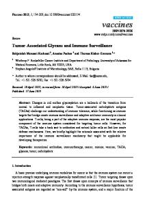
6. IL-10 a cytokine with dual effect on tumors Although IL-10 is commonly regarded as an antiinflammatory and immunosuppressive cytokine that favors tumor escape from immune surveillance, accumulating evidence indicates that IL-10 also possesses some proinflammatory properties [60]. It is now clear that depending on the dose and route of administration, IL-10 may display either pro or anti-inflammatory effects (Fig. 1). For example, while IL-10 prevents type-1 diabetes development in NOD mice when administered systemically, [61] this cytokine accelerates the onset rate of the disease when transgenically expressed in the pancreas [62]. In addition, while intravitreally injected IL-10 effectively reduces LPSinduced uveitis in rabbits, intraperitoneal administration
Fig. 1. Paradoxical pro-tumoral and anti-tumoral activities of IL-10. IL-10 exerts different effects depending on tissue location and concentration. For example, at the tumor site low concentrations of IL-10 can be secreted by tumor cells or by infiltrating T cells, and in this context IL-10 dowregulates MHC class I expression on the tumor surface, rendering it less sensitive to T CD8+ mediated cytotoxicity and more sensitive to NK cell-mediated lysis. In lymph nodes (LN) IL-10 can be secreted by TCD4+ (Th2) cells, Treg cells and Tr1 cells. In this context IL-10 may work as a tolerogenic cytokine, inhibiting the anti-tumor response. On the contrary, in the periphery, IL-10 produced by injected DC or the administration of the recombinant cytokine acts as an immunological adjuvant by activating NK cells to produce IFN-g thus inducing DC maturation. Both IL-10 and its functional domain peptide IT9302 need to be tested in vivo for antitumor responses.
Please cite this article in press as: Salazar-Onfray F, et al., Paradoxical effects of cytokines in tumor immune surveillance and tumor immune escape, Cytokine Growth Factor Rev (2007), doi:10.1016/j.cytogfr.2007.01.015
CGFR-433; No of Pages 12
6
F. Salazar-Onfray et al. / Cytokine & Growth Factor Reviews xxx (2007) xxx–xxx
exacerbates LPS-induced uveitis in mice [63]. IL-10treatment has also been shown to aggravate experimental autoimmune myasthenia gravis through induction of Th2 and B-cell responses to the acetylcholine receptor [64]. Finally, neutralizing monoclonal antibodies against IL-10 ameliorates the course of murine systemic lupus erythematosus in NZB/ NZW F1 mice [65]. In fact, IL-10 has the pleiotropic ability of influencing positively or negatively the function of innate and adaptive immunity in different experimental models. Therefore, this cytokine might be considered either a target of antiimmune escape therapeutic strategies or rather, an immunological adjuvant in the fight against cancer. However, until now, IL-10 is the only cytokine that has been shown to posses an inhibitory effect on the MHC class I antigen presentation. This has led us to propose that its expression, in certain tumors, would be related to mechanisms of tumor evasion [56]. This cytokine has been associated to immunosuppression, tumor growth, and inhibition of Th1 responses, because it inhibits the production and secretion of IL-2, IFN-g and a variety of other pro-inflammatory cytokines such as TNF-a, IL-1, IL-6 and IL-12 in different immune cell populations, including lymphocytes and monocytes [53,54]. Several authors have reported the presence of IL-10 in fresh tumors biopsies of patients with different types of cancer such as melanoma, renal carcinoma, ovarian tumors, and several types of adenocarcinoma [66–69]. It has been demonstrated that solid tumors express IL10 mRNA [70] and that patients with different types of adenocarcinoma show high levels of IL10 in plasma [56]. It has been proposed that IL-10 increases the metastatic potential of tumors since the expression of this cytokine is more frequent in metastatic tumors than in primary tumors [71].
7. IL-10 inhibits MHC class I antigen presentation in tumors Studies from our laboratory, have demonstrated that melanoma cell lines pre-treated with recombinant IL-10 (rIL-10) are less sensitive to tumor-specific CTLs. This effect is directly correlated with the ability of IL-10 to downregulate MHC class I expression [55]. These findings were confirmed by studies describing a negative regulation of the MHC class I and II and the adhesion molecule ICAM1 in melanoma cell lines [72]. These results, added to the multiple inhibitory effects of IL-10, have led the proposition that secretion of this cytokine might be a mechanism employed by tumors to evade the immune response [56]. Subsequently, we have demonstrated that the effect of IL10 in antigen presentation is associated with a decrease in MHC class I expression and inhibition of the expression and functionality of TAP-1 and TAP-2 molecules in murine tumors transfected with IL-10, such as the RMA lymphoma and the P815 plasmacytoma [73,74]. Other groups have observed similar effects in human B lymphomas [75]. On the
other hand, tumors that constitutively express IL-10, like J558L plasmacytoma or lymphoma YAC-1, when transfected with IL-10 antisense, significantly increase their cell surface expression of MHC class I and reduce their sensitivity to NK-cell mediated lysis [73,76]. Treatment of the RMA murine lymphoma cell line with different doses of rIL-10 or transfection with the gene that encodes for IL-10, provide a phenotype similar to the RMAS mutant [73] which has a TAP-2 peptide mutation and shows a deficient antigen presentation. A strong correlation can be observed between expression of IL-10; increase in tumor sensitivity to NK cell-mediated lysis, and diminished tumor sensitivity to specific CTLs [55,73,74].
8. In vitro and in vivo effects of IL-10 in an experimental melanoma model The in vivo effects of the local expression of IL-10 in tumor rejection are variable and contradictory. There are evidences that show that the dose of IL-10 used, the route of administration and the local or systemic application of the cytokine are fundamental in determining its effects [60] (Fig. 1). High doses of IL-10 would have a stimulatory effect on the immune response, mainly through modulation of T lymphocyte and NK cell activities [77]. This effect has been demonstrated by a more effective rejection of certain tumors in presence of high levels of IL-10 [77]. For example, transfected TSA mammary murine tumor cells that produce high levels of IL-10 are more rapidly eliminated than controls in BALB/c mice. This effect can be explained by an increase of the lytic activity mediated by CTLs and NK cells [78]. Similar effects have been observed, using different immune competent murine strains and tumors transfected to produce high IL-10 levels, such as the murine CL8-1 and B16 melanoma [79], the mammary carcinoma 410-4 [80] and the sarcoma MCA [81]. The predominant role of NK cells in these systems is corroborated by results obtained in T lymphocyte-deficient mice. In fact, Chinese hamster ovary cells transfected with mouse IL-10 are rapidly rejected in severe combined immunodeficient (SCID) mice [82]. Unpublished results obtained by our laboratory indicate that lymphoma RMA cells and human BL melanoma cells transfected with IL-10 are more efficiently rejected than their controls in SCID mice, and that this effect can be reversed by depleting NK cells using an anti-asialo antibody. On the contrary, there are several evidences of the immunosuppressive effects of IL-10 in relation to antitumoral immunity [56]. For example, it has been observed that transgenic mice that overexpress IL-10, exhibit a reduced ability to reject tumors [83]. In the case of murine fibrosarcoma WP4, that expresses lower amounts of IL-10, a major tumorigenicity is observed, which is explained by an inhibition in the generation of CTL and T CD4+ lymphocytes [84].
Please cite this article in press as: Salazar-Onfray F, et al., Paradoxical effects of cytokines in tumor immune surveillance and tumor immune escape, Cytokine Growth Factor Rev (2007), doi:10.1016/j.cytogfr.2007.01.015
CGFR-433; No of Pages 12
F. Salazar-Onfray et al. / Cytokine & Growth Factor Reviews xxx (2007) xxx–xxx
The plasmacytoma J558L cell line, that constitutively expresses low doses of IL-10, also shows similar effects which are mediated by an inhibition of antigen presenting cells [74]. It has been also demonstrated that B16 melanoma transfected with the IL-10 gene to produce low concentrations of IL-10, escapes the immune system control by virtue of the inhibition of MHC class I expression and the induction of an angiogenic phenotype [77]. Melanoma cells treated with IL-10 were shown to have decreased, but peptide inducible, expression of MHC class I, decreased sensitivity to MHC class I-restricted CTLs and increased sensitivity NK cell-meditated lysis [55–73]. These findings could be explained, at least partially, by a downregulation of TAP1/TAP2 expression. In addition IT9302, a nanomeric peptide (AYMTMKIRN), homologous to the Cterminal functional domain of the human IL-10 sequence, was demonstrated to mimic these previously described IL10 effects on MHC class I-related molecules [85]. Besides the down-regulation of MHC class I at the cell surface, the IL-10 homologue peptide also caused a dose-dependent inhibition of IFN-g-induced cell surface expression of MHC class I in melanoma cells together with an increase in melanoma cell sensitivity to lysis by NK cells [85]. These results demonstrate that a small synthetic peptide derived from IL-10 can mimic the antigen presentationrelated effects mediated by this cytokine in human melanomas and increase tumor sensitivity to NK cells; these effects are critical in the design of future strategies for cancer immunotherapy.
9. The effect of recombinant cytokines in cancer immunotherapy In human cancer therapy, recombinant cytokines are generally used to enhance immunity. Cytokines can be used to activate the innate immune system: NK cells, macrophages, and neutrophils. They can also regulate the adaptive immune system, including T and B cell-mediated responses. In the immune system, cytokines function in cascades. Thus clinical trials of individual cytokines are rarely useful, since cytokines tend not to work individually, but rather act in concert. Among the individual cytokines that have been tested and found ineffective for cancer treatment is IL-1b, which nevertheless can still be indirectly useful because it helps to decrease the severe toxicity of IL-2 [86]. TNF-a certainly sounded promising for a period of time, but in fact caused severe hypotension when used systemically. IL-4 showed minimal anti-cancer activity and was toxic. IL-6 had some activity against cancer cells, but turned out to be a growth factor for myeloma cells [87]. Other cytokines such as GM-CSF, which was primarily used in stem cell transplantation to reconstitute the myeloid lineage, has been studied in melanoma showing controversial results. IL-2 and IFN-a2b are two cytokines approved by the FDA for the treatment of cancer. IL-2 has demonstrated
7
activity against renal cell carcinoma, melanoma, lymphoma, and leukemia. IFN-a2b has activity in those tissues but also in Kaposi’s sarcoma, chronic myelogenous leukemia, and hairy cell leukemia. Overall, cytokines are molecules that appear to have applications in the treatment of hematological malignancies or immunogenic tumors. Here, we will examine the major cytokines currently in use or under evaluation for cancer therapy: IFN-a, IL-2, GM-CSF, and IL-12.
10. Interferon-a Initially described for their antiviral activities, type I-IFN are now recognized as central regulatory elements of the immune response. IFN-a is definitely active in hairy cell leukemia, where complete and partial responses have been documented [88]. In addition, a significant increase in survival was observed in different clinical trial of patients with chronic myelogenous leukemia suggesting that IFN can be also active in this disease [89]. In kidney cancer, few but consistent results have been shown in phase 2 clinical trials, where reported response rates average 11% (ranges from 8 to 29%). Currently, IFN is included in the treatment of kidney cancer, but unlike IL-2, it is not FDA-approved, and is considered a palliative secondline therapy. IFN-a has a long history in metastatic melanoma. A series of phase 2 trials with high-dose IFN showed response rates similar to existing chemotherapies (20–22%) [90]. There is much more promise in the use of IFN as an adjuvant in melanoma treatment [91]. In one randomized trial, stage 3 or 2B patients were observed or given high-dose IFN for 1 year (n = 287). The IFN-treated group showed disease-free and relapse-free benefits, and an overall survival medium of 3.8 years (versus 2.78), which ultimately led to FDA approval and the acceptance of interferon as a standard adjuvant therapy for stage III resected melanoma.
11. Interleukin-2 IL-2 is a T-cell growth factor, that has been used with relative success in melanoma and renal cell carcinoma. In dose escalation studies, patients treated with high doses of IL-2 showed clinical responses. Patients with renal cell carcinoma showed high response rates to IL-2 (24% at the highest dose of IL-2), and the median survival for all patients was 16.3 months, with 10–20% survival 5–10 years after treatment [92]. Melanoma patients [92] who have successfully responded to IL-2 treatment are alive and their disease did not progress for 5–15 years later. Nevertheless the toxicity of the treatment is a limiting factor. The administration of high doses of IL-2 can be compared to inducing a controlled and reversible state of septic shock.
Please cite this article in press as: Salazar-Onfray F, et al., Paradoxical effects of cytokines in tumor immune surveillance and tumor immune escape, Cytokine Growth Factor Rev (2007), doi:10.1016/j.cytogfr.2007.01.015
CGFR-433; No of Pages 12
8
F. Salazar-Onfray et al. / Cytokine & Growth Factor Reviews xxx (2007) xxx–xxx
Administration of IL-2 at low-doses in patients with renal cancer and melanoma has also shown benefits [93]. In this case objective responses rates of 18–23% were reported, without high levels of toxicity. It has been also proposed that patient selection is critical for IL-2 therapy. As expected, melanoma patients with high performance status do well, as do patients who develop vitiligo or autoimmune thyroiditis. These findings support the idea that IL-2 promotes an autoimmune T-cell mediated response against melanocytes.
12. Granulocyte–monocyte colony stimulating factor and interleukin-12 GM-CSF has been shown to stimulate an intense inflammatory reaction, suggesting that this cytokine creates an advantageous intracellular environment for tumor antigen presentation [24]. Several phase I/II clinical trials investigating GM-CSF-secreting tumor vaccines have been conducted in several types of cancers. Expression of the GM-CSF gene within tumor cell populations has demonstrated evidence of anti-tumor activity particularly in prostate cancer and non-small cell lung cancer [94]. IL-12 is a heterodimeric protein that promotes NK and T cell activity and is considered to be a growth factor for B cells. Its anti-tumor activity has been demonstrated in mouse models, but IL-12 has shown minimal therapeutic effect in patients. However, IL-12 may have a potential value as an adjuvant in vaccines based on immunogenic peptides as observed in patients with resected melanoma (Stage III and IV) [95].
13. Interleukin-10 as a therapeutic agent The powerful immunomodulatory properties of IL-10 and the promising results obtained from experimental models in the course of several inflammatory diseases have supported the application of IL-10 in clinical trials. So far human recombinant IL-10 (Ilodecakin/Tenovil; ScheringPlough Research, Kenilworth, NJ) has been tested in healthy volunteers, patients with Crohn’s disease, rheumatoid arthritis, psoriasis, hepatitis C infection, HIV infection, as well as in the inhibition of therapy-associated cytokine storm in organ transplantation and the Jarisch Herxheimer reaction. In phase-I clinical trials, safety, tolerance, pharmacokinetics, pharmacodynamics, immunological, and hematological effects of single or multiple doses of IL-10 administered by intravenous (i.v.) or subcutaneous (s.c.) route have been investigated in various settings in healthy volunteers [96]. Overall, these studies have shown that IL-10 is well tolerated without serious side effects at doses of up to 25 g/kg, and mild to moderate flu-like symptoms are observed in a fraction of recipients at doses up to 100 g/kg
[97]. Single i.v. or s.c. doses of IL-10 may result in transient neutrophilia, lymphocytopenia, and monocytosis and a delayed decrease in platelet counts was observed following a single s.c. dose of IL-10 [96]. Administration of IL-10 did not produce adverse effects on the studied patient population [98]. Generation of neutralizing antibodies was not observed in any of the studies. In vivo administration of IL-10 inhibited LPSinduced production of IL-6, IL-1, and TNF-a in whole blood cell assays and decreased proliferative responses and IFN-g production following phytohemagglutinin stimulation of peripheral blood mononuclear cells (PBMC), indicating that IL-10 retains immunomodulatory activity when administered in vivo. IL-10 has not been tested yet in any type of cancer, probably because expected anti-inflammatory effects may favor tumor growth. However, a large body of pre-clinical evidence is in contrast with the abovementioned forecast. Besides the increased immunogenicity and reduced tumorigenicity of IL-10-transfected melanoma and carcinoma cell lines, exogenous IL-10 administration has been shown to mediate regression of established melanoma and breast cancer metastases in various preclinical in vivo models [78–80]. Remarkably, while viral IL10 can inhibit tumor rejection, cellular IL-10 favors the elimination of cancer cells, confirming that the double function of this cytokine can be split and associated to different domains of the molecule [99]. For this reason, the potential use of an IL-10 functional domain homologous peptide such as IT9302 may result interesting from a therapeutic point of view. Many authors, including our group, have linked the anti-tumor capacity of IL-10 to an enhanced NK cell activity [73–85] and others have demonstrated that this anti-tumor effect depends on CD8+ [100] or CD4+ T cells [101]. Regarding DC vaccines, it is now very clear that mature monocyte-derived DC express both IFN-g and IL-10, thus inducing in vivo DTH reactions [102]. Although administration of IL-10 before anti-tumor immunization results in immunosuppression and tumor progression, in line with the well-known IL-10 inhibitory activity on DC-mediated antigen presentation [100], injection of IL-10 immediately after immunization significantly augmented anti-tumor immunity and vaccine efficacy [60]. In addition, the number of antigen-specific CTLs is higher in animals treated with vaccine plus IL-10 rather than those treated with vaccine alone, suggesting that IL-10 might act as an immunological adjuvant maintaining the number of antigen-activated CTLs during vaccineinduced tumor rejection [101,102]. A follow-up study in melanoma patients showed a correlation between clinical regression and IL-10 protein levels in tumor cells from samples obtained before therapy [60]. Therefore, it is possible that the presence of high in situ levels of IL-10 may condition the tumor microenvironment to the anticancer effect mediated by anti-tumor vaccines and this outcome might be associated with an increased NK cell activity against the tumor.
Please cite this article in press as: Salazar-Onfray F, et al., Paradoxical effects of cytokines in tumor immune surveillance and tumor immune escape, Cytokine Growth Factor Rev (2007), doi:10.1016/j.cytogfr.2007.01.015
CGFR-433; No of Pages 12
F. Salazar-Onfray et al. / Cytokine & Growth Factor Reviews xxx (2007) xxx–xxx
14. Conclusions It is not surprising that the cytokine network profoundly affects the growth of tumors in vivo and plays a significant role in the immunosurveillance against newly generated malignant cells. The role of cytokines in checking the formation of new tumors is mediated by their ability to modulate antitumor antigen-specific responses, according to the original interpretation of immunosurveillance, and by the activation of non-specific mechanisms, including those based on the processes of inflammation and innate resistance. Due to the large variety of functionally diverse members of the various families of cytokines, these molecules may influence both positively and negatively growth and metastasis. Many cytokines act directly on tumor cells, as growth promoting or inhibiting factors. Certain cytokines act directly on the growth, differentiation, or survival of endothelial cells, while others act by attracting inflammatory cell types and affecting angiogenesis. Pro-inflammatory and chemotactic cytokines influence the tumor environment and control the quantity and nature of infiltrating hematopoietic effector cells, with inhibiting or enhancing effects on tumor growth. Due to the potent and complex activity of cytokines against tumor growth, a great effort has been made in recent years, following the cloning and availability of recombinant forms of a large number of cytokines, to exploit their antitumor potential for cancer immunotherapy. Many cytokines have proven to have a potent therapeutic anti-tumor effect in preclinical models; however with only some exceptions, translation to clinical practice has been disappointing. If one considers the complexity of the cytokine network, with an intricate tangle of positive and negative feedbacks, synergies and antagonisms, it is not surprising that the use of a single cytokine in tumor therapy will exhibit serious difficulties. In addition, physiologic differences between experimental animal models and patients as well as the inadequacy of most preclinical cancer models makes it very difficult to determine the most efficient treatment protocols in patients. Because, in the large majority of the cases, the cytokine toxicity is due to the same physiologic function responsible for the anti-tumor effect, it is very difficult to dissociate these two effects. Structural modifications of the cytokine molecule may in some cases augment the anti-tumor effects while limiting cytokine toxicity. An example can be illustrated by peptides containing a functional domain of a cytokine, such as IT9302; this peptide is homologous to the functional domain of IL-10, which has been demonstrated to increase tumor sensitivity to NK-cell mediated destruction. However, the poor clinical performance observed in the majority of the immunotherapeutic approaches might also be attributed to tumor evasion mechanisms mediated not only by cytokines such as TGF-b, IL-10 or PGE2 [56], but also by the tumor-induced acidic microenvironment that
9
favors tumor cell survival and contributes to impairment of immune cells [103]. Tumor-immune escape may also involve, among others [104], tumor counterattack strategies including tumor-induced T cell deletion by Fas–Fas ligand interactions [105] or direct phagocytic activity by tumor cells (cannibalism) [106]. Taken together, these observations support the notion that the design of novel therapeutic strategies should also consider the blockade of critical inhibitory signals and tumor counterattack strategies at the tumor site.
Acknowledgments This work was supported by grants from the Fund for the Promotion of Scientific and Technological Development (FONDEF DO2I1088), the National Fund for Scientific and Technological Development, Chile (FONDECYT 1060935), Nu´cleo Milenio de Inmunologı´a e Inmunoterapia P04/030-F, and the Research Support Office, University of Chile Clinical Hospital (OAIC). Competing interest statement: The authors declare that they have no competing financial interests.
References [1] Burnet FM. Cancer—a biological approach. BMJ 1957;1:841–7. [2] Thomas L. In: Lawrence HS, editor. Cellular and Humoral Aspects of the Hypersensitive States, New York: Hoeber-Harper; 1959. 529– 532. [3] Dunn GP, Bruce AT, Ikeda H, Old LJ, Schreiber RD. Cancer immunoediting: from immunosurveillance to tumor escape. Nat Immunol 2002;3:991–8. [4] Gallucci S, Lolkema M, Matzinger P. Natural adjuvants: endogenous activators of dendritic cells. Nat Med 1999;5:1249–55. [5] Dranoff G. Cytokines in cancer pathogenesis and cancer therapy. Nat Rev Cancer 2004;4:11–22. [6] Falcone M, Sarvetnick N. Cytokines that regulate autoimmune responses. Curr Opin Immunol 1999;11:670–6. [7] Bach EA, Aguet M, Schreiber RD. The IFN gamma receptor: a paradigm for cytokine receptor signaling. Annu Rev Immunol 1997;15:563–91. [8] Boehm U, Klamp T, Groot M, Howard JC. Cellular responses to interferon-gamma. Annu Rev Immunol 1997;15:749–95. [9] Munder M, Mallo M, Eichmann K, Modolell M. Murine macrophages secrete interferon gamma upon combined stimulation with interleukin (IL)-12 and IL-18: A novel pathway of autocrine macrophage activation. J Exp Med 1998;187:2103–8. [10] Harris DP, Haynes L, Sayles PC, et al. Reciprocal regulation of polarized cytokine production by effector B and T cells. Nat Immunol 2000;1:475–82. [11] Janeway CA, Travers P, Walport M, Shlomchik M. Immunobiology. New York: Garland Publishing; 2001, 732. [12] Ikeda H, Old LJ, Schreiber RD. The roles of IFN gamma in protection against tumor development and cancer immunoediting. Cytokine Growth Factor Rev 2002;13:95–109. [13] Dighe AS, Richards E, Old LJ, Schreiber RD. Enhanced in vivo growth and resistance to rejection of tumor cells expressing dominant negative IFN gamma receptors. Immunity 1994;1:447–56. [14] Kaplan DH, Shankaran V, Dighe AS, et al. Demonstration of an interferon gamma-dependent tumor surveillance system in immunocompetent mice. Proc Natl Acad Sci USA 1998;95:7556–61.
Please cite this article in press as: Salazar-Onfray F, et al., Paradoxical effects of cytokines in tumor immune surveillance and tumor immune escape, Cytokine Growth Factor Rev (2007), doi:10.1016/j.cytogfr.2007.01.015
CGFR-433; No of Pages 12
10
F. Salazar-Onfray et al. / Cytokine & Growth Factor Reviews xxx (2007) xxx–xxx
[15] Shankaran V, Ikeda H, Bruce AT, et al. IFNgamma and lymphocytes prevent primary tumour development and shape tumour immunogenicity. Nature 2001;410:1107–11. [16] Street SE, Trapani JA, MacGregor D, Smyth MJ. Suppression of lymphoma and epithelial malignancies effected by interferon gamma. J Exp Med 2002;196:129–34. [17] Brunda MJ, Luistro L, Hendrzak JA, Fountoulakis M, Garotta G, Gately MK. Role of interferon-gamma in mediating the antitumor efficacy of interleukin-12. J Immunother Emphasis Tumor Immunol 1995;17:71–7. [18] Enzler T, Gillessen S, Manis JP, et al. Deficiencies of GM-CSF and interferon gamma link inflammation and cancer. J Exp Med 2003;197:1213–9. [19] Yao L, Sgadari C, Furuke K, et al. Contribution of natural killer cells to inhibition of angiogenesis by interleukin-12. Blood 1999;93: 1612–21. [20] Wolf SF, Temple PA, Kobayashi M, et al. Cloning of cDNA for natural killer cell stimulatory factor, a heterodimeric cytokine with multiple biologic effects on T and natural killer cells. J Immunol 1991;146:3074–81. [21] Manetti R, Parronchi P, Giudizi MG, et al. Natural killer cell stimulatory factor (interleukin 12 [IL-12]) induces T helper type 1 (Th1)-specific immune responses and inhibits the development of IL4-producing Th cells. J Exp Med 1993;177:1199–204. [22] Nastala CL, Edington HD, McKinney TG, et al. Recombinant IL-12 administration induces tumor regression in association with IFNgamma production. J Immunol 1994;153:1697–706. [23] Mu J, Zou JP, Yamamoto N, et al. Administration of recombinant interleukin 12 prevents outgrowth of tumor cells metastasizing spontaneously to lung and lymph nodes. Cancer Res 1995;55: 4404–8. [24] Dranoff G, Jaffee E, Lazenby A, et al. Vaccination with irradiated tumor cells engineered to secrete murine granulocyte-macrophage colony-stimulating factor stimulates potent, specific, and long-lasting anti-tumor immunity. Proc Natl Acad Sci USA 1993;90:3539–43. [25] Markowicz S, Engleman EG. Granulocyte-macrophage colony-stimulating factor promotes differentiation and survival of human peripheral blood dendritic cells in vitro. J Clin Invest 1990;85: 955–61. [26] Nathan C. Points of control in inflammation. Nature 2002;420:846– 52. [27] Coussens LM, Werb Z. Inflammation and cancer. Nature 2002;420: 860–7. [28] Balkwill F, Mantovani A. Inflammation and cancer: back to Virchow? Lancet 2001;357:539–45. [29] O’Byrne KJ, Dalgleish AG. Chronic immune activation and inflammation as the cause of malignancy. Br J Cancer 2001;85:473–83. [30] Balkwill F. Tumor necrosis factor or tumor promoting factor? Cytokine Growth Factor Rev 2002;13:135–41. [31] Huang C, Li J, Ma WY, Dong Z. JNK activation is required for JB6 cell transformation induced by tumor necrosis factor-alpha but not by 12-O-tetradecanoylphorbol-13-acetate. J Biol Chem 1999;274: 29672–6. [32] Pollard JW. Tumour-educated macrophages promote tumour progression and metastasis. Nat Rev Cancer 2004;4:71–8. [33] Micheau O, Tschopp J. Induction of TNF receptor I-mediated apoptosis via two sequential signaling complexes. Cell 2003;114: 181–90. [34] Ogata A, Chauhan D, Teoh G, et al. IL-6 triggers cell growth via the Ras-dependent mitogen-activated protein kinase cascade. J Immunol 1997;159:2212–21. [35] Hodge DR, Peng B, Cherry JC, et al. Interleukin 6 supports the maintenance of p53 tumor suppressor gene promoter methylation. Cancer Res 2005;65(11):4673–82. [36] Yu CY, Wang L, Khaletskiy A, et al. STAT3 activation is required for interleukin-6 induced transformation in tumor-promotion sensitive mouse skin epithelial cells. Oncogene 2002;21:3949–60.
[37] Mullen CA, Coale MM, Levy AT, et al. Fibrosarcoma cells transduced with the IL-6 gene exhibit reduced tumorigenicity, increased immunogenicity, and decreased metastatic potential. Cancer Res 1992;52:6020–4. [38] Sun WH, Kreisle RA, Phillips AW, et al. In vivo and in vitro characteristics of interleukin 6-transfected B16 melanoma cells. Cancer Res 1992;52:5412–5. [39] Khong HT, Restifo NP. Natural selection of tumor variants in the generation of tumor escape phenotypes. Nat Immunol 2002;3:999– 1005. [40] Massague J. TGF-beta signal transduction. Annu Rev Biochem 1998;67:753–91. [41] Letterio JJ, Roberts AB. Regulation of immune responses by TGFbeta. Annu Rev Inmunol 1998;16:137–61. [42] Leveen P, Larsson J, Ehinger M, et al. Induced disruption of the transforming growth factor beta type II receptor gene in mice causes a lethal inflammatory disorder that is transplantable. Blood 2002;100: 560–8. [43] Dang H, Geiser AG, Letterio JJ, Nakabayashi T, Kong L, Fernandes G, et al. SLE-like autoantibodies and Sjogren’s syndrome-like lymphoproliferation in TGF-beta knockout mice. J Immunol 1995;155:3205–12. [44] Gorelick L, Flavell RA. Transforming growth factor-b in T cell biology. Nat Rev Immunol 2002;2:46–53. [45] Sakaguchi S. Regulatory T cells in immune surveillance and treatment of cancer. Semin Cancer Biol 2006;16:115–23. [46] Nishimura E, Sakihama T, Setoguchi R, et al. Induction of antigenspecific immunologic tolerance by in vivo and in vitro antigenspecific expansion of naturally arising Foxp3+CD25+CD4+ regulatory T cells. Int Immunol 2004;16:1189–201. [47] Lopez M, Aguilera R, Perez C, Mendoza-Naranjo A, Pereda C, Ramirez M, et al. The role of regulatory T lymphocytes in the induced immune response mediated by biological vaccines. Immunobiology 2006;211:127–36. [48] Nomura T, Sakaguchi S. Naturally arising CD25+CD4+ regulatory T cells in tumor immunity. Curr Top Microbiol Immunol 2005;293:287–302. [49] Dudley ME, Wunderlich JR, Yang JC, Sherry RM, Topalian SL, Restifo NP, et al. Adoptive cell transfer therapy following nonmyeloablative but lymphodepleting chemotherapy for the treatment of patients with refractory metastatic melanoma. J Clin Oncol 2005;23:2346–57. [50] Li MO, Sanjabi S, Flavell RA. Transforming growth factor-beta controls development, homeostasis, and tolerance of T cells by regulatory T cell-dependent and -independent mechanisms. Immunity 2006;25:455–71. [51] Jarnicki AG, Lysaght J, Todryk S, Mills KH. Suppression of antitumor immunity by IL-10 and TGF-beta-producing T cells infiltrating the growing tumor: influence of tumor environment on the induction of CD4+ and CD8+ regulatory T cells. J Immunol 2006;177:896–904. [52] Veldhoen M, Hocking RJ, Atkins CJ, et al. TGF beta in the context of an inflammatory cytokine milieu supports de novo differentiation of IL-17-producing T cells. Immunity 2006;24:179–89. [53] de Waal-Malefty R, Abrams J, Bennett J, Figdor C, y de Vries J. Interleukin 10 (IL-10) inhibits cytokine synthesis by human monocytes: an autoregulatory role of IL-10 produced by monocytes. J Exp Med 1991;174:1209–20. [54] Fiorentino D, Bond M, y Mosmann T. Two types of mouse T helper cells. IV. Th2 clones secrete a factor that inhibits cytokine production by Th1 clones. J Exp Med 1989;170:2080–95. [55] Matsuda M, Salazar F, Petersson M, Masucci G, Hansson J, Pisa P, et al. Interleukin-10 pre-treatment protects target cells from tumorand allo-specific cytotoxic T-cells and down-regulates HLA class I expression. J Exp Med 1994;180:2371–6. [56] Salazar-Onfray F. Interleukin-10: a cytokine used by tumors to escape immunosurveillance. Med Oncol 1999;16:86–94.
Please cite this article in press as: Salazar-Onfray F, et al., Paradoxical effects of cytokines in tumor immune surveillance and tumor immune escape, Cytokine Growth Factor Rev (2007), doi:10.1016/j.cytogfr.2007.01.015
CGFR-433; No of Pages 12
F. Salazar-Onfray et al. / Cytokine & Growth Factor Reviews xxx (2007) xxx–xxx [57] Chen W. Dendritic cells and (CD4+)CD25+ T regulatory cells: crosstalk between two professionals in immunity versus tolerance. Front Biosci 2006;11:1360–70. [58] Mills KH. Regulatory T cells: Friend or for in immunity to infection? Nat Rev Immunol 2004;4:841–55. [59] Suffia IJ, Reckling SK, Piccirillo CA, Goldszmid RS, Belkaid Y. Infected site-restricted Foxp3+ natural regulatory T cells are specific for microbial antigens. J Exp Med 2006;203:777–88. [60] Mocellin S, Marincola FM, Young HA. Interleukin-10 and the immune response against cancer: a counterpoint. J Leuk Biol 2005;78:1043–51. [61] Pennline KJ, Roque-Gaffney E, Monaham R. Recombinant human IL-10 prevents the onset of diabetes in the nonobese diabetic mouse. Clin Immunol Immunopathol 1994;71:169–75. [62] Hill N, Sarvertnick N. Cytokines: promoters and dampeners of autoimmunity. Curr Opin Immunol 2002;14:791–7. [63] Rosenbaum JT, Angell E. Paradoxical effects of IL-10 in endotoxininduced uveitis. J Immunol 1995;155:4090–4. [64] Zhang GX, Xiao BG, Yu L, van der Meide PH, Link H. Interleukin 10 aggravates experimental autoimmune myasthenia gravis through inducing Th2 and B cell response to AchR. J Neuroimmunol 2001;113:10–8. [65] Ishida H, Muchamuel SS, Sakaguchi S, Andrade S, Menon S, Howard M. Continuous administration of anti-interleukin-10 antibodies delays the onset of autoimmunity in NZB/W FI mice. J Exp Med 1994;179:305–10. [66] Gastle G, Abrams J, Nanus D, Osterkamp R, Silver J, Liu F, et al. Interleukin-10 production by human carcinoma cell lines and its relationship to interleukin-6 expression. Int J Cancer 1993;55:96. [67] Huang M, Wang J, Lee P, Sharma S, Mao H, Meissner H, et al. Human non-small lung cancer cells express a type 2 cytokine pattern. Cancer Res 1995;55:3847. [68] Kruger-Krasagakes S, Krasagakis K, Garbe C, Schmitt E, Huls C, Blankenstein T, et al. Expression of interleukin 10 in human melanoma. Br J Cancer 1994;70:1182. [69] Gotlieb WH, Abrams JS, Watson JM, Velu TJ, Berek JS, Martı´nezMaza O. Presence of interleukin 10 (IL-10) in the ascites of patients with ovarian and other intra-abdominal cancers. Cytokine 1992;4:385. [70] Pisa P, Halapi E, Pisa E, Gerdin E, Hising C, Bucht A, et al. Selective expression of interleukin 10, interferon-g and granulocyte-macrophage colony-stimulating factor in ovarian cancer biopsies. Proc Natl Acad Sci USA 1992;89:7708. [71] Dummer W, Bastian BC, Ernst N, Schanzle C, Schwaaf A, Brocker EB. Interleukin-10 production in malignant melanoma: preferential detection of IL-10-secreting tumor cells in metastatic lesions. Int J Cancer 1996;66:607. [72] Yue FY, Dummer R, Geertsen R, Hofbauer G, Laine E, Manolio S, et al. Interleukin-10 is a growth factor for human melanoma cells and down-regulates HLA class-I, HLA class-II and ICAM-1 molecules. Int J Cancer 1997;71:630. [73] Salazar-Onfray F, Petersson M, Franksson L, Matsuda M, Blankenstein T, Ka¨rre K, et al. IL-10 converts mouse lymphoma cells to a CTL-resistant, NK sensitive phenotype with low but peptideinducible MHC class I expression. J Immunol 1995;154: 6291. [74] Salazar-Onfray F, Charo J, Petersson M, Freland S, Noffz G, Qin Z, et al. Down-regulation of the expression and function of TAP in murine tumor cell lines expressing Interleukin-10. J Immunol 1997;159:3195. [75] Zeidler R, Eissner G, Meissner P, Uebel S, Tampe R, Lazis S, et al. Downregulation of TAP1 in B lymphocytes by cellular and EpsteinBarr virus-encoded interleukin-10. Blood 1997;90:2390. [76] Petersson M, Charo J, Salazar-Onfray F, Noffz G, Qin Z, Klein G, et al. Constitutive IL-10 production accounts for the high NK sensitivity, low MHC class I expression and poor TAP1/2 function in the prototype NK target YAC-1. J Immunol 1998;161:2099.
11
[77] Garcia-Hernandez ML, Hernandez-Pando R, Gariglio P, Berumen J. Interleukin-10 promotes B16-melanoma growth by inhibition of macrophage functions and induction of tumour and vascular cell proliferation. Immunology 2002;105:231. [78] Giovarelli M, Musiani P, Modesti A, Dellabona P, Casorati G, Allione A, et al. Local release of IL-10 by transfected mouse mammary adenocarcinoma cells does not suppress but enhances anti tumor reaction and elicits a strong cytotoxic lymphocyte and antibodydependent immune memory. J Immunol 1995;155:3112. [79] Zheng LM, Ojcius DM, Garaud F, Roth C, Maxwell E, Li Z, et al. Interleukin-10 inhibits tumor metastasis through an NK cell-dependent mechanism. J Exp Med 1996;184:579–84. [80] Kundu N, Beaty TL, Jackson MJ, Fulton AM. Antimetastatic and antitumor activities of interleukin 10 in a murine model of breast cancer. J Natl Cancer Inst 1996;88:536–41. [81] Kundu N, Fulton AM. Interleukin-10 inhibits tumor metastasis, downregulates MHC class I, and enhances NK lysis. Cell Immunol 1997;180:55–61. [82] Richter G, Kruger-Krasagakes S, Hein G, Huls C, Schmitt E, Diamantstein T, et al. Interleukin 10 transfected into chinese hamster ovary cells prevents tumor growth and macrophage infiltration. Cancer Res 1993;53:4134. [83] Hagenbaugh A, Sharma S, Dubinett SM, Wei SHY, Aranda R, Cheroutre H, et al. Altered immune responses in interleukin 10 transgenic mice. J Exp Med 1997;185:2101. [84] Barth Jr RJ, Coppola MA, Green WR. In vivo effects of locally secreted IL-10 on the murine antitumor immune response. Ann Surg Oncol 1996;3:381–6. [85] Kurte M, Lo´pez M, Aguirre A, Escobar A, Aguillo´n JC, Charo J, et al. A synthetic peptide homologous to the functional domain of human interleukin 10 downregulates the expression of the MHC class I alpha chain and TAP1/2 in human melanoma cells. J Immunol 2004;173:1731–7. [86] Veltri S, Smith JW. Interleukin 1 trials in cancer patients: a review of the toxicity, antitumor and hematopoietic effects. Stem Cells 1996;14:164–76. [87] Abroun S, Ishikawa H, Tsuyama N, Liu S, Li FJ, Otsuyama K, et al. Receptor synergy of interleukin-6 (IL-6) and insulin-like growth factor-I in myeloma cells that highly express IL-6 receptor alpha. Blood 2004;103(6):2291–8. [88] Quesada JR, Hersh EM, Manning J, et al. Treatment of hairy cell leukemia with recombinant alfa-interferon. Blood 1986;68:493–7. [89] Allan NC, Richards SM, Shepherd PC. UK Medical Research Council randomized, multicenter trial of interferon alfa n1 for chronic myeloid leukaemia: improved survival irrespective of cytogenetic response. Lancet 1995;345:1392–7. [90] Sertoli MR, Bernego MG, Ardizzoni A, et al. Phase II trial of recombinant alfa-2b interferon in the treatment of metastatic skin melanoma. Oncology 1989;46:96–8. [91] Kirkwood JM, Strawderman MH, Ernstoff MS, Smith TJ, Borden EC, Blum RH. Interferon alfa-2b adjuvant therapy of high-risk resected cutaneous melanoma: the Eastern Cooperative Oncology Group Trial EST 1684. J Clin Oncol 1996;14:7–17. [92] Rosenberg SA, Yang YC, Topalian SL, et al. Treatment of 283 consecutive patients with metastatic melanoma or renal cell cancer using high-dose bolus interleukin-2. JAMA 1994;271:907–13. [93] Tourani JM, Lucas V, Mayeur D, et al. Subcutaneous recombinant interleukin-2 (rIL-2) in outpatients with metastatic renal cell carcinoma. Results of a multicenter SCAPP1 trial. Ann Oncol 1996;7:525–8. [94] Eager R, Nemunaitis J. GM-CSF gene-transduced tumor vaccines. Mol Ther 2005;12:18–27. [95] Lee P, Wang F, Kuniyoshi J, et al. Effects of interleukin-12 on the immune response to a multipeptide vaccine for resected metastatic melanoma. J Clin Oncol 2001;19:3836–47. [96] Huhn RD, Radwanski E, O’Connell SM, Sturgill MG, Clarke L, Cody RP, et al. Pharmacokinetics and immunomodulatory properties
Please cite this article in press as: Salazar-Onfray F, et al., Paradoxical effects of cytokines in tumor immune surveillance and tumor immune escape, Cytokine Growth Factor Rev (2007), doi:10.1016/j.cytogfr.2007.01.015
CGFR-433; No of Pages 12
12
[97]
[98]
[99]
[100]
[101] [102]
[103]
[104]
[105]
F. Salazar-Onfray et al. / Cytokine & Growth Factor Reviews xxx (2007) xxx–xxx of intravenously administered recombinant human interleukin-10 in healthy volunteers. Blood 1996;87:699–705. Moore K, de Waal-Malefyt R, Coffman L, Mosmann T, y O’Garra A. Interleukin-10 and Interleukin-10 receptor. Annu Rev Immunol 2001;19:683–765. Andersen SR, Lambrecht LJ, Swan SK, Cutler DL, Radwanski E, Affrime MB, et al. Disposition of recombinant human interleukin-10 in subjects with various degrees of renal function. J Clin Pharmacol 1999;39:1015–20. Suzuki T, Tahara H, Narula S. Viral interleukin 10 (IL-10), the human herpes virus 4 cellular IL-10 homologue, induces local anergy to allogeneic and syngeneic tumors. J Exp Med 1995;182: 477–86. Fujii S, Shimizu K, Shimizu T, Lotze M. Interleukin-10 promotes the maintenance of antitumor CD8(+) T-cell effector function in situ. Blood 2001;98:2143–51. Segal B, Glass D, Shevach E. Cutting edge: IL-10-producing CD4+ T cells mediate tumor rejection. J Immunol 2002;168:1–4. Escobar E, Lo´pez M, Serrano A, Ramirez M, Pe´rez C, Aguirre A, et al. Dendritic cell immunizations alone or combined with low doses interleukin-2 induce specific immune responses in melanoma patients. Clin Exp Immunol 2005;142:555–68. Gatenby RA, Gawlinski ET, Gmitro AF, Kaylor B, Gillies RJ. Acidmediated tumor invasion: a multidisciplinary study. Cancer Res 2006;66:5216–23. Rabinovich GA, Gabrilovich D, Sotomayor EM. Immunosuppressive strategies that are mediated by tumor cells. Annu. Rev. Immunol. 25 (doi:10.1146/annurev.immunol.25.022106.141609), Epub ahead of print. Andreola G, Rivoltini L, Castelli C, Huber V, Perego P, Deho P, et al. Induction of lymphocyte apoptosis by tumor cell secretion of FasL-bearing microvesicles. J Exp Med 2002;195(10): 1303–16.
[106] Lugini L, Matarrese P, Tinari A, Lozupone F, Federici C, Iessi E, et al. Cannibalism of live lymphocytes by human metastatic but not primary melanoma cells. Cancer Res 2006;66:3629–38. Dr. Salazar Onfray is a scientist who obtained his PhD at the Karolinska Institute in Sweden in 1998. He returned to Chile in 1999, to the Disciplinary Program of Immunology at the Institute of Biomedical Science and successfully established a productive research group. Dr. Salazar major interest is focused in several aspects of tumor immunology, from basic research to the clinic. Dr. Salazar described the effect of a cytokine IL-10 on tumor antigen presentation and how the expression of IL-10 could be used by tumors to evade the immune system. Using a short peptide, homologous to IL-10 functional domain he has shown that the peptide can affect antigen presentation in tumors and maturation of dendritic cells, a finding that is relevant, not only for tumor immunology, but also for autoimmunity. He has also identified and characterized new melanoma antigens derived from the Melanocortin 1 receptor, which he demonstrated to be overexpressed in primary and metastatic melanoma. Recently, Dr. Salazar has organized and lead a multidisciplinary group with specialties in oncology, immunology, biology and biochemistry in the first experience in Chile of immunotherapy based on dendritic cells for the treatment of melanoma. Dr. Salazar has been advisor to several undergraduate students and is highly committed to the training of young immunologists. Dr. Salazar has been invited as speaker to several international and national conferences, and he has become a national authority, recognized by basic and clinical scientist and physicians, in immunotherapy of cancer. He has participated in several scientific societies and he is acting as President of the Chilean Society of Immunology and Vice-president of the Latinoamerican Association of Immunology Societies (ALAI).
Please cite this article in press as: Salazar-Onfray F, et al., Paradoxical effects of cytokines in tumor immune surveillance and tumor immune escape, Cytokine Growth Factor Rev (2007), doi:10.1016/j.cytogfr.2007.01.015