OTOSCLEROSIS Timothy C. Hain, MD and Alan Micco, MD.
Please read our disclaimer Return to Index. November 23, 2003 Goto online hearing screener (Handtronix site)
Search this site
Page last modified:
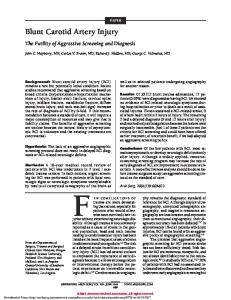
Figure 1. Cross section of ear. Otosclerosis involves the small bones of the middle ear, the malleus (2), the incus (3) and the stapes (4), as well as the bone that surrounds the inner ear, which is called the otic capsule.
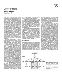
Otosclerosis is a disease of the bones of the inner ear. These are labeled the malleus, incus and stapes (2-4) in figure 1, and are also known in aggregate as the "ossicles". The ossicles become knit together into an immovable mass, and do not transmit sound as well as when they are more flexible. An illustration of one form of this called "stapes fixation" is shown in figure 2. Otosclerosis can also affect the other ossicles (malleus and incus) and the otic capsule -- the bone that surrounds the inner ear.
Figure 2. Stapes fixation in otosclerosis. A bony ankylosis (knee) knits the bone of the middle ear to the stapes, preventing normal transmission of sound from the eardrum into the inner ear. Otosclerosis is usually inherited in an autosomal dominant pattern with variable penetrance. This means that you have a 50-50% chance of getting the gene for otosclerosis if one parent has it, but that not everyone with the gene develop symptoms. Hearing loss usually begins between the ages of 11-30. The hearing loss can be of two types. When otosclerosis involves the small bones of the middle ear, a conductive type loss is found. This type of hearing loss can be corrected both by a hearing aid as well as a surgical procedure called stapedectomy. When otosclerosis significantly involves the bone which surrounds the inner ear, called the "otic capsule", a sensory type hearing loss occurs. This type of hearing loss is not correctable by stapedectomy. While hearing aids are usually worth trying, they also may be ineffective. When otosclerosis involves both the small bones and the cochlea, a "mixed" type hearing loss occurs. Tinnitus is common in otosclerosis NATURAL COURSE OF THE DISEASE Hearing loss generally begins between the ages of 10 and 30. Early on the disease is called "otospongiosis". During this time there is active remodeling of bone of the otic capsule. There may be no conductive hearing loss at this point but rather there may be a "sensory" type hearing loss. The sensory type hearing loss, at this point, is attributed to leakage of enzymes from bone into the inner
ear (Grayeli et al, 2003). It is this process that is thought to possibly be inhibited by medical treatment (sodium Fluoride). Sensory hearing loss is also attributed to atrophy of the spiral ligament caused by involvement of lamellar bone at the inner surface of the cochlear capsule. A third cause is vascular due to degeneration of the stria vascularis. In early stages, treatment with fluoride may be helpful. The sensory component of the disease may eventually lead to complete deafness, but fortunately, this is unusual Later on, a conductive pattern of hearing loss appears. The conductive type of otosclerosis usually progresses up to a maximum in the 30's. After this, it rarely progresses. Dizziness and imbalance is a feature of otosclerosis in roughly 25% of cases. In about half of all patients, there is a family history of similar problems. Women are affected twice as often as men. People of African-American descent only rarely have otosclerosis -- it is usually a condition found in persons of Caucasian or Oriental descent. Pregnancy often has an adverse effect. Otosclerosis is often discovered during or just after pregnancy. The effect of hormone supplements post-menopause is unknown. Usually both ears are affected, although in about 10-15% of patients, hearing loss occurs on one side only.
CAUSE OF OTOSCLEROSIS: Most feel that otosclerosis is an inherited, an autosomal dominant disease with variable penetrance. As there is also also evidence of viral influences in otosclerosis, a recent hypothesis is that otosclerosis requires a combination of a specific gene with exposure to a specific virus (e.g. measles) for it to be expressed and hearing loss to occur (McGuirt et al, 1998). The gene that predisposes to otosclerosis may be similar to the gene that causes osteogenesis imperfecta -- a generalized bone disease. Persons with osteogenesis imperfecta often develop conductive hearing loss. The genes that confer susceptibility to otosclerosis may also provide some protection against otitis media (Manolidis et al, 2003). Some feel that chronic measles infection in bone predispose patients to otosclerosis. Viral materials can be found in osteoblasts in otosclerotic lesions (Nadol, 1998). Variants of otosclerosis exist in which there is a mutation in the NOG gene (Brown et al, 2003) Pathologically, otosclerosis occurs only in human temporal bones, and is considered to be a disorder of new bone formation. Histologic otosclerosis, meaning that it is found only on section but has no symptoms, is found in one of every 2.5 to 10% of whites in the United States (Declau et al, 2001). Clinical otosclerosis, with involvement of hearing, occurs in one in every 10 patients with histologic otosclerosis (Nadol, 1998). This results in the conclusion that .25% to 1% of the population should exhibit clinical otosclerosis. DIAGNOSIS OF OTOSCLEROSIS Diagnosis is usually made by a combination of family history, conductive hearing loss pattern, and CT scan of the temporal bone. Hearing tests may initially show a sensory pattern and later show the typical conductive loss pattern. Acoustic reflexes may eventually be absent, but early on may show the "on-off" effect. Tympanometry often shows stiffening of the ossicular chain. The CT scan is specific but insensitive. A reason for doing this, however, is to detect patients with superior canal
dehiscence, as they can also show a conductive hearing loss. VEMP testing can also be used for this purpose. Dizziness can occur in otosclerosis and was reported in one study to occur in 15% of patients. Pathologically there is degeneration of the vestibular ganglion (Scarpa's ganglion). The mechanism for dizziness is unknown, although there is speculation that it derives from release of enzymes from metabolically active bone into the inner ear(Causse et al, 1982).. Other possibilities might be a "halo" effect -- persons visiting ear doctors may be more likely to attribute dizziness to their ear than others, occlusion of fluid pathways within the inner ear from bony overgrowth, or another effect on the ear caused by the same underlying (unknown) cause as otosclerosis.
TREATMENT OF OTOSCLEROSIS There are four treatment options: 1. Do nothing -- Otosclerosis does not have to be treated. It is usually advisable to have a hearing test repeated once a year (or earlier if hearing drops). 2. Hearing aid -- aids are usually effective for conductive hearing loss. 3. Medical treatment -- an early proposed medical treatment wassodium fluoride, which is a dietary supplement (not a drug). This treatment is not widely accepted, and has not been proven to be effective. A large uncontrolled study of about 1500 patients by Dr. Shambough and associates suggested that it was effective. The idea of using fluoride is not unlike that of using it for teeth -fluoride speeds up hardening of bone. Other more rigorous trials have reported similar results (Bretlau et al, 1989). Side effects of fluoride (Florical and Monocal are the two preparations available over the counter) include occasional stomach upset, allergic itching, and increased joint pains. If aggravation of arthritis occurs, the Fluoride is stopped and the joints return to their previous state in a few weeks. In such a situation, patients can "pace themselves", taking as much of the medication as can be tolerated. Typical doses are one tablet three times a day (florical) and one-two tablets three times a day (Monocal). After two years of fluoride treatment, the dose of fluoride is reduced from three times a day to once a day. Once the otospongiosis phase of otosclerosis is over and there is a clear cut otosclerosis documented by conductive hearing loss, fluoride may be stopped. In theory, avoidance of estrogens or use of estrogen blockers might be helpful in individual with otosclerosis as otosclerosis frequently worsens during pregnancy, suggesting hormonal modulation. To our knowledge, this hypothesis has not been studied. There has recently been use of medications designed for osteoporosis, the diphosphonate family (e.g. Brookler, 1997). A double blinded study found no significant difference (Kennedy et al, 2003). Some studies even report worsening of hearing on these drugs (Yasil et al, 1998), which is would be expected if the drug was ineffective. At this writing (2003), it is not clear whether these drugs are helpful. 4. Surgical treatment.
For conductive hearing loss, in 1957, Dr. John Shea invented the procedure of stapedectomy, which produced excellent hearing results, which remain good for many years after the surgery. This procedure may allow avoidance of hearing aids. It, however, does not help the sensory component of the hearing loss and at best, may close the "air-bone" gap. It also does not affect the vertigo that is sometimes associated with otosclerosis. According to Nadol, stapedectomy is indicated in patients with good bilateral inner-ear function, and conductive hearing loss ranging from 25-30 dB in elected frequencies. Stapedectomy is unreasonable if discrimination scores are lower than 65% as this indicates that there is a substantial sensory component. Patients with stapedectomy may attain better results with hearing aids because of the need for lessor amplification. Stapedectomy may fail for a number of reasons. It is a somewhat difficult and delicate procedure. There may be displacement of the prosthesis, enclosure of the fenestra (window), or erosion of the incus. Disease may progress so that correction of the conductive component is inadequate. An earlier procedure was the fenestration operation (Pulec, 2002). This operation is rarely offered. A small fenestrata stapedotomy is still done in some institutions (House et al, 2002).
Acknowledgements: Images marked as copyright Northwestern University were developed with the support of an NIH grant to Northwestern University Dept. of Otolaryngology, and are used with permission.