Original Article Article original
ORTHOPEDIC MANAGEMENT IN AUTOSOMAL RECESSIVE SPASTIC ATAXIA OF CHARLEVOIX-SAGUENAY Marc Bouchard, MD;* Gaëtan Langlois, MD† OBJECTIVE: To review the orthopedic management of choice in patients having autosomal recessive spastic ataxia of Charlevoix-Saguenay (ARSACS). DESIGN: A retrospective study from April 1978 to April 1997. SETTING: Centre hospitalier de la Sagamie, Chicoutimi, Que. PATIENTS: A review of the records of patients having ARSACS who were identified in the registry of the Neuromuscular Diseases Clinic at the Centre hospitalier de la Sagamie revealed 26 patients who received surgical orthopedic treatment. Initially, the patients were offered conservative treatment, which consisted of physiotherapy sessions, the wearing of an ankle-foot orthosis or serial casting. When this was unsuccessful, foot surgery was considered. RESULTS: During the study period, 49 orthopedic procedures were done, including 24 triple arthrodeses; of these, 9 were combined with lengthening of the Achilles tendon. Most triple arthrodeses were done in patients between the ages of 30 and 49 years. The surgical options evolved during the study from Lambrinudi arthrodesis through arthrodesis of the ankle to triple arthrodesis with lengthening of the Achilles tendon. CONCLUSIONS: As a complement to conservative treatment, surgery has a place in the care of patients with ARSACS. Clinically, the most effective surgical procedures are triple arthrodesis with percutaneous lengthening of the Achilles tendon and adductor and psoas tenotomies combined with neurectomy of the obturator nerve for perineal hygiene. OBJECTIF : Revoir l’approche orthopédique favorisée chez des patients atteints d’ataxie récessive spastique de Charlevoix-Saguenay (ARSACS). CONCEPTION : Étude rétrospective, d’avril 1978 à avril 1997. CONTEXTE : Centre hospitalier de la Sagamie, Chicoutimi (Québec). PATIENTS : Une étude des dossiers des patients atteints d’ARSACS identifiés dans le registre de la Clinique des maladies neuromusculaires du Centre hospitalier de la Sagamie a révélé que 26 patients avaient été traités par chirurgie orthopédique. Au début, on a offert aux patients un traitement conservateur qui consistait en séances de physiothérapie et port d’une orthèse cheville-pied ou de plâtres d’inhibition séquentiels. Lorsque le traitement a échoué, on a envisagé une chirurgie du pied. RÉSULTATS : Au cours de la période d’étude, on a réalisé 49 interventions orthopédiques, y compris 24 triples arthrodèses dont 9 ont été associées à un allongement du tendon d’Achille. La majorité des triples arthrodèses ont été pratiquées chez des patients âgés de 30 à 49 ans. Au cours de l’étude, les options chirurgicales ont évolué de l’arthrodèse de Lambrinudi à la triple arthrodèse avec allongement du tendon d’Achille, en passant par l’arthrodèse de la cheville. CONCLUSIONS : En complément au traitement conservateur, l’intervention chirurgicale a sa place dans le traitement des patients atteints d’ARSACS. Sur le plan clinique, les interventions chirurgicales les plus efficaces sont la triple arthrodèse avec allongement percutané du tendon d’Achille et ténotomie des adducteurs et du psoas associées à une neurectomie du nerf obturateur pour l’hygiène du périnée.
From the Department of Surgery, Université Laval, Quebec, Que. Presented at the Journée Scientifique du programme d’orthopédie de l’Université Laval, Quebec, Que., Mar. 13, 1998. *Resident in orthopedic surgery, Université Laval †Orthopedic surgeon, Centre Universitaire de Santé d’Estrie, Sherbrooke, Que. Accepted for publication Apr. 18, 1999. Correspondence to: Dr. Marc Bouchard, 3230, rue Beaurepaire, Sainte-Foy, Québec (Québec) G1X 1H4; fax 418 658-7451,
[email protected] © 1999 Canadian Medical Association (text and abstract/résumé)
440
JCC, Vol. 42, No 6, décembre 1999
SPASTIC ATAXIA OF CHARLEVOIX-SAGUENAY
A
utosomal recessive spastic ataxia of Charlevoix-Saguenay (ARSACS) is a progressive neurologic disorder first descibed as a distinct pathologic entity in 1978 by Bouchard and colleagues.1 Since then, studies have characterized the epidemiologic,2 clinical,3 biochemical,4 electrophysiologic,5–8 radiologic9 and genetic10 features of ARSACS. In this paper we describe the clinical manifestations of the disease and the management of musculoskeletal damage. There are more than 300 patients living with ARSACS today. All are gathered in two specific geographic areas in the Province of Quebec: Charlevoix and Saguenay-Lac SaintJean, where Charlevoix’s immigrants have contributed to almost 70% of the present population.2 ARSACS is inherited by recessive autosomal transmission. In 1995, Heyer11 hypothesized that it could be transmitted by the mitochondria. Genetic research has excluded ARSACS from linkage to human chromosone 9, which is associated with Friedreich’s ataxia,12 another spinocerebellar progressive disorder similar to ARSACS. However, similar neurologic diseases associated with different chromosomes must be ruled out when exploring the genetic cause of ARSACS: chromosome 5q of spinocerebellar ataxia type 1, 6p of spinal muscular ataxia and 11q of ataxia telangiectasia.10 Recently, Richter and colleagues13 observed “excess shared homozygosity at 13q11” among patients with ARSACS. Clinically, ARSACS becomes symptomatic very early in life. When a child is learning to walk, an abnormal number of falls may be noted.14 With aging, ataxia progresses. This process accelerates as the patients reach the third decade of life. Then, secondary to lesions of the central nervous system, nystagmus, dysarthria and spasticity appear. Lesions in the peripheral nervous system add to these clinical features:
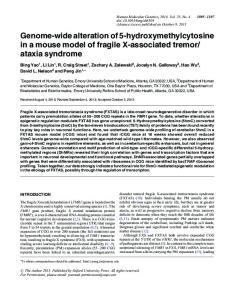
muscle weakness, atrophy and musculoskeletal deformation take place. The musculoskeletal changes seen in ARSACS include, in the upper limb, atrophy of the interosseous muscles of the hand causing claw hands. More important changes occur in the lower limb. The hips contract in adduction and flexion greatly compromising perineal hygiene. Knee flexure appears secondary to spasticity of flexor muscles. An equinus of the ankles occurs, which in adults is refractive to conservative management. A combination of changes can be observed in the foot: calcaneovarus deformity associated with accentuation of the plantar arch, metatarsus adductus and claw toes (Fig. 1). All of the lesions can be asymmetric. Differences among patients vary depending on the genetic penetrance. Ataxia, muscle weakness and spasticity threaten the patient’s ability to walk, so the orthopedic surgeon must consider muskuloskeletal interventions that will help to improve or maintain locomotion, prevent and control lower limb deformity and facilitate tendon transfer. Therefore we reviewed the orthopedic management of patients having a diagnosis of AR-
SACS between April 1978 and April 1997, who were registered with the Neuromuscular Diseases Clinic at the Centre Hospitalier de la Sagamie (Chicoutimi, Que.).
PATIENTS AND METHOD Of the 203 cases of ARSACS registered, all were supervised by a multidisciplinary team (neurologist, pediatrician, orthopedic surgeon, physical therapist, occupational therapist and prosthetist), and 26 of them were closely followed up by an orthopedic surgeon. The 26 files were reviewed with respect to orthopedic diagnosis and management. The information included date of birth, sex, date of diagnosis, surgical procedures done and the indications for them.
FINDINGS A conservative treatment program was established at the follow-up Neuromuscular Diseases Clinic. Physical therapy was directed at preventing equinus of the foot and maintaining good range of motion by stretching and muscle strenghtening. Patients
FIG. 1. Foot deformity in autosomal recessive spastic ataxia of Charlevoix-Saguenay (ARSACS). Calcaneovarus is associated with plantar arch accentuation, a metatarsus adductus and clawing of the toes. CJS, Vol. 42, No. 6, December 1999
441
49 8 5 4
2
6
8
1
1
2
5
2
2
2
1 3
2 2 2 3 2 2
3 1 Total
Adductor and psoas muscle tenotomies and obturator nerve neurectomy
Staple removal
Correction of claw toes
2 Pseudarthrosis revision (talonavicular and calcaneocuboidal)
Plantar aponeurectomy
Ankle arthrodesis
Achilles tendon lengthening only
Triple arthrodesis and Achilles tendon lengthening
Triple arthrodesis, extensor digitorum and hallucis longus transfer
Triple arthrodesis only
JCC, Vol. 42, No 6, décembre 1999
2
1
3
1984 1983
wore a fixed or articulated ankle-foot (AF) orthosis between periods of exercise. When those treatments were insufficient, serial casting was used for 2 to 4 weeks. When conservative treatment failed, corrective surgery was planned to reposition the foot in its most plantigrade position to allow proper locomotion. Some patients required hip surgery to improve perineal hygiene. The surgical procedures varied (Table I). Eighteen (12 men and 6 women) of the 26 patients had at least 1 operation for a total of 49 surgical procedures. The patients ranged in age from 12 to 59 years at the time of surgery. The mean follow-up from the time of diagnosis was 11.5 years (range from 1 month to 19 years). Among the 49 surgical interventions, there were 24 triple arthrodeses, 9 of them associated with Achilles tendon lengthening. When surgical procedures were classified by age groups (Table II), the majority of these triple arthrodeses were performed on patients between the ages of 30 and 49 years. Patients who had hip surgery were older, and all but 3 patients had only 1 operation.
DISCUSSION
1978 Procedure
1
2
3 1
1994 1992 1990 1989 1988 1986 1985
1987
Year Surgical Procedures Performed During the Study Period by Year
Table I 442
4
2 1 1
3
2
1
10 2 6 2
9
1996
2 2 1
5
10
1997
Total procedures
BOUCHARD AND LANGLOIS
The surgical procedures for foot deformity in ARSACS evolved during the study period. Initially, a triple arthrodesis of Lambrinudi associated with a transfer of the extensor hallucis longus and the extensor digitorum longus was preferred.5 The technique was difficult and the clawing toes were worsened by the transfer. Tendon transfers were therefore abandoned. One arthrodesis of the ankle was performed in a patient with ARSACS. This surgical option was not repeated because it had the disadvantage of not correcting the equinus of the forefoot. The resulting accentuated arch later gave rise to serious difficulties with shoe wear and ambulation.
SPASTIC ATAXIA OF CHARLEVOIX-SAGUENAY
After evaluation of these outcomes, a new procedure was needed to treat the orthopedic problems associated with ARSACS. The next step was the triple arthrodesis associated with a percutaneous split lengthening procedure of the Achilles tendon. This lengthening was performed through 3 small incisions (2 median and 1 lateral) and attempted to diminish the spasticity causing the equinus of the foot. The triple arthrodesis was performed through a standard lateral approach.
Articular cartilage was cut and slices were used as graft material to arthrodese the 3 joints. The subtalar joint was fixed with a cannulated 7.3-mm lag screw. The calcaneocuboidal joint was fixed with a staple and the talonavicular joint was only supplemented with graft (Figs. 2 and 3). The arthrodesis was immobilized in a cast for 3 to 4 weeks postoperatively followed by application of an AF orthosis fixed at 90° until fixation was obtained. Later, the AF orthosis was set to articulate.
Complications of this procedure included 3 pseudarthroses (12.5%), 1 talonavicular and 2 calcaneocuboidal, which necessitated revision surgery. These results can be classified as satisfying considering the spasticity due to ARSACS, which is an important risk factor for pseudarthrosis.15 Other complications encountered were 3 cases of bursitis caused by staples, which forced their removal. Our series has the limits of any retrospective study of a selected popula-
Table II Surgical Procedures (N = 49) Performed According to Patient Age and Sex (M/F) Patient age, yr Procedure Triple arthrodesis only
10–19
20–29
30–39
40–49
2/M
1/M, 1/F
4/M
2/M
2/M, 2/F
1/M
2/M
4/M
3/M
2/M, 2/F
4/F
Triple arthrodesis, extensor digitorum and hallucis longus transfer Triple arthrodesis and Achilles tendon lengthening Achilles tendon lengthening only Ankle arthrodesis Plantar aponeurectomy
2/F
1/M 2/M
Pseudarthrosis revision (talonavicular and calcaneocuboidal)
3/M
Correction of claw toes
2/F
Staple removal
1/M
Adductor and psoas muscle tenotomies and obturator nerve neurectomy Total
50–59
4
FIG. 2. Management by triple arthrodesis. The subtalar joint is fixed with a cannulated lag screw; the calcaneocuboidal joint is fixed with a staple and the 3 joints are grafted.
9
22
2/M 2/F
2/F
10
4
FIG. 3. Radiograph 8 weeks after a triple arthrodesis performed according to the technique shown in Fig. 2. The arthrodesis appears to be consolidated. CJS, Vol. 42, No. 6, December 1999
443
BOUCHARD AND LANGLOIS
tion. Subjectivity is present, but the findings provide clues on how the foot problems caused by ARSACS can be managed when conservative treatment has reached its limits.
CONCLUSIONS The treatment of spasticity and foot changes in ARSACS will evolve. It is possible that transfer or hemitransfer of the tibialis posterior muscle could be utilized to counterbalance the equinus. We believe that muscle lengthening as performed in patients with cerebral palsy will gain popularity for the treatment of orthopedic problems in ARSACS. Experiments with botulism toxin to evaluate what is to be earned by surgical lengthening seem promising.16 This toxin is being used on a larger scale by neurologists. This treatment should help to decide for whom and when the lengthening procedure should be done. Future research will dictate the proper management of the uncommon problems encountered in this rare but severely debililating disease.
References 1. Bouchard JP, Barbeau A, Bouchard R, Bouchard RW. Autosomal recessive spastic ataxia of Charlevoix-Saguenay. Can J Neurol Sci 1978;5(1):61-9. 2. De Braekeleer M, Giasson F, Mathieu J, Roy M, Bouchard JP, Morgan K. Genetic epidemiology of autosomal recessive spastic ataxia of Charlevoix-
444
JCC, Vol. 42, No 6, décembre 1999
Saguenay in northeastern Quebec. Genet Epidemiol 1993;10(1):17-25. 3. Bouchard JP. Recessive spastic ataxia of Charlevoix-Saguenay. In: de Jong JM, editor. Handbook of clinical neurology. vol 16. Amsterdam: Elsevier; 1991. p. 451-9. 4. Melançon SB, Potier M, Dallaire P, Rollin G, Fontaine G, Grenier B. Pyruvate dehydrogenase, lipoamide dehydrogenase and citrate synthase activity in fibroblasts from patients with Friedreich’s and Charlevoix-Saguenay ataxia. Can J Neurol Sci 1979;6(2): 241-2. 5. Peyronnard JM, Charron L, Barbeau A. The neuropathy of CharlevoixSaguenay ataxia: an electrophysiological and pathological study. Can J Neurol Sci 1979;6(2):199-203. 6. Bouchard RW, Bouchard JP, Bouchard R, Barbeau A. Electroencephalographic findings in Friedreich’s ataxia and autosomal recessive spastic ataxia of Charlevoix-Saguenay (ARSACS). Can J Neurol Sci 1979;6(2): 191-4. 7. Bouchard JP, Barbeau A, Bouchard R, Bouchard RW. Electromyography and nerve conduction studies in Freidreich’s ataxia and autosomal recessive spastic ataxia of Charlevoix-Saguenay (ARSACS). Can J Neurol Sci 1979;6 (2):185-9. 8. Dionne J, Wright G, Barber H, Bouchard R, Bouchard JP. Oculomotor and vestibular findings in autosomal recessive spastic ataxia of CharlevoixSaguenay. Can J Neurol Sci 1979;6(2): 177-84.
9. Langelier R, Bouchard JP, Bouchard R. Computed tomography of posterior fossa in hereditary ataxias. Can J Neurol Sci 1979;6(2):195-8. 10. Richter A, Morgan K, Bouchard JP, Poirier J, Mercier J, Gosselin F, et al. Clinical and molecular genetic studies on autosomal recessive spastic ataxia of Charlevoix-Saguenay (ARSACS). Adv Neurol 1993;61:97-103. 11. Heyer E. Mitochondria and nuclear genetic contribution of female founders to a contemporary population in northeast Quebec. Am J Hum Genet 1995; 56(6):1450-5. 12. Richter A, Bouchard JP, Poirier J, Mercier J, Melançon SB. Autosomal recessive spastic ataxia of CharlevoixSaguenay: exclusion of the ARSACS gene from linkage to human chromosome 9 [abstract]. Am J Hum Genet 1991;49:A357. 13. Richter A, Rioux JD, Bouchard JP, Mercier J, Mathieu J, Ge B, et al. Location score and haplotype analyses of the locus for autosomal recessive ataxia of Charlevoix-Saguenay, in chromosome region 13q11. Am J Hum Genet 1999;64(3):768-75. 14. Pineault D. L’ataxie recessive spastique de Charlevoix Saguenay [information document]. Chicoutimi (Que.): Centre Hospitalier de la Sagamie; 1995. 15. Holt ES, Hansen ST, Mayol KA, Sangeorzan BJ. Ankle arthrodesis using internal screw fixation. Clin Orthop 1991;268:21-8. 16. Kessler KR, Benecke R. Botulinum toxin: from poison to remedy. Neurotoxicology 1997;18(3):761-70.