MASTER THESIS
Orthodontic movement of root canal treated or traumatized teeth: Is there a greater risk of root resorption?
Svetlana Kobets ODO-07
Supervisors: Heidi Kerosuo, Professor (Orthodontics), IKO Eero Kerosuo, Professor (Endodontics), IKO
1
CONTENTS
Summary (Sammendrag på norsk)
p3
Introduction
p4
Classification of the different types of tooth resorptions Biology of tooth movement Root resorption associated with orthodontics
Aims of the Master Thesis
p9
Literature Review
p9
Methods Orthodontic movement of root canal treated teeth Orthodontic tooth movement of teeth with a history of trauma
Questionnaire among dentists in IKO/TkNN
p 14
General discussion and conclusion
p 17
List of literature (References)
p 20
Appendix (spørreskjema)
p 23
2
SAMMENDRAG Det finnes ulike oppfatninger angående ortodontisk behandling av tenner som er tidligere rotfylt eller tidligere traumatisert. Noen mener at det finnes mer risiko for resorpsjon ved ortodontisk forflytning av slike tenner enn levende "friske" tenner. Det første målet med dette litteraturstudiet var å se om det finnes vitenskapelige bevis for eventuell risiko for resorpsjon ved ortodontisk forflytning av rotfylte/tidligere traumatiserte tenner. Det andre målet var å finne ut hva praktiserende tannleger mener om temaet. 23 artikler var samlet inn gjennom PubMed og veiledere, samt 6 bøker var valgt fra det Helse- og Naturvitenskapelige Biblioteket ved UiT. Et spørreskjema med påstander ang ortodontisk forflytning av rotfylte tenner og tidligere traumatiserte tenner var delt ut til tannleger/spesialister på Studentklinikken og TkNN. Antall spørreskjema utdelt var 41,og 21 var besvart. Resultatene fra litteraturen var at rotfylte tenner (hvis godt renset og tettet) kan forflyttes med samme sikkerhet som vitale tenner. Når det gjelder tenner med tidligere historie av trauma, så sa mesteparten av litteraturen at det er noe større risiko for resorpsjon ved ortodontisk behandling av slike tenner. Besvarelsene av spørreskjema viste at meningen er delt (50/50) ang mer risiko for resorpsjon av rotfylte tenner. Derimot var de fleste deltakere (95%) enige om at det er per risikabelt å forflytte tenner med tidligere trauma enn tenner uten trauma. Hovedkonklusjonen er at på grunn av lite tilgjengelige artikler og ingen RCT-studier (randomiserte kontrollerte studier) angående tema diskutert i denne masteroppgaven, trenges mer det mer forskning for å gjøre opp en sikker mening. Større antall deltakere i spørreundersøkelsen er også ønskelig for å føre resultatene over på allmenheten av praktiserende tannleger i Norge.
3
INTRODUCTION There are divided opinions among dental practitioners and specialists about performing orthodontic tooth movement on teeth that have previously been endodontically treated or have a history of moderate/severe trauma. The opinions are of that orthodontic movement of these teeth may generally have a higher risk of apical root resorption, or less predictive tooth movement may occur than with healthy vital teeth.
Already 70 years ago it was suggested by John Steadman in Angle Orthodontist (1) that a previously endodontically treated tooth may not endure the force used in orthodontic tooth movement as well as a "healthy" tooth because the PDL has been damaged and thus the tooth is “weakened”, and acts like a foreign body. The same skepticism has later existed for the traumatized tooth.
It is a very relevant topic, since the teeth being moved, especially central incisors and to some degree lateral incisors, may often have a history of trauma. They might even have been root filled after the trauma. Also, more and more adults are having appliances nowadays because they didn't have the opportunity to do so at a younger age, ore did not have access to information about orthodontic treatment or in relation to advanced restorative procedures. In adults referred to orthodontic treatment there is a high probability that some of the teeth have been root canal treated (2).
Classification of the different types of tooth resorptions: Tooth resorption is a common sequela following injuries to or irritation of the periodontal ligament and/or tooth pulp. The course of tooth resorption involves an elaborate interaction among inflammatory cells, resorbing cells, and hard tissue structures. The key cells involved in resorption are of the clastic type, which include osteoblasts and odontoclasts. Types of tooth resorption include internal resorption, external resorption and a combination of those two. Tooth resorption is the loss of tooth tissue (i.e. cementum and dentine) as a result of odontoclastic action (3).
4
Andreasen (6) classified tooth resorption as follows: Tooth resorption can be divided in to Internal and External resorption. Internal resorption can then be divided into inflammatory and replacement resorption. External resorption can be divided into surface, inflammatory and replacement resorption. External resorption often occurs after chronic injury affecting the PDL such as orthodontic treatment, traumatic occlusion and pressure from developing cysts, apical granulomas or ectopic erupting teeth (4). Trope (5) classified tooth resorption on the basis of etiology: Root resorption can be divided into external and internal resorption. Internal root resorption is inflammation in the pulp/root canal due to e,g, an infection, traumatic injury or extreme heat produced when cutting on dentin without adequate water spray. External root resorption is then divided into; Type 1: Attachment damage alone (root resorption caused by traumatic injury); Type 2: Infection alone (root resorption caused by an infection in the root canal or sulcus); Type 3: Attachment damage plus infection (root resorption caused by a traumatic injury and infection in the root canal or sulcus). Ne and coworkers-workers (3) further classified the external resorption into four categories by its clinical and histologic manifestations: external surface resorption, external inflammatory root resorption, replacement resorption, and ankylosis. The difference between ankylosis and replacement resorption being that in the latter, there is always a connective tissue present between the bone and the tooth. External inflammatory root resorption can be further categorized into cervical resorption with or without a vital pulp (invasive cervical root resorption) and external apical root resorption. Other variations of resorption include combined internal and external resorption and transient apical breakdown (TAB). Heithersay (6) presented an alternative classification of tooth resorption as follows: Trauma induced resorption: Pressure (trauma) induced resorption that is central to this thesis, can for example be caused by natural exfoliation (permanent teeth pressing on the primary), in relationship to ectopic eruption (e.g. canines that press on the laterals) or due to orthodontic movement forces as well as a result of different traumatic injuries to teeth/alveolar bone. Pressure induced resorption is then further divided into; 1) Surface resorption - a shallow resorption of cementum often with involvement of a small amount of underlying dentine. This type of resorption is self-limiting. In the absence of superimposed infection, it will heal uneventfully with reparative cementum; 2) Transient apical resorption – a resorptive process following luxation injuries and associated with a transient apical breakdown. A transient process, the internally resorbed apex will close uneventfully; 3) Inflammatory resorption – 5
caused by the response of the dento-alveolar apparatus to an infection. That leads to an inflammation that results in tooth resorption. These resorptions may occur as internal, external or combined internal-external lesions. It will stabilize after the infection is gone, but the tooth tissue will not heal/grow back. Inflammatory resorption can be related to for example periodontal disease, orthodontic movement and internal bleaching; 4) Replacement resorption – involves the progressive replacement of tooth structure by bone and ultimately tooth loss. This kind of resorption follows the death of PDL-cells due to factors such as compression or drying (as in the case of delaying replantation of an avulsed tooth) and eventually leads to ankylosis. Infection induced dental resorption: The response of the dento-alveolar apparatus to infection is characterized by inflammation which may result in tooth resorption. This may be a consequence of infective endodontic pathosis alone or superimposed on trauma induced resorption. The resorption process will arrest when the infection is removed. Hyperplastic invasive resorption: The resorbing tissue invades the hard tissue of the tooth in a destructive and apparently uncontrolled fashion, akin to the nature of some fibro-osseous lesions such as fibrous dysplasia. Unlike the first two types of resorption, simple elimination of the cause of the lesion is ineffective in arresting their progress. Moderate and severe injuries like for example crown-root fractures, root fractures, avulsions, alveolar socket fractures and some types of luxations injuries may lead to both internal/external inflammatory resorption and/or to ankylosis resorption (replacement resorption) (6, 7).
Biology of tooth movement Orthodontic tooth movement is based on force applied on the tooth and the periodontal ligament (PDL) and consequent alveolar bone remodeling. When the PDL is subjected to mechanical load, it responds by initiating the complex biologic process which results in remodeling of the alveolar bone and translocation of the tooth. The PDL consists of a number of cell types embedded in an extracellular matrix composed mainly of type I collagen fibers with ground substance (proteoglycans and glycoproteins) and oxytalin fibers. The main cell types are osteoblasts, osteoclasts, fibroblasts and macrophages. These cells are responsible for the bone remodeling and tooth movement. The fibroblasts and osteoblasts are always present in the PDL, while macrophages and osteoclasts are "summoned" when needed. 6
The exact cellular and molecular biological events that occur during orthodontic tooth movement are not yet fully understood, but several theories exist, of which the pressure tension theory is the most well-known. The pressure-tension theory is based on an alteration in blood flow within the PDL produced by sustained pressure that causes the tooth to shift position within the PDL space, compressing the ligament in pressure areas and stretching on the opposite end. Blood flow is decreased where the PDL is compressed, while it usually is maintained or increased where the PDL is under tension (8, 9). When a force is placed on a tooth, bone is laid down on the tension side of the PDL (osteoblastic activity) and resorbs on the pressure side (osteoclastic activity) (10). The cells of the PDL react because of deformation of their cytoskeleton. There is evidence that the shape of a cell can influence its activity; rounded cells tend to be catabolic whereas flattened cells are anabolic (9). The osteoblasts are the cells responsible for bone formation, and also for activating the osteoclasts, which are the bone resorbing cells. The activation is a complex mechanism involving secretion of different types of intra-/extracellular messengers and growth factors. On the tension side, the osteoblasts are flattened. Second messengers and intracellular messengers are released in response to tension. Fibroblasts differentiate into osteoblasts, and osteoblastic activity and bone production increases (8). On the pressure side, the osteoblasts change their form from flattened to round, and thereby exposing the osteoid for osteoclasts to resorb (8). However, if the forces are high and exceed the capillary blood pressure, cell death will occur in the bone on the pressure side and a cellfree (necrotic) bone area will form. This is described as hyalinization, due to the glass-like appearance of these regions when viewed with light microscopy resembling hyaline cartilage. When this happens, remodeling of bone bordering the necrotic area of the PDL must be accomplished by cells derived from adjacent undamaged areas. After a delay of several days, cellular elements begin to invade the necrotic (hyalinized) area. Osteoclasts appear within the adjacent bone marrow spaces and begin to attack the underside of the bone immediately adjacent to the necrotic PDL area. This process is called undermining resorption and will result in a stepwise movement of tooth (8, 9). Even forces as light as 30g can produce some areas of hyalinization, if directed to a small area. This tends to occur more with tipping and intrusive movement than bodily movement of teeth, presumably because the force is dissipated more evenly though the PDL during bodily movement. Therefor lighter forces are used in tipping movement as compared to bodily movement (10).
7
Root resorption associated with orthodontics: Orthodontic force application induces a local process that includes all of the characteristics of inflammation: redness, heat, swelling and pain. This inflammation which is essential for tooth movement is actually the fundamental component also behind the process of root resorption. The specific causes of root resorption during orthodontic treatment are not well understood, but it seems that some degree of external root resorption occurs in all the teeth undergoing orthodontic movement. Factors like duration of the treatment, the type and amount of force used, root/tooth anatomy and genetic susceptibility are reported to play a role (4). There are also discussions about whether previous trauma or endodontic treatment of the tooth being moved may increase the risk of root resorption (11). Also resorption prior to orthodontic movement may be a risk for the treatment. Root resorption following orthodontic treatment is considered as surface resorption or transient inflammatory resorption. When the pressure is stopped, spontaneous healing (in case of surface resorption) will take place, or just a stabilization of the tooth tissue/termination of the resorption process in case of inflammatory resorption (4, 12). Pressure (trauma) induced resorption, can for example be caused by natural eruption (permanent teeth pressing on the primary), and is found in relationship to ectopic eruption (e.g. canines that press on the laterals) or due to orthodontic forces as well as a result of different traumatic injuries to teeth/alveolar bone (7).
Replacement resorption, which sometimes can occur after trauma to the tooth, ultimately leads to ankylosis. When a tooth is ankylosed, it cannot be orthodontically moved and the treatment will fail (6). Replacement resorption is a continuous process whereby the tooth is gradually resorbed and subsequently replaced by bone. Ankylosis is a dental situation in which the roots of teeth lose their normal attachment to the bone (PDL) and become fused directly to the bone (no connective tissue between the tooth and the bone (3). Ankylosis can be diagnosed radiographically. Ankylosis can occur in only a small root area of the tooth and might not always be easy to diagnose beforehand (6).
8
AIMS OF THE MASTER THESIS The aims of this master thesis were twofold. First, to find out whether there is an increased risk for root resorption during orthodontic movement of traumatized and/or root filled teeth. Secondly, to find out the opinions of the dentists in the student clinic and TkNN about the risk of resorption when orthodontically treating traumatized/RCT-teeth, and compare the answers with the present evidence.
LITERATURE REVIEW Methods: Before searching for evidence from the literature, following questions were formulated: 1) Can root canal treated teeth be orthodontically moved as readily as vital teeth, or is there a greater risk of root resorption? 2) Can previously traumatized teeth be orthodontically moved as readily as nontraumatized teeth, or is there a greater risk of root resorption?
Articles were collected using following keywords and combinations in PubMed: -
Orthodontic movement + previously traumatized teeth (5 hits)
-
Orthodontic movement + previously traumatized incisors (4 hits)
-
Orthodontic movement + endodontically treated teeth (30 hits)
-
Orthodontic movement + root filled teeth (14 hits)
-
Orthodontic movement + root canal treated teeth (23 hits)
-
Orthodontic treatment + previously traumatized teeth (7 hits)
-
Orthodontic treatment + previously traumatizes incisors (6 hits)
-
Orthodontic treatment + endodontically treated teeth (65 hits)
-
Orthodontic treatment + root canal treated teeth (50 hits)
Based on those hits, articles that had the subject of root resorption after orthodontic movement of endodontically treated teeth or previously traumatized teeth as the main theme or part of the main theme were included in this master thesis. There were 20 articles specific to the study question that were chosen and then printed out from the web, or copied from the 9
journals that they’ve been published in, and 3 articles were given by the supervisors. I have also searched the University’s Nature- and Health Library (Natur- og Helsebibliotek ved UiT), and 6 textbooks that had some relevant info were identified.
The articles included were 3 prospective studies (4, 13-14), 6 retrospective studies (15-20), 9 literature reviews (3, 5, 6, 12, 21-24, ), 1 case report (25) and 4 animal studies (11, 26-28).
Orthodontic movement of root canal treated teeth There was only one prospective study dealing with this theme; Llamas-Carrera and coworkers (4) had a study sample of 77 patients with a mean age of 32,7+/- 10,7 years, who each had one root filled tooth before completion of multiband/bracket orthodontic therapy for at least 1 year. The conclusion of the study was that there was no significant difference in the amount of severity of root resorption during orthodontic tooth movement between root filled teeth and their contralateral teeth with vital pulps.
In their retrospective study, Esteves and coworkers (16) had a sample of 16 patients (age not specified). The patients had a maxillary central incisor treated endodontically before orthodontic treatment, and a vital homologous tooth for control. Similarly, Spurrier and coworkers (19) had a sample of 43 patients (age 13-16y) who had one or more anterior teeth treated endodontically before orthodontic therapy. In both studies, measurements were made by comparing the pre- and post-treatment (with brackets/bands) radiographs. Esteves and coworkers (16) came to a conclusion that there was no difference in apical root resorption between maxillary central incisors treated endodontically and untreated homologs when subjected to orthodontic treatment. Surprisingly, Spurrier and coworkers (19) even found that endodontically treated incisors resorbed with less frequency and severity than vital control teeth. Mirabella & Årtun (18) examined periapical radiographs of 343 patients age 20,0-70,1 years who were treated for 0,5-5,2 years with fixed orthodontic appliance. The conclusion was in keeping with Spurrier and coworkers (19), that endodontically treated teeth were more resistant to root resorption than vital teeth.
The article by Wickwire and coworkers from 1974 was the one with contrasting results. They found in their radiographic survey of 45 orthodontic patients (children, age not specified), that the incidence of root resorption in the endodontically treated teeth was greater compared to their adjacent vital controls. But most of the teeth examined in this study have had a previous 10
traumatic injury prior to the RCT. This may have caused a bias in the results of root resorption after orthodontic movement of root canal treated teeth
The evidence value from case reports is low. Therefore, as an example, Bender and coworkers (25) reviewed 2 cases, one with a 7,3yr-old female with orthodontic treatment after a traumatic accident to her left maxillary central incisor, and other with a 15yr-old male who had a traumatic accident to his right maxillary central incisor at an age of 7. In both cases the patients received standard root canal treatment on the traumatized tooth, and were followed up with radiographic examinations. The result was that during orthodontic movement, endodontically treated teeth had the radiographic appearance of less severe apical resorption and with lesser frequency than the vital teeth. From 1990’s and 2000’s literature reviews such as Kindelan and coworkers (22), Hamilton & Gutman (12) and Steiner & West (23) have come to the same conclusion that root canal treatment at such is not a risk of orthodontic treatment and that endodontically treated teeth can be moved as readily as vital teeth.
The evidence from animal studies is in line with the majority of the mentioned studies. As an example, Mah and coworkers (26) used 12 young male ferrets in their study. Mandibular canines were treated endodontically in each ferret. Then, bilateral movement of the canines was induced with an orthodontic spring. Mattison and coworkers (11) used 11 vital and endodontically treated canines of 6 adult cats and subjected them to orthodontic forces. Both studies came to a conclusion that there was no significant difference between root resorption of endodontically treated and vital teeth when both were subjected to orthodontic movement.
Suggestions exist for why endodontically treated teeth might experience less resorption than vital teeth during orthodontic movement. One is presented by Bender and coworkers (25), who suggested an explanation based namely on the absence of a pulp in a root filled tooth. Since root canal treated teeth do not have pulp tissue (thereby missing the blood supply and the nervous system), they naturally react in a different way to the orthodontic forces. Several neuropeptides are released when the dental nerve is stimulated by the moving forces, with the most important being Substance P (SP), Calcitonin gene-related peptide (CGRP), Neurokinin A (NKA), Vasoactive intestinal polypeptide (VIP), and Neuropeptide Y (NPY). These substances act as neurogenic vasodilators/vasoconstrictors, and play a role in bone 11
metabolism (growth and remodeling). CGRP-immunoreactive fibers play an important role in the pulp response to inflammation and wound healing. It is suggested that the loss of the release of neuropeptides from a pulp that has been removed, would result in a decrease of the CGRP-immunoreactive fibers, and thus a reduction in the amount of resorption seen in endodontically treated teeth. So if a root canal is well-cleaned, shaped and threedimensionally obturated, minimal (or no) resorptive/remodeling changes may occur apically during tooth movement (12, 22).
Orthodontic tooth movement of teeth with a previous history of trauma Fonseca and coworkers (29) stated that the teeth most commonly traumatized by root fractures are the maxillary incisors (75%). Therefore, orthodontic movement of previously traumatized teeth represents a common problem in routine orthodontic treatment. The risk, incidence and type of root resorption that can occur following all types of tooth trauma have been clearly described. The assessment of the effects of orthodontic tooth movement on previously traumatized teeth however, has received little attention in the literature (12).
In a prospective study, Brin and coworkers (13) studied 139 children (mean age of 13,7y), who were divided in 4 groups. Group T were 56 children who encountered trauma to their maxillary incisors, group O with 29 orthodontic patients with intact incisors, group TO with 28 previously traumatized orthodontic patients, and group C with 26 children serving as the control group. Orthodontic treatment was restricted to tipping movement. All patients were examined clinically and radiographically. The time that elapsed from the trauma to the latest examination was about 3 years in most of the children. The result was that traumatized incisors that were subjected to orthodontic movement were more susceptible to root resorption than teeth injured only or those subjected to orthodontic movement only. Malmgren and coworkers (14) studied 55 previously traumatized maxillary incisors (both central and lateral) prospectively in 27 patients (boys and girls age 7-15y). Intraoral radiographs of the incisors before and after orthodontic treatment were examined. The result was that teeth with slight or moderate trauma and an intact PDL after an observation period of at least 4 to 5 months could be moved with a prognosis comparable to that of uninjured teeth. Nevertheless, the authors concluded that when it came to the prognosis for orthodontic movement of teeth with severe trauma, further studies are required.
12
Pulpal condition clinically and radiologically after orthodontic extrusion of previously traumatizes (n=77) and non-traumatized (n=400) teeth, and after previous dental trauma without subsequent orthodontic treatment (n=193) has been examined retrospectively by Bauss and coworkers (15). The age range of the patients was 6,6-27,3 years. Also in an earlier study with similar study design but different subjects (17), 559 patients were examined. The mean age of patients at the trauma was 9,5y; the mead age of the patients at the end of orthodontic treatment was 15y. The teeth studied were maxillary central and lateral incisors. Both articles reported that orthodontically treated teeth with severe periodontal injuries have a higher susceptibility to pulp necrosis and root resorption than non-traumatized teeth.
Orthodontic treatment of traumatized teeth as also been studied on animals. Graupner (28) performed orthodontic treatment on 4 female Mocaca rhesus monkeys. One of the maxillary central incisors was extracted and replanted in 3 monkeys prior to the orthodontic treatment. The 4th one served as a control. The teeth were retracted with brackets for 30, 60 and 90 days. The monkeys were followed up radiographically, and then sacrificed. The findings revealed that resorption corresponded to the treatment time, regardless of the type of movement of the teeth. Only a small measure of resorption was observed in the orthodontic control monkey, a bit less than in the experimented subjects.
Review articles have come to somehow different conclusions. Earlier studies were reviewed by 4 articles (12, 21-22, 24), and it was concluded that root resorption occur more readily in orthodontic treatment of previously traumatized teeth than in non-traumatized teeth. However, in their review article, Medeiros & Mucha (21) did not think that the findings referred presented sure evidence of that previously traumatized teeth resorbed more. The authors stated that the most likely cause of the root resorption might be the traumatic impact itself, and that these complications can happen regardless the use of orthodontic force.
13
RISK OF ORTHODONTIC TREATMENT IN PREVIOUSLY ENDO-TREATED TEETH OR TEETH WITH A HISTORY OF TRAUMA A QUESTIONNAIRE SURVEY Aim: To find out if dentists consider endodontic treatment or trauma history of a tooth to be a risk factor for orthodontic treatment. Subjects: The subjects of the survey were all dentists/specialists in the Student Clinic and TkNN (Tannhelsetjenestens kompetansesenter for Nord-Norge) in Tromsø (N=41). A questionnaire was designed and distributed to the subjects in the premises of TkNN and the student clinic. Twenty-one dentists (21/41) returned a completed questionnaire. There were 11 participants from the Student Clinic and 10 participants from TkNN. The questionnaire was answered anonymously.
Methods: Statements regarding orthodontic treatment of teeth with endodontic treatment and/or previous history of trauma were included in the questionnaire as follows: 1. In general, orthodontic movement of previously endo-treated teeth should be avoided or performed with extra care: a) because of a higher risk of internal/external root resorption b) because orthodontic movement of endodontically treated teeth is more unpredictable than movement of vital teeth 2. Orthodontic treatment of incisors with previous history of trauma involves a higher risk of internal/external root resorption in the following cases: a) In teeth with previous history of fractures that involve the pulp (ex: complicated crown fractures, crown-root fractures, root fractures) b) In teeth with previous history of luxations
14
c) In teeth with previous history of successful replantation after avulsion (= no resorption 1 year after the replantation)
The participants were asked to answer each statement, if it was True or False in their opinion. If they were not sure, they would leave the statement unanswered. Questions about the work place and place of education of the participant (Norway or abroad) were also included.
Results:
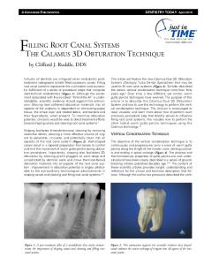
Figure 1: Distribution of the respondents’ answers to different statements in the questionnaire (1a-2c)
Approximately half of the participants (10) had their dental degree from Norway, and other half (11) outside Norway.
Regarding the question about teeth that had been root filled, 57% of the practitioners (12 out of 21) didn’t think that those teeth have a higher risk of internal/external root resorption (Fig1, column 1a.) The answers were similarly distributed regarding the predictability of the outcome of orthodontic movement (Fig 1, 1b). It was not the same person who did not give any answer in questions 1b-2c. 15
As to teeth with a previous history of trauma, all participants except one thought that those teeth have a higher risk of resorption when orthodontically moved than vital teeth without any past trauma. 71% (15/21) thought it was a higher risk of moving teeth with a history of fractures involving the pulp, 76% (16/21) thought the same about teeth with a history of luxations, and 80% (17/21) about teeth with a history of successful replantation (Figure 1, columns 2a-c).
Discussion: 41 dentists/specialists received the questionnaire, and 21 answers were returned. The 50% response rate can still be considered acceptable as such, but since the sample is very limited, it cannot be generalized to dentists’ opinion in general. The low response rate might be due to that a lot of personnel, both in the Student Clinic and TkNN, work part time in Tromsø and part time abroad, resulting in that they simply forget to fill out the questionnaire, or are away for a too long period of time. Another reason may be that they feel that they do not have the time, or the theme is too far away from their area of expertise that they do not have the knowledge or the desire to answer. A bigger sample and response rate would be preferable to make a certain conclusion on the practitioners’ opinion on the subject.
The opinions among the participants deviated most regarding root canal treated teeth and orthodontic treatment. Approximately half of the practitioners thought that it’s not a problem to move those, and others would not recommend orthodontic movement (Figure 1). The bigger portion of practitioners were unanimous about the bigger risk of root resorption when moving previously traumatized teeth. Both the deviation in opinions regarding orthodontic treatment of root filled teeth, and the (almost) unanimous opinion of the resorption risk when moving traumatized teeth regardless of type of trauma, may be due to that the general practitioners at Studentklinikken have the possibility to refer the patients needing orthodontic treatment to the specialists, and do not themselves have much clinical experience with these issues.
16
GENERAL DISCUSSION The first aim of this literature study was to find out if there was more risk of apical root resorption when orthodontically moving teeth that had previously been root canal treated or have a history of moderate/severe trauma. To accomplish that, 20 articles were chosen through PubMed, 3 articles provided by the supervisors, as well as some references concerning the theme in 6 textbooks from the University’s Nature- and Health Library. The second aim was to find out what colleagues thought about the studied subject. A questionnaire was handed out to the dentists and specialists in the Student Clinic and TkNN to get an answer.
Orthodontic movement of previously root canal treated teeth: Most of the literature embedded in this work concerning orthodontic movement of root filled teeth, came to a conclusion that endodontically treated teeth can be moved without more risk of apical root resorption than vital teeth. As long as the PDL is intact and looks normal in a radiograph and the teeth are cleaned, shaped and sealed properly, endodontically treated teeth respond to orthodontic force in the same way as teeth with vital pulps (9). It is even suggested that root filled teeth may resorb less than vital teeth. The main theory supporting that seems to emphasize the absence of a pulp, resulting in less production of several neuropeptides (neurotransmitters) and thus less resorption (25). The results from the questionnaire showed that half of the practitioners regarded orthodontic movement of root canal treated teeth to involve higher risk of root resorption, which contradicts with the majority of the literature reviewed. The discrepancy may be due to that there is not much research on the subject and also maybe the existence of skepticism due to lack of knowledge about RCT and biology of tooth movement in the past.
Nevertheless, a definitive conclusion cannot be drawn about this subject because of the low number of articles found in PubMed. Neither were there any RCT-studies (Randomized Controlled Trials), which are regarded as the "gold standard" with the highest value of evidence. Several of the studies also had small samples of patients and controls, and some studies are performed on animals. Thus, more research is needed in this area.
17
Orthodontic movement of previously traumatized teeth: Most of the literature agreed on that teeth that have a history of moderate or severe trauma might have a higher risk of apical root resorption during/after orthodontic tooth movement. The more severe the traumatic injury, the poorer the prognosis for the tooth being moved (12, 13, 15, 17, 21-22, 24, 27-28). Also, traumatized teeth with signs of root resorption prior to orthodontic movement may be more prone to root resorption during movement (30). Most of the participants in the questionnaire, answered that they would be reluctant to orthodontically move teeth with previous history of trauma. This seems to be in accordance to the literature, and it also shows that the practitioners were well aware of the long-term risks of trauma also regarding orthodontic treatment. Also, the association between traumatized teeth and orthodontic movement has been studied more intensively than ortho-endo association. There was surprisingly little difference between answers to the different trauma categories. One would have expected that coronal fractures involving the pulp would present a lesser risk to orthodontic treatment than for example luxations.
Because of the small sample of literature found in PubMed and the library, plus the absence of RCT-studies on this subject as well, a certain conclusion cannot be drawn. Definitely, more research is needed, especially on orthodontic movement of teeth with previous moderate/severe trauma.
CONCLUSIONS Most of the literature reviewed in this study show that root canal treated teeth can be orthodontically moved as readily as vital teeth, and may even resorb less than vital teeth during orthodontic treatment. However, half of the dentists associated an endodontically treated tooth with more risk of root resorption and would either avoid orthodontic movement or perform it with extra care. More research is required to make a more definitive conclusion on the matter. There is evidence that teeth with a history of severe/moderate trauma may have a higher risk of tooth resorption during the orthodontic treatment, and the majority of 18
the dentists at IKO seemed to be aware of this increased risk. More evidence is needed to make definitive conclusions.
19
REFERENCES 1. Steadman R. Resume of the literature on root resorption. Angle Ortho 1942;12:28-36 (as summarized by Hamilton R.S, Gutman J.L., see ref #12) 2. Buttke T.M, Proffit W.R. Referring adult patients for orthodontic treatment. The J Am Dent Assoc. 1999; 130: 1 73-79. 3. NE Rita F, Witherspoon D.E, Gutman J.L. Tooth Resorption. Quintessence International. 1999; 30. 4. Llamas-Carreras J.M, Amarilla A, Solano E, Velasco-Ortega E, Rodriguez-Varo L, Segura-Egea J.J. Study of external root resorption during orthodontic treatment in root filled teeth compared with their contralateral teeth with vital pulps. DOI. 2010; 10.1111/j.1365-2591.2010.01722. 5. Trope M. Root resorption of dental and traumatic origin: Classification based on etiology. Pract Periodont Aesthet Dent 1998;10(4):515-522 6. Heithersay GS. Management of tooth resorption. ADJ Supplement. 2007;52:(1 Suppl):S105-S121 7. www.dentaltraumaguide.org - ©Dental Trauma Guide 2010 - produced in cooperation with the Resource Centre for Rare Oral Diseases and Department of Oral and MaxilloFacial Surgery at the University Hospital of Copenhagen 8. Mitchell L. An introduction to orthodontics (3rd Edition) 2007: 41-45. Oxford University Press Inc., New York. 9. Proffit W.R, Fields Jr H.W, Sarver D.M. Contemporary orthodontics (4th Edition) 2006: 318, Chapter 9. Mosby Elsevier Inc., Michigan. 10. Cobourne M.T, DiBiase A.T, Handbook of Orthodontics. 2010; Chapter 5. Mosby Elsevier Inc., Edinburgh, London, New York, Oxford, Philadelphia, St Louis, Sydney, Toronto.
20
11. Mattison G.D, Delivanis H.P, Delivanis P.D, Johns P.I. Orthodontic root resorption of vital and endodontically treated teeth. JOE. 1984; 10: 354-358. 12. Hamilton R.S, Gutman J.L. Endodontic-orthodontic relationships: a review of integrated treatment planning challenges. IEJ. 1999; 32: 343-380. 13. Brin I, Ben-Bassat Y, Heling L, Endelberg A. The influence of orthodontic treatment on previously traumatized permanent incisors. EJO. 1991; 372-377 14. Malmgren O. Goldson L, Hill C, Orwin A, Petrini L, Lundberg M. Root resorption after orthodontic treatment of traumatized teeth.AJO. 1982; 82: 487-491. 15. Bauss O, Schäfer W, Sadat-Khonsari R, Knösel M. Influence of Orthodontic Extrusion on Pulpal Vitality of Traumatized Maxillary Incisors. JOE. 2010; 36: 203-207. 16. Esteves T, Ramos A.L, Pereira C.M, Hidalgo M.M. Orthodontic root resorption of endodontically treated teeth. J Endod. 2007 Feb;33(2):119-22. Epub 2006 Nov 22 17. Bauss O, Röhling J, Sadat-Khonsari R, Kiliaridis S. Influence of orthodontic intrusion on pulpal vitality of previously traumatized maxillary permanent incisors. Am J Orthod Dentofacial Orthop. 2008 Jul;134(1):12-7. 18. Mirabella A.D, Årtun J. Prevalence and severity of apical root resorption of maxillary anterior teeth in adult orthodontic patients. EJO. 1995; 17:) 93-99. 19. Spurrier S.W, Hall S.H, Joondeph D.R, Shapiro D.R, Riedel R.A. A comparison of apical root resorption during orthodontic treatment in endodontically treated and vital teeth. AM J Othod Dentofac Orthop 1990;97:130-4. 20. Wickwire N.A, McNeil M.H, Norton L.A, Duell R.C. The Effects of Tooth Movement Upon Endodontically Treated Teeth. The Angle Orthodontist.1974; 44: No. 3, pp. 235242. 21. Medeiros R.B, Mucha J N. Immediate vs late orthodontic extrusion of traumatically intruded teeth. Dental Traumatology. 2009; 25: 380-385. 22. Kindelan S.A, Day P.F, Kindelan J.D, Spencer J.R, Duggal M.S. Dental trauma: an overview of its influence on the management of orthodontic treatment. Part 1 Journal of Orthodontics. 2008; 35: 68–78. 21
23. Steiner D.R, West J.D. Orthodontic-endodontic treatment planning of traumatized teeth. Seminars in Orthodontics. 1997; 3: 39-44. 24. Hines F.B. A radiographic evaluation of the response of previously avulsed teeth and partially avulsed teeth to orthodontic movement. AJO. 1979; 75: 1-19. 25. Bender I.B, Byers M.R, Mori K. Periapical replacement resorption of permanent, vital, endodontically treated incisors after orthodontic movement: Report of two cases. JOE. 1997; 23: 768-773. 26. Mah R, Holland G.R, Pehowich E. Periapical changes after orthodontic movement of root-filled ferret canines. JOE. 1996; 22: 298-303. 27. Gordon N.S. Effects of orthodontic force upon replanted teeth: A histologic study. AJO. 1972; 62: 544. 28. Graupner J.G. The effects of orthodontic force on replanted teeth: A radiographic survey. AJO. 1972; 62: 544-545. 29. Fonseca, Walker, Betts, Barber, Powers. Oral and Maxillofacial Trauma (3rd Edition). 2005; 1: 446.
30. Gutman J.L, Dumsha T.C, Lovdahl P.E. Problem Solving in Endodontics. Prevention, Identification and Management (4th Edition) 1997: 485. Mosby Elsevier Inc., Maryland Heights, Missouri. 31. Graham, Vanarsdall, Vig. Orthodontics, Current principles & Techniques (4th Edition). 2005: 209.
22
APPENDIX
Spørreskjema ang ortodontisk behandling på tenner med tidligere Endobehandling/traumatiserte tenner Arbeidsplass: Studentklinikk __ Landet du gjennomførte utdannelsen din i:
Norge __
TkNN __ Utfor Norge __
Vær snill og se på påstandene nedenfor, og ta standpunkt til disse: 1) Generelt, bør ortodontisk forflytning av tidligere endo-behandlede tenner unngås eller utføres med omhu; a) pga økt risiko for intern/ekstern rotresorpsjon.
Sant/Usant?
b) pga at ortodontisk forflytning av endo-behandlede tenner er mer uforutsigbart enn behandling av vitale tenner. Sant/Usant?
2) Ortodontisk behandling av insisiver med tidligere historie av trauma involverer økt risiko for intern/ekstern rotresorpsjon disse tennene i følgende tilfeller: a) Tidligere historie med fraktur som involverer pulpa (eks: komplisert kornefraktur, krone-rot fraktur, rot fraktur). Sant/Usant?
b) Tidligere historie med luksasjoner Sant/Usant?
c) Tidligere historie med vellykket reimplantasjon etter avulsjon (= no resorption 1year after the trauma). Sant / Usant?
23