Int J Clin Exp Pathol 2016;9(12):12861-12865 www.ijcep.com /ISSN:1936-2625/IJCEP0041245
Original Article Basal cell carcinoma arising from the epidermoid cyst Seok Joo Lee1, Hyemin Kim1, Junjeong Choi2, Sang Kyum Kim1 Department of Pathology, Yonsei University College of Medicine, Seoul, Republic of Korea; 2College of Pharmacy, Yonsei Institute of Pharmaceutical Sciences, Yonsei University, Incheon, Republic of Korea 1
Received October 6, 2016; Accepted October 20, 2016; Epub December 1, 2016; Published December 15, 2016 Abstract: Basal cell carcinoma and epidermoid cysts are common skin afflictions that arise in the epidermis and hair follicles of the skin. Only a small number of cases have been reported in which a basal cell carcinoma has arisen within an epidermoid cyst. We present the clinical features of this unusual condition in this report and believe that the information will prove useful to researchers of basal cell carcinoma. Keywords: Basal cell carcinoma, epidermoid cyst, carcinogenesis
Introduction
Materials and methods
Basal cell carcinoma is the most common malignant tumor of keratinocytes [1]. Genetic studies of mice in which cell lineages were traced indicate that basal cell carcinomas arise from several cells types [2], specifically from stem cells of the interfollicular epidermis, infundibulum, isthmus, the bulge of the hair follicle, and from the lineages of these cells.
Cases
Epidermoid cysts are the most common cysts of the epidermis [3]. They are also referred to as infundibular cyst and epidermal inclusion cyst. Most epidermoid cysts originate from the follicular infundibulum, so the term infundibular cyst is apt. An epidermal inclusion cyst specifically refers to implantation of epidermal elements into the dermis, which is one way that an epidermoid cyst can be induced. Most epidermal inclusion cysts do, in fact, originate from the infundibular portion of the hair follicle, perhaps explaining why these terms are used interchangeably. Basal cell carcinomas that arise from an epidermoid cyst are theoretically possible but are, in practice, uncommon, with just a few reported cases. We present and describe two cases of this unusual scenario and present the clinicopathological characteristics of the tumor in the context of the existing literature.
Two cases of basal cell carcinoma arising within an epidermoid cyst were diagnosed at Severance Hospital, Yonsei University College of Medicine, Seoul, Republic of Korea. Representative sections were excised, stained with hematoxylin and eosin (H&E), and reviewed by three pathologists (S. K. Kim, S. J. Lee, and H. M. Kim). Immunohistochemistry Tumors were fixed in formalin and embedded in paraffin. Briefly, 5-μm thick sections were cut using a microtome, transferred onto adhesive slides, and dried at 62°C for 30 min. Immunohistochemistry with an antibody against Ber-EP4 (Catalog# OP187, Calbiochem, San Diego, CA, USA) was performed with an automated immunohistochemical staining instrument (Ventana Discovery® XT, Ventana Medical System, Inc., Oro Valley, AZ, USA). Results Cases Case 1: This patient in this case was a 51-yearold man with no history of skin cancer. He presented with a growth that had been on his left knee for 5 years. The mass was excised and the specimen was examined by the hospital’s
Basal cell carcinoma arising from the epidermoid cyst
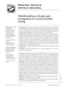
Figure 1. Images of microscopic and immunohistochemical analysis of basal cell carcinoma arising from the epidermoid cyst. Tissue from Case 1. A. The excised specimen was a well-defined oval mass revealed a unilocular cyst containing keratin material with a thick wall on its cut surface. B, C. Image from H&E staining shows epidermoid cyst lined with mature squamous epithelium. (Magnification 10× and 100×, respectively). D. Image from H&E staining taken from the basal cell carcinoma component of the tissue that was associated with the epidermoid cyst. (Magnification 100×). E. Proliferating germinative cells with a characteristic outer palisade of cells associated with a surrounding loose fibromucinous stroma. (Magnification 200×). F. Immunohistochemical staining of from the basal cell carcinoma component with antibody to Ber-EP4. (Magnification 200×).
Pathology Department. The 2×1.9-cm welldefined oval mass revealed a cyst containing keratin material with a thick wall on its cut surface (Figure 1A). Microscopic examination revealed that the cyst was lined with squamous epithelium (Figure 1B). Granular layer was present and the cyst was filled with keratinaceous material (Figure 1C). These histological features are compatible with epidermoid cysts. In parts of the cyst, lobules of germinative cells were present in the cyst wall and were connected with the basal layer of the squamous epithelium (Figure 1D). A characteristic outer palisade of cells associated with a surrounding loose fibromucinous stroma was detected, permitting us to diagnose basal cell carcinoma (Figure 1E). The results from immunohistochemical staining showed the presence of Ber-EP4 in the germinative cells of the basal cell carcinoma (Figure 1F). The patient was discharged without complication, and with no evidence of metastasis or local recurrence at a follow up examination 12 months after the surgery.
cal examination and imaging study, the nodule was diagnosed as an epidermoid cyst (Figure 2A). It was histologically examined after excisional biopsy. Microscopic examination revealed cystic changes lined with mature squamous epithelium that had a granular layer (Figure 2B, 2C). There was germinative cell proliferation with features of basal cell carcinoma on the cyst wall which was connected with the basal layer of the squamous epithelium (Figure 2D, 2E). Immunohistochemical staining revealed the presence of Ber-EP4 in the germinative cells of the basal cell carcinoma (Figure 2F). The basal cell carcinoma extended to the surgical margins as demonstrated by Mohs surgery. This surgery was followed by radiation therapy for 1 month. Clinical presentation of basal cell carcinoma arising from the epidermoid cyst
Case 2: The patient in Case 2 was an 85-yearold man with a history of basal cell carcinoma on his nose, with two recurrences. He presented with a nodule on the lower border of the scar remaining from a previous procedure. On physi-
The cases of basal cell carcinoma arising from the epidermoid cyst that we collected from a literature search are presented in Table 1. If the two cases identified here are included, we conclude there are 29 reported cases of this condition. Patients ranged from 30 to 85 years old, with a mean age of 58 years. There were
12862
Int J Clin Exp Pathol 2016;9(12):12861-12865
Basal cell carcinoma arising from the epidermoid cyst
Figure 2. Images of microscopic and immunohistochemical analysis of basal cell carcinoma arising from the epidermoid cyst. Tissue from Case 2. A. Computer Tomography reveals approximately 21-mm mass in the face between right medial canthus and nasal bone. (Upper: sagittal section, lower: axial section). B, C. Image from H&E staining shows epidermoid cyst lined with mature squamous epithelium. (Magnification 10× and 100×, respectively). D. Image from H&E staining taken from the basal cell carcinoma component of the tissue that was associated with the epidermoid cyst. (Magnification 100×). E. Proliferating germinative cells with a characteristic outer palisade of cells associated with a surrounding loose fibromucinous stroma. (Magnification 200×). F. Immunohistochemical staining of from the basal cell carcinoma component with antibody to Ber-EP4. (Magnification 200×).
more males than females: 18 cases and 11 cases, respectively. The lesions ranged 0.3 to 6.0 cm in area, with a mean area of 2.2 cm. Most lesions were on the head and neck (21 out of 29), but seven were located on the trunk or extremities. The lesions had been present from 2 months to 56 years, with a mean of 13.8 years. Discussion The epidermoid cyst is the most common cystic lesion of the skin. It originates from the infundibular portion of hair follicle (or buried epidermis) and it has keratohyaline granules in its squamous lining, similar to the stratum granulosum of the epidermis. These cysts are usually benign and malignancies related to them are rare. The incidence of malignancy arising in cysts of the skin has been reported to range from slightly more than 1% to 9% [4]. In an evaluation of 2245 epidermoid cysts, there was only a single case of carcinoma, of squamous cells, and the incidence rate was estimated at 0.045% [5]. In an evaluation of 77 malignant tumors that had arisen from keratinous cysts, there were 18 cases of basal cell carcinoma and eight of 12863
these had arisen from an epidermoid cyst [6]. This study was done in 1977, and subsequently, several additional cases of basal cell carcinoma arising from the epidermoid cyst have been reported. Tanaka et al. concluded that long-standing epidermoid cysts may have the potential for malignant transformation [7]. As we presented in Table 1, it was 13.8 years in average before basal cell carcinoma was detected in epidermoid cyst. As we mentioned above, epidermoid cysts can be induced by implantation of epidermal elements into the dermis or they may originate from the infundibular portion of the hair follicle. Because stem cells of the interfollicular epithelium or the infundibulum of the hair follicle, and the lineages of these cells, are considered the cells of origin of basal cell carcinoma, basal cell carcinoma arising from the epidermoid cyst is theoretically possible, although it is apparently uncommon. Therefore, detailed analysis is required for a histological diagnosis of the epidermoid cyst to determine if basal cell carcinoma components are present, as they could be easily overlooked. There is, at present, no information on the pathogenesis of basal cell carcinoma arising Int J Clin Exp Pathol 2016;9(12):12861-12865
Basal cell carcinoma arising from the epidermoid cyst Table 1. Cases of basal cell carcinoma arising from epidermoid cysts Case No. 1 2 3 4 5 6 7 8 9 10 11 12 13 14 15 16 17 18 19 20 21 22 23 24 25 26 27 28 29
Sex
Age
Location
Size (cm)
M M F M F M M M M F M M M M M F F F F M M F M F F M F M M
59 55 72 50s 46 76 61 78 63 54 67 80 43 30 55 35 39 74 40 31 42 74 65 67 47 79 65 51 85
Lower neck Forehead Eyelid Shoulder Nose Knee Face Eyelid Nose Nose Nose Nose Eyelid Forearm Back Scalp Nose Chest Neck Eyebrow Forehead Forehead Cheek Chin Nose Shoulder Knee Nose
0.3 3.5 1.0 4.5 1.5, 2.2 2.2 1.0 0.7 6.0 4.0 2.0 2.5 0.5 2 to 3 2.2 0.8
from the epidermoid cyst. This is in contrast to basal cell carcinoma that occurs in the epidermis, which has been attributed to sun exposure. We hope the information provided here will prove useful to furthering our understanding of basal cell carcinomas, particularly as those arising from epidermoid cysts are unrelated to sun exposure.
Duration (year) 2 months 15.0 56.0 18.0 34.0 2.0 1.0 3.0 15.0 20.0 9.0 1.0 5.0 -
Disclosure of conflict of interest None. 12864
Reference [8] [8] [9] [10] [11] [7] [12] [13] [14] [15] [16] [17] [18] [19] [20] [21] [22] [23] [24] [24] [24] [4] [4] [4] [4] [4] [6] Case 1 Case 2
Address correspondence to: Dr. Sang Kyum Kim, Department of Pathology, Yonsei University College of Medicine, 50 Yonsei-ro, Seodaemun-gu, Seoul 120-752, South Korea. Tel: 82-2-2228-6751; Fax: 82-2-362-0860; E-mail:
[email protected]
References [1]
Acknowledgements This study was supported by a faculty research grant of Yonsei University College of Medicine (6-2015-0087 and 6-2016-0034).
Recurrence (follow up period) No (8 months) No (8 months) No (2 years) No (12 years) No (18 years) No (4 years) No (9 years) No (6 months) No (2 years) No (6 months) No (12 months) Yes (14 months)
[2] [3] [4] [5]
Weedon D, Morgan MB, Gross C, Nagore E and Yu LL. Pathology and Genetics of Skin Tumours. Lion, France: IARC; 2006. Blanpain C. Tracing the cellular origin of cancer. Nat Cell Biol 2013; 15: 126-134. Scott GA. Dermatopathology. McGraw-Hill; 2010. McDonald LW. Carcinomatous change in cysts of skin. Arch Dermatol 1963; 87: 208-211. Cameron DS and Hilsinger RL Jr. Squamous cell carcinoma in an epidermal inclusion cyst:
Int J Clin Exp Pathol 2016;9(12):12861-12865
Basal cell carcinoma arising from the epidermoid cyst
[6] [7]
[8]
[9]
[10]
[11] [12] [13] [14] [15]
case report. Otolaryngol Head Neck Surg 2003; 129: 141-143. Delacretaz J. Keratotic basal-cell carcinoma arising from an epidermoid cyst. J Dermatol Surg Oncol 1977; 3: 310-311. Tanaka M, Terui T, Sasai S and Tagami H. Basal cell carcinoma showing connections with epidermal cysts. J Eur Acad Dermatol Venereol 2003; 17: 581-582. Udovenko O, Guo Y, Connelly T and Mones JM. Basal-Cell Carcinoma Occurring in Cutaneous Infundibular Cysts: Report of 2 Cases and Review of the Literature. Am J Dermatopathol 2015; 37: 635-638. Jakobiec FA, Zakka FR and Hatton MP. Eyelid basal cell carcinoma developing in an epidermoid cyst: a previously unreported event. Ophthal Plast Reconstr Surg 2010; 26: 491-494. Liau JL, Altamura D, Ratynska M and Verdolini R. Basal cell carcinoma arising from an epidermal cyst: when a cyst is not a cyst. Case Rep Dermatol 2015; 7: 75-78. Terada T. Basal cell carcinoma arising from epidermal cyst. J Cutan Med Surg 2015; 19: 105-106. Fukui Y. Hyouhiyounoushu yori hassei shitato kangaerareru kiteisaiboujouhishu no ichi rei. Jpn J Dermatol 1986; 96: 752. Ikeda I and Ono T. Basal cell carcinoma originating from an epidermoid cyst. J Dermatol 1990; 17: 643-646. Takahashi C. Basal cell carcinoma arising from epidermal cyst. Jpn J Clin Dermatol 1992; 46: 310-311. Gonda K. Hyouhinoushu kara hassei shitato kanngaerareru bihaibu no kiteisaibougan. Hifubyoh-Shinryoh 1998; 20: 1053-1056.
12865
[16] Nakamura A, Sumikawa M and Tani M. Ruihyouhinoushu yori hassei shita kiteisaiboujouhishu. Skin Research 1999; 41: 299. [17] Sakai M, Kunisada M, Adachi A and Hayashi K. Ruihyouhinoushu yori shoujitato kanngaerareru kiteisaiboujouhishu no ichi rei. Jpn J Dermatol 2000; 110: 718. [18] Saito M, Yamamoto M, Kato Y, Ohoi T and Koga M. Ruihyouhinoushuheki ni shinjun wo mitometa BBC no ichi rei. Jpn J Dermatol 2000; 110: 332. [19] Ban M and Kitajima Y. Hyouhinoushu kara shoujita kiteisaibougan. Jpn J Dermatol 2003; 113: 812. [20] Dini M, Innocenti A and Romano GF. Basal cell carcinoma arising from epidermoid cyst: a case report. Dermatol Surg 2001; 27: 585586. [21] George SM, Al-Hilli F and Ratnakar KS. Basal cell carcinoma arising in an epidermal cyst. The Gulf J of Dermatol Venereol 2002; 9: 4546. [22] Aneiros-Fernandez J, Husein-ElAhmed H, Siendones IeA, Aneiros-Cachaza J and Arias-Santiago S. Basal cell carcinoma arising from an epidermal cyst. JAAD 2011; 64: AB127. [23] Oztek I, Bas L, E U, Dogru6z K and Yavuz F. Carcinoma changes in cysts of skin. Eur J Plast Surg 1994; 17: 252-257. [24] Mehregan DA, al-Sabah HY and Mehregan AH. Basal cell epithelioma arising from epidermoid cyst. J Dermatol Surg Oncol 1994; 20: 405406.
Int J Clin Exp Pathol 2016;9(12):12861-12865