MEDICINE
REVIEW ARTICLE
Orbital Tumors: Diagnosis and Surgical Treatment Werner Hassler, Renate Unsöld, Uta Schick
SUMMARY Introduction: A broad variety of tumors can involve the small, circumscribed orbit. Two thirds of the lesions are benign and one third are malignant. The proportion of malignant tumors, e. g. metastases and lymphomas, increases with age. In childhood dermoid cysts and hemangiomas are commoner. Methods: Selective literature review and personal clinical experience. Results: In most cases a proptosis develops. Patients often report impaired vision and double vision. Meningiomas are the most prevalent, benign, and slow growing tumors. The choice of operative access depends on localization, size, boundaries, and the type of process. These rare lesions should be treated only in specialized centers able to offer minimal invasive surgical approaches. Dtsch Arztebl 2007; 104(8): A 496–501. Key words: orbital tumor, hemangioma, cancer diagnosis, cancer treatment, surgery
M
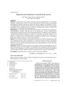
any types of primary and secondary tumor can arise in the orbit (table 1). Orbital processes are rare; accurate figures on their frequency are hard to obtain because of differences between the groups of patients presenting with orbital processes to different types of physicians. Orbital surgery lies in a border area between the various surgical specialties concerned with the anatomically adjacent structures. This article concerns orbital tumors and their clinical presentation and treatment. Particular emphasis is laid on the choice of the best surgical approach. Minimally invasive approaches are now increasingly used. One of the more recently developed approaches, such as the transconjunctival (figure 1), pterional (frontotemporal) contralateral, or pterional extradural approach, can be used as alternative to a classic extracranial approach (lateral orbitotomy) or a more extensive transcranial approach (1, 2, 3). Some specialists may choose to operate by way of the neighboring paranasal sinuses (transethmoidal or transmaxillary approach). Part of this article is devoted to the special topic of orbital meningiomas, which are classified by location (4). The treatment options in this particular area have been dramatically transformed since 2002; these lesions are increasingly being treated with stereotactic radiosurgery (5, 6, 7). Unfortunately, the overall incidence of non-infectious, non-inflammatory lesions in the orbit is so small that there are as yet no major randomized studies concerning their treatment (i.e., studies of evidence level A1 or A2). The available data are derived from comparative studies with small numbers of patients (evidence level B) and from non-comparative studies (evidence level C). The authors discuss selected papers from the literature and their own clinical experience. Infectious and inflammatory disorders, such as endocrine orbitopathy, will not be discussed.
Symptom-oriented differential diagnosis Orbital tumors frequently impair vision. Visual acuity is often reduced by malignant and inflammatory/infectious processes (79%), as well as by optic nerve gliomas (67%); it is less commonly reduced by meningiomas (45%) and other benign processes (39%). The patient's quality of life can also be substantially impaired by pain, which is a prominent feature of inflammatory/infectious disorders (96%) and meningiomas (70%). Proptosis can be caused by nearly any type of orbital process and impairs cosmesis (table 2). Neurochirurgische Klinik, Wedau Kliniken Duisburg (Prof. Dr. med. Hassler, PD Dr. med. Schick); Neuroophthalmologische Praxis Düsseldorf (Prof. Dr. med. Unsöld)
Dtsch Arztebl 2007; 104(8): A 496–501 ⏐ www.aerzteblatt.de
1
MEDICINE
TABLE 1 Orbital processes in the authors' clinical series (583 patients, 1991–2005).
Diagnosis Meningioma Spheno-orbital Optic sheath Anterior clinoid process
Number 116 95 41
Cavernoma
44
Aneurysm of ophthalmic artery
44
Metastasis
41
Inflammation/abscess/pseudotumor
31
Lymphoproliferative/leukemic processes
25
Foreign body/trauma
22
Optic nerve glioma
20
Pleomorphic adenoma
16
Epidermoid or dermoid cyst
12
Pneumatosis dilatans
9
Hemangiopericytoma
9
Osteoma
9
Neurinoma
9
Venous malformation
8
Fibrous dysplasia
8
Mucocele
7
Capillary hemangioma
4
Rhabdomyosarcoma
2
Neurogenic sarcoma
2
Adenoid cystic carcinoma
2
Giant cyst
2
Dysontogenetic cyst
1
Osteopetrosis
1
Lipoma
1
Sarcoidosis
1
Leiomyoma of the optic nerve
1
The mean duration of symptoms until diagnosis is 4.2 months for malignant tumors, 15 months for gliomas, and 28 months for meningiomas. The mean age at the initial manifestation of disease is 19 years for optic gliomas, which is considerably lower than that for neurinomas (42 years) or meningiomas (54 years) (table 3) (8).
Topographical distribution The intraconal space is delimited by the conus, which connects the four rectus muscles to each other. The most common intraconal processes affecting the optic nerve are optic nerve glioma, optic sheath meningioma, and lymphoma. Other intraconal processes include cavernoma, neurinoma, and metastases. The extraconal compartment takes up only a small amount of space within the orbit, surrounding the muscular conus like a tube. The most common extraconal processes are dermoid tumor (dermoid cyst) and pleomorphic adenoma of the lacrimal gland (8). The subperiosteal compartment is defined as the potential space between the periosteum and the bony orbit. Mucocele is the most common process affecting this compartment. The bony orbit and the sphenoid wing can be the site of an osteoma, a malignant tumor, or Dtsch Arztebl 2007; 104(8): A 496–501 ⏐ www.aerzteblatt.de
2
MEDICINE
fibrous dysplasia, but the most common process affecting these structures is a sphenoid wing meningioma. Meningiomas are often found at the orbital apex, the superior orbital fissure, and the cavernous sinus. The intracranial periorbital dura mater heading toward the optic nerve can be infiltrated by an optic sheath meningioma or by other types of meningioma of variable size and extent. Pleomorphic adenoma and carcinoma arise in the lacrimal gland and duct system. Lymphoma and infections can affect the preseptal segment or the lids (8).
Histology The most common orbital tumor is a benign, slowly-growing meningioma arising from the medial portion of the sphenoid wing, the anterior clinoid process, or the optic nerve sheath. The peak incidence of orbital meningioma is between the ages of 30 and 50. Women are more commonly affected than men. Clinically, meningiomas manifest themselves with gradual loss of visual acuity, accompanied by optic nerve atrophy or proptosis. Sphenoorbital meningiomas, i.e., tumors arising from the bone of the sphenoid wing and also involving the orbit, involve bone and possibly also muscle over a wide area and may require a correspondingly extensive resection (9, 10, 11). Tumors of this type account for only about 4% of all intracranial meningiomas. Most meningiomas grow along the medial portion of the sphenoid wing and enter the orbit from there. On the other hand, optic sheath meningiomas often grow along the optic canal into the cranial cavity (figure 2). 35% of all optic nerve tumors are optic sheath meningiomas. These lesions account for Figure 1: Transconjunctival approach: at the end of the operation, the conjunctival flap is brought back into position and secured with a 10/0 suture.
TABLE 2 Common symptoms of orbital tumors (8)Diagnose
Symptom
A
Frequency (%)
Proptosis
92
Visual field defect, loss of visual acuity
74
Diplopia, strabismus
66
Pain
34
Lacrimation
23
Conjunctival edema
22
Inflammation
13
1% to 2% of all meningiomas and 1.7% of all orbital tumors. The authors' classification scheme distinguishes intraorbital (type 1) from intracanalicular (type 2) optic sheath meningiomas and from those that are both intraorbital and intracanalicular (type 3). When an intraorbital tumor (type 1) begins to affect visual acuity, it should be treated with radiation at first (5, 6, 7), and any large exophytic tumor mass should be resected. For type 2 tumors, surgical decompression of the optic canal can stave off the impending loss of vision. Type 3 tumors growing toward the intracranial compartment are best treated with extradural Dtsch Arztebl 2007; 104(8): A 496–501 ⏐ www.aerzteblatt.de
3
MEDICINE
decompression of the optic canal, intracranial tumor resection, and postoperative irradiation of the intraorbital portion (4). The long-term prognosis is poor for vision, but good for patient survival. Vascular processes are the second most common type of mass affecting the optic nerve; capillary hemangioma accounts for most cases in children (12, 13, 14). The eyelids are often involved as well. As long as visual acuity is unimpaired, these lesions can be treated expectantly, as they sometimes regress spontaneously in 1 to 2 years. Cavernoma, the most common vascular malformation in the orbit, is the second most common type of orbital process in patients aged 31 to 40 and the third most common type in patients aged 41 to 50 (15). Orbital cavernomas account for 4% of all orbital processes and 9% to 13% of all cranial cavernomas of the head. These intraconally situated lesions cause slowly progressive, painless proptosis and, later, impairment of visual acuity. They rarely present with an acute, clinically significant hemorrhage. At surgery, a cavernoma can be collapsed by electrocoagulation but must then be completely removed because of the danger of regrowth if it is left in place. If surgery is performed early, the patient's vision may recover. "Developmental venous anomalies" (DVAs) are a heterogeneous group of venous malformations; different subtypes of DVA were previously called varices, varicoceles or venous angiomas. Unlike intracranial DVAs, orbital DVAs can bleed, exert mass effect, and produce proptosis that worsens with the Valsalva maneuver. DVAs should be treated if they cause severe pain or significant cosmetic disturbance. The treatment can be endovascular, surgical, or a combination of both (8). Hemangiopericytoma is a rare (< 1%) vascular wall tumor of adulthood. It behaves rather aggressively, is locally invasive, and undergoes malignant degeneration in 30% of cases, possibly giving off distant metastases. The prognosis, however, is good as long as the tumor is diagnosed early and completely resected. Lymphangioma is a benign, hamartomatous, cystic tumor of childhood, sometimes associated with facial or palatal cysts. It accounts for up to 4% of orbital processes, depending on the case series. Unlike a capillary hemangioma, a lymphangioma does not enlarge if the patient performs a Valsalva maneuver. These tumors grow diffusely, crossing anatomical boundaries, and they form extensive cysts that are often the site of hemorrhage (chocolate cysts). A lymphangioma may enlarge during an episode of respiratory infection because of lymphoid proliferation of connective tissue. Either of these two processes can manifest itself as acute proptosis. Acute worsening of vision or progressive proptosis are the only indications for surgical resection or for the puncture and aspiration of large cysts. Optic nerve glioma (pilocytic astrocytoma) usually arises in childhood, particularly in children with neurofibromatosis (12). It accounts for 4% of all orbital tumors, 66% of all primary tumors of the optic nerve, and 1% to 5% of all gliomas in childhood. The initial manifestation is usually visual impairment. Small children may present with amblyopic strabismus or nystagmus. The treatment of optic nerve glioma depends on its location. Tumors involving the optic chiasm and hypothalamus are usually treated with external beam irradiation or seed implantation. The goal of treatment in such cases is the reduction, or at least stabilization, of tumor size. Children below the age of 5 years are treated at first with chemotherapy only. The authors favor surgical debulking of large suprasellar tumors. Intraorbital optic nerve tumors that have grown into the cranial cavity should be resected only when the patient's vision is severely impaired or proptosis is severe. The optic nerve is transected behind the
TABLE 3 Frequency of orbital processes by age (8)
0–10 years
11–20 years
21–30 years
31–40 years
41–50 years
51–60 years
> 60 years
Capillary hemangioma
Rhabdomyosarcoma
Mucocele
Meningioma
Meningioma
Lymphoma
Lymphoma
Optic nerve glioma Dermoid cyst
Inflammation
Cavernoma
Mucocele
Meningioma
Metastasis
Rhabdomyosarcoma
Adenoid cystic carcinoma
Adenoid cystic carcinoma
Cavernoma
Mucocele
Meningioma
neurofibroma
Dtsch Arztebl 2007; 104(8): A 496–501 ⏐ www.aerzteblatt.de
4
MEDICINE
globe, at the orbital apex, and intracranially just anterior to the optic chiasm through a combined extra- and intradural pterional approach. The clinical course and prognosis of optic nerve glioma also depend on its location. Involvement of the optic tract or other postchiasmatic structures increases the likelihood of visual impairment and also increases mortality. If the hypothalamus is involved, the mortality rises from 5% to 50%. Neurinoma usually arises in adulthood, neurofibroma in childhood. Neurinoma accounts for 1% to 4% of all orbital tumors. Patients under 30 years old with multiple neurofibromas are suffering from neurofibromatosis. They cause progressive exophthalmos, while the visual acuity is preserved. Because these tumors tend to develop on the sensory branches of peripheral nerves, ocular motility also remains intact for a long time. The motility of the globe is restricted only when the tumor becomes large enough to impede its movement. These well-demarcated tumors can often be completely neurosurgically removed in their early stage, with a good prognosis. Neurofibromas rarely undergo secondary degeneration. Fibrous dysplasia most commonly involves the bone of the orbital roof (< 1% of all orbital tumors). These lesions grow slowly, often compromising the optic canal. In symptomatic cases, the optic nerve is decompressed in its canal. Osteomas most commonly arise from bone of the paranasal sinuses (< 1% of all orbital tumors). They only rarely primarily involve the orbital bone itself. They are removed only if symptomatic. Pleomorphic adenoma, a histologically mixed, benign tumor, is the most common benign epithelial tumor of the lacrimal gland (1% to 4%). It usually arises in middle age. It may involve bone. These lesions can be cured by total resection with preservation of the capsule. Residual tumor fragments can undergo secondary degeneration. Rhabdomyosarcoma is the most common malignant orbital tumor of the first decade of life (1% to 3% of orbital processes). It is usually found extraconally in the superior nasal quadrant and may additionally involve the conjunctiva, uvea, and lid. Orbital rhabdomyosarcoma has the best prognosis among all locations, provided that the paranasal sinuses are not involved. The 3-year recurrence-free survival rate is 91%. For most patients, the initial manifestation is massive proptosis with inferotemporal displacement of the globe, accompanied by lid edema and conjunctival injection. Anteriorly situated rhabdomyosarcomas can be totally resected. Posteriorly situated ones are biopsied for diagnostic purposes and then treated with a combination of radiation and chemotherapy (17). Adenoid cystic carcinoma of the lacrimal gland arises in middle age (< 1%). Infiltration of sensory nerves by the tumor causes pain. These tumors have a poor prognosis, even when they are radically resected; they tend to recur locally and to give off distant metastases (the presence of metastases worsens the prognosis). Brachytherapy can be used for local tumor control as an alternative to enucleation of the globe. Metastases are more common with advancing age (18). In various studies, orbital metastases account for 1% to 13% of all orbital tumors. The primary tumor may be in the breast, lung, prostate, gastrointestinal tract, or kidney. Metastasis occurs by way of the bloodstream; thus, by the time an orbital metastasis becomes apparent, the cancer is often widespread, and there are not uncommonly intracranial tumor deposits as well. Diplopia and pain accompany orbital metastases more commonly than they accompany primary orbital tumors. A multiplicity of suddenly arising symptoms is typical of orbital metastases. Their treatment is palliative; above all, the creation of new deficits should be avoided. The diagnosis of an orbital metastasis must nonetheless be confirmed by biopsy, because these lesions have an extensive differential diagnosis. Total excision should be attempted only if the lesion is small, well circumscribed, and easily accessible. Symptomatic improvement for six to twelve months can be achieved with radiotherapy. Even if the orbital tumor can be locally controlled, however, the overall prognosis with respect to systemic disease progression and survival is generally poor. The different varieties of lymphoma are diseases of old age. Lymphoma can be either intra- or extraconal, and may arise in the conjunctiva (< 8%). Non-Hodgkin lymphoma is extranodal in 40% of cases; 5% to 14% of these affect the orbit. The symptoms include proptosis, restrictive limitation of ocular motility, and, later, visual impairment. Non-Hodgkin lymphoma is difficult to distinguish radiologically from inflammatory syndromes or lymphatic hyperplasia. Among the MALT (mucosa-associated lymphatic tissue) syndromes, those more commonly found in the orbit ( e.g., in the conjunctiva or lacrimal gland) are Dtsch Arztebl 2007; 104(8): A 496–501 ⏐ www.aerzteblatt.de
5
MEDICINE
Figure 2: Intraoperative view of an optic sheath meningioma (type 2a, with extension via the optic canal into the cranial cavity). Surgery was performed through a pterional approach, with decompression of the canal, and the intraorbital portion of the tumor was irradiated.
classified as extranodal marginal zone lymphomas (EMZL). These diseases have a good prognosis; once the diagnosis is confirmed by biopsy, they can be effectively treated with moderately intense radiotherapy. The most common type of orbital lymphoma, which is also the most aggressive and prognostically least favorable type, is diffuse large-cell B-cell lymphoma (DLCL). Aggressive lymphomas must be treated primarily with chemotherapy (19, 20, 21, 22). 15% to 20% of EMZL and 50% of DLCL become systemic at some point in their course; these types of lymphoma must therefore be followed up at close intervals. Dermoid cysts are the most common type of orbital cyst (2% of all orbital processes). They are of congenital origin and become clinically evident in childhood or young adulthood. They should not be opened during surgical excision so as not to produce an inflammatory reaction. They should be completely removed, as they otherwise tend to recur. Aneurysmal bone cysts are benign destructive lesions of bone that enlarge by local extension; they are rarely found in the orbit (0.25% of orbital lesions). Their optimal treatment is currently a matter of debate. The options include radiotherapy, surgery, and embolization.
Operative approaches The choice of approach depends on the location, size, demarcation, and histological type of the lesion (please see additional table at the end of this article). The least traumatic approach should be chosen. The procedures that can be performed range from open biopsies for confirmation of a diagnosis to the subtotal resection of diffusely infiltrating processes with preservation of function and the complete excision of well-circumscribed lesions. Fineneedle biopsies are suitable for confirmation of the diagnosis when the primary lesion is known (e.g., in metastatic solid tumors or lymphoma) but do not provide enough tissue for more thorough immunohistochemical studies. Very extensive surgery, with wide excision of bone followed by complicated reconstructive techniques, is rarely necessary. In general, open operations in the orbit are performed by one of two types of approach: transcranial or direct extracranial (3, 8). A transcranial approach is preferable for processes extending behind the orbit into the cranial cavity or located in the optic canal or the superior orbital fissure. A classic pterional approach is standard and is used by the authors for orbital apex tumors as well. If the tumor involves the posteromedial portion of the orbit, inferomedial to the optic nerve, a pterional approach from the opposite side will be needed. An exclusively extradural pterional approach can be used to decompress the optic nerve in the optic canal and superior orbital fissure, as well as for tumor biopsy or resection (2). The supraorbital approach occupies an intermediate position between the extra- and transcranial approaches. Well-circumscribed processes superior to the optic nerve can be reached either intra- or extraconally through an eyebrow incision. The supraorbital approach is particularly useful for the removal of cavernomas and neurinomas (15, 16). Processes located exclusively within the orbit can be reached by a wide variety of relatively noninvasive extracranial approaches, including lateral orbitotomy, the transethmoidal and transconjunctival approaches (figure 1), and the frontal trans-sinusoidal approach (1). Lateral orbitotomy is useful for well-circumscribed periorbital and intraconal tumors and for tumors located dorsally, basally, and laterally to the optic nerve, as well as for tumors of Dtsch Arztebl 2007; 104(8): A 496–501 ⏐ www.aerzteblatt.de
6
MEDICINE
the lacrimal gland. The authors favor a transconjunctival approach for basal, medial intra- and extraconal tumors, and particularly for biopsies of intraconal processes. Even if the tumor is larger than the globe itself, it can still be removed by this approach without any sacrifice of bone (1).
Recommendations > Any patient with visual loss, proptosis, or impaired ocular motility should undergo MRI to detect or exclude an intraorbital or intracranial process. > Patients found to have one of these rare tumors should be referred to a specialized center for further treatment. > Orbital surgery is a difficult and delicate undertaking that requires microsurgical technique.
Summary The surgical treatment of orbital processes occupies a border area between different surgical specialties, including otorhinolaryngology, oral/maxillofacial surgery, ophthalmology, and neurosurgery. The spectrum of pathology is broad, and the potential operative approaches are numerous. A good understanding of the variety of disease processes in the orbit, and of the available interdisciplinary approaches, is needed so that the optimal mode of treatment can be determined. Collaboration across specialties improves the chances of successful treatment. Conflict of Interest Statement The authors declare that they have no conflict of interest as defined by the guidelines of the International Committee of Medical Journal Editors. Manuscript received on 11 April 2006; final version accepted on 14 July 2006. Translated from the original German by Ethan Taub, M.D.
REFERENCES 1. Hejazi N, Hassler W, Farghaly F: Transconjunctival microsurgical approach to the orbit: a retrospective preliminary review and analysis of experience with the first 15 operative cases. Neurosurg Quart 1999; 9: 197–208. 2. Hassler WE, Eggert H: Extradural and intradural microsurgical approaches to lesions of the optic canal and the superior orbital fissure. Acta Neurochir (Wien) 1985;74: 87–93. 3. Mauriello JA, Flanagan JC: Surgical approaches to the orbit. In: Mauriello JA, Flanagan JC (Hrsg.): Management of orbital and ocular adnexal tumors and inflammations. Berlin, Heidelberg, New York: Springer 1990; 149–525. 4. Schick U, Dott U, Hassler W: Surgical management of meningiomas involving the optic nerve sheath. J Neurosurg 2004; 101: 951–9. 5. Miller NR: Primary tumours of the optic nerve and its sheath. Eye 2004; 18: 1026–37. 6. Pitz S, Becker G, Schiefer U, Wilhelm H, Jeremic B, Bamberg M: Stereotactic fractionated irradiation of optic nerve sheath meningioma: a new treatment alternative. Br J Ophthalmol 2002; 86: 1265–8. 7. Turbin RE, Pokorny K: Diagnosis and treatment of orbital optic nerve sheath meningioma. Cancer Control 2004; 11: 334–41. 8. Hassler W, Schick U: Orbitachirurgie aus neurochirurgischer Sicht. In: Moskopp D, Wassmann H (Hrsg.): Neurochirurgie – Fachwissen in einem Band. Stuttgart, New York: Springer 2004: 263–73. 9. De Jesús O, Toledo MM: Surgical management of meningioma en plaque of the sphenoid ridge. Surg Neurol 2001; 55: 265–9. 10. Maroon JC, Kennerdell JS, Vidovich DV, Abla A, Sternau L: Recurrent spheno-orbital meningiomas. J Neurosurg 1994; 80: 202–8. 11. Schick U, Bleyen J, Bani A, Hassler W: Management of meningiomas en plaque of the sphenoid wing. J Neurosurg 2006; 104: 208–14. 12. Gunalp I, Gunduz K: Pediatric orbital tumors in Turkey. Ophthal Plast Reconstr Surg 1995; 11: 193–9. 13. Gunalp I, Gunduz K: Vascular tumors of the orbit. Doc Ophthalmol 1995; 89: 337–45. 14. Shields JA, Shields CL: Vascular and hemorrhagic lesions. In: Shields JA, Shields CL (Hrsg.): Atlas of Orbital Tumors. Philadelphia, Baltimore, New York: Lippincott Williams&Wilkins 1999; 45–3. 15. Schick U, Dott U, Hassler W: Surgical treatment of orbital cavernomas. Surg Neurol 2003; 60: 234–44. 16. Schick U, Bleyen J, Hassler W: Treatment of orbital schwannomas and neurofibromas. Br J Neurosurg 2003; 17: 541–5. 17. Christ WM, Anderson JR, Meza JL et al.: Intergroup rhabdomyosarcoma study IV: results for patients with nonmetastatic disease. J Clin Oncol 2001; 19: 3091–102.
Dtsch Arztebl 2007; 104(8): A 496–501 ⏐ www.aerzteblatt.de
7
MEDICINE
18. Shields JA, Shields CL, Brotman H, Carvalho C, Perez N, Eagle RC Jr.: Cancer metastatic to the orbit. The 2000 Robert M. Curts Lecture. Ophthal Plast Reconstr Surg 2001; 17: 346–54. 19. Lee S, Suh CO, Kim GE, Yang WI, Lee SY, Hahn JS, Park JO: Role of radiotherapy for primary orbital lymphoma. Am J Clin Oncol 2002; 25: 261–5. 20. Nutting CM, Jenkins CD, Norton AJ, Cree I, Rose GE, Plowman PN: Primary orbital lymphoma. The Hematology Journal 2002; 3: 14–6. 21. Schick U, Lermen O, Unsöld R, Hassler W: Treatment of primary orbital lymphomas. Eur J Haematol 2004; 72: 186–93. 22. Shields CL, Shields JA, Carvalho C, Rundle O, Smith AF: Conjunctival lymphoid tumors: clinical analysis of 117 cases and relationship to systemic lymphoma. Ophthalmology 2001; 108: 979–84. Corresponding author PD Dr. med. Uta Schick Neurochirurgische Klinik, Wiedau Kliniken Universitätsklinikum Giessen und Marburg Zu den Rehwiesen 9 D-47055 Duisburg, Germany
[email protected]
ADDITIONAL MATERIAL SEE NEXT PAGE
Dtsch Arztebl 2007; 104(8): A 496–501 ⏐ www.aerzteblatt.de
8
MEDICINE
Additional Material ADDITIONAL TABLE Approaches to the orbit
Approach
Indications
Contraindications
Advantages
Disadvantages
Processes lying medial or mediobasal to the optic nerve; limited access to the apex
Good exposure, well-tolerated procedure
Visible surgical scar
Extracranial approaches to the orbit Lateral orbitotomy
Well-circumscribed periorbital and intraconal tumors, dorsal, basal, and lateral to the optic nerve; lacrimal gland tumors
Transethmoidal
Extraconal tumors medial to Superior, basal, and lateral the optic nerve, traumatic injury tumors, intraconal processes of the optic canal
Well tolerated, usually used by ENT surgeons, no brain retraction
Limited exposure, approach through unsterile sinuses, more bleeding
Frontal transsinusoidal
Tumors or trauma of the frontal Extensive frontobasal injury, sinus, retention cysts involving intraconal tumors the orbit, extraconal processes extending into the frontal sinus
Minimally invasive, particularly for retention cysts
Visible surgical scar, risk of infection, limited indications
Transmaxillary
Basal lesions, whether intraor (preferably) extraconal, contacting the maxillary sinus
Lesions above the optic nerve
Well tolerated, performed in collaboration with ENT surgeons
Limited exposure, hemorrhage, approach through unsterile sinus
Transconjunctival
Basal, medial intra- and extraconal tumors, biopsy of intraconal processes
Very small lesions, orbital apex tumors, lateral, superior, and extraconal tumors
Minimally invasive, ideal for cavernomas, excellent cosmetic result
Only for highly experienced surgeons (collaboration with ophthalmologists is recommended)
Processes below the optic nerve
Minimally invasive extradural approach with minimal manipulation of the orbital structures and brain, no limitation by the size of the lesion, excellent cosmetic result
Permanent hypesthesia of the forehead
Orbital apex tumors, tumors below the optic nerve, complex tumors infiltrating bone
Good general exposure
Traumatizing approach requiring brain medial retraction
Combined extra- and intracranial approach Supraorbital via eyebrow incision
Well-circumscribed intraand extraconal processes above the optic nerve
Transcranial approaches to the orbit Subfrontal
Intraorbital optic nerve glioma growing into the cranial cavity, and lateral tumors of the optic nerve
Frontolateral, pterional
Ideal approach for tumors Tumors medial and basal of the superior orbital fissure, to the optic nerve, lacrimal optic canal, orbital apex, and gland tumors intraorbital optic nerve; tumors located dorsal to the optic nerve; and lateral extra- and intraconal tumors
Broad exposure, minimal brain retraction, good access to the extra- and intradural and intraorbital compartments
No disadvantages
Pterional extradural
Ideal for decompression of the Processes in the anterior optic nerve in the optic canal, portion of the orbit suitable for periorbital tumors and for tumors near the superior and inferior orbital fissures and the cavernous sinus
Well tolerated, no brain retraction, clear exposure
Detailed anatomical knowledge is essential
Pterional contralateral
Tumors of the medial orbital apex, aneurysms of the ophthalmic artery
Direct approach to medial processes
Difficult without navigation, injury to the olfactory n. is possible, may involve extensive brain retraction
Not suitable for other processes
Dtsch Arztebl 2007; 104(8): A 496–501 ⏐ www.aerzteblatt.de
9