MRI of the Knee
Jennifer Swart, M.D. Musculoskeletal Radiology South Texas Radiology Group
Financial Disclosure • I have no relevant financial relat...
Jennifer Swart, M.D. Musculoskeletal Radiology South Texas Radiology Group
Financial Disclosure • I have no relevant financial relationships with commercial interests to disclose.
Outline • Coils, Patient Positioning • Acquisition Parameters, Planes and Pulse Sequences • Knee Arthrography • Normal Anatomy • Abnormal Anatomy (Injury Patterns) • High Field MRI (3.0T Magnets)
This presentation is the intellectual property of the author. Contact them for permission to reprint and/or distribution.
Imaging Details • Supine Positioning • Slight external rotation • Dedicated knee coil – 8 channel • 14 to 16 cm field of view • 2.5 to 5 mm slice thickness • Rarely use intravenous gadolinium • Exam time 15 minutes
MRI Pulse Sequences • T1 weighted Sequences – Fat sensitive – Good anatomic resolution • Proton Density Sequences – Fat and fluid sensitive – Best anatomic resolution • T2 Fat Saturated Sequences – Fluid sensitive, all else dark – Pathology sequence – Poor anatomic resolution
MRI Acquisition Planes • Scout Image – Find the knee in the magnetic field • Axial Images – Parallel to tibial plateau • Coronal Images – Parallel to posterior margin of femoral condyles • Sagittal Images – Perpendicular to sagittal plane
This presentation is the intellectual property of the author. Contact them for permission to reprint and/or distribution.
Axial Images
Axial MPGR
Axial T2 FS
Coronal Images
Coronal T1
Coronal T2 FS
Sagittal Images
Sagittal PD
Sagittal T2 FS
This presentation is the intellectual property of the author. Contact them for permission to reprint and/or distribution.
MR Knee Arthrography • Infrequently Performed • Allows T1 weighted imaging for best spatial resolution • Mainly used in cartilage and postoperative meniscus assessment • Fluoroscopically guided • Anterior approach with 25 g needle • 20-30cc Dilute Gadolinium injected • MR performed within 45 minutes after exercise
MR Arthrogram Images • Distended joint, gadolinium fills tears in structures that line the joint • Sequences: T1 axial, coronal, sagittal with fat saturation
– Only bright structure is gadolinium • Coronal T1 no fat saturation • Sagittal T2 with fat saturation
MR Arthrogram Knee Loose Osteochondral Lesion
Coronal T2 Fat Sat
Coronal T1 Post Gad Fat Sat
Sagittal T1 Post Gad Fat Sat
This presentation is the intellectual property of the author. Contact them for permission to reprint and/or distribution.
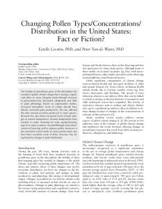
Normal Anatomy: Medial Collateral Ligament (MCL)
Coronal T1
Coronal T2 Fat Sat
Normal Anatomy: Lateral Collateral Ligament (LCL or FCL)
Coronal T1
Coronal T2 Fat Sat
Normal Anatomy: Anterior Cruciate Ligament (ACL)
Sagittal PD
Sagittal T2 Fat Sat
This presentation is the intellectual property of the author. Contact them for permission to reprint and/or distribution.
Normal Anatomy: Posterior Cruciate Ligament (PCL)
Sagittal PD
Sagittal T2 Fat Sat
Normal Anatomy: Medial Meniscus
Sagittal PD
Sagittal T2 Fat Sat
Normal Anatomy: Lateral Meniscus
Sagittal PD
Sagittal T2 Fat Sat
This presentation is the intellectual property of the author. Contact them for permission to reprint and/or distribution.
Normal Anatomy: Coronal Plane Menisci
Coronal T1
Coronal T2 Fat Sat
Interpreting Knee MR • Systematic, disciplined approach is crucial
– Don’t go for the money • Structured Report
– – – – – – –
Menisci Cruciates Extensor Mechanism Collaterals Cartilage Fluid Bone Marrow
• Look for Injury Patterns • Address the clinical question
Grade 2 MCL Sprain
Coronal T1
Coronal T2 Fat Sat
This presentation is the intellectual property of the author. Contact them for permission to reprint and/or distribution.
Grade 3 LCL Sprain
Coronal T2 fat sat
Acute Interstitial ACL Tear
Sagittal PD
Sagittal T2 Fat Sat
Segond Fracture Associated ACL injuries Posterolateral Corner Injury
Sagittal T2 Fat Sat
Sagittal T2 Fat Sat
This presentation is the intellectual property of the author. Contact them for permission to reprint and/or distribution.
ACL Avulsion
Sagittal T2 Fat Sat
Coronal T2 Fat Sat
PCL Avulsion
Coronal T1
Coronal T2 fat sat
ACL Graft Tear
Intact ACL Graft Sagittal PD
Torn ACL Graft Sagittal PD
This presentation is the intellectual property of the author. Contact them for permission to reprint and/or distribution.
Chronic ACL Tear
Sagittal PD
Sagittal T2 Fat Sat
Acute PCL Tear
Sagittal PD
Sagittal T2 Fat Sat
Radial Lateral Meniscus Tear
Axial MPGR
Sagittal PD
This presentation is the intellectual property of the author. Contact them for permission to reprint and/or distribution.
Complex Medial Meniscus Tear
Sagittal PD
Sagittal T2 Fat Sat
Bucket Handle Medial Meniscus Tear
Coronal T2 Fat Sat
Sagittal PD
Flipped Locked Lateral Meniscus Tear
Sagittal PD
Sagittal T2 Fat Sat
This presentation is the intellectual property of the author. Contact them for permission to reprint and/or distribution.
Discoid Lateral Meniscus Tear
Sagittal PD
Sagittal T2 Fat Sat
Parameniscal Cyst presenting as mass - percutaneous aspiration and rupture
Medial and Lateral Bucket Handle Tears
Sagittal T2 Fat Sat
Coronal T2 Fat Sat
Axial MPGR
This presentation is the intellectual property of the author. Contact them for permission to reprint and/or distribution.
Cartilage Defects
Sagittal T2 Fat Sat
Coronal T2 Fat Sat
Sagittal T2 Fat Sat
Post Intervention Cartilage Assessment Pre-Microfracture
Post-Microfracture
Axial T1 Post Arthrogram
Axial T1 Post Arthrogram
Baker’s Cysts
Sagittal PD
Sagittal T2 Fat Sat
This presentation is the intellectual property of the author. Contact them for permission to reprint and/or distribution.
Baker’s Cyst Rupture
Sagittal PD
Sagittal T2 Fat Sat
IT Band Friction Syndrome
Coronal T1
Coronal T2 Fat Sat
Transient Patellar Dislocation
Axial T2 Fat Sat
Coronal T2 Fat Sat
This presentation is the intellectual property of the author. Contact them for permission to reprint and/or distribution.
Jumper’s Knee (Infrapatellar Tendonopathy)
Sagittal PD
Sagittal T2 Fat Sat
Quadriceps Tendon Rupture
Sagittal PD
Sagittal T2 Fat Sat
Infrapatellar Tendon Rupture
Sagittal PD
Sagittal T2 Fat Sat
This presentation is the intellectual property of the author. Contact them for permission to reprint and/or distribution.
Chronic hemorrhagic bursitis
Recent Advances: High Field MRI 3.0 Tesla versus 1.5 Tesla MRI • Twice the magnetic field strength • Twice the signal to noise in a given pixel – Increase matrix / decrease pixel size (increase spatial resolution) – Decrease slice thickness (increase spatial resolution)
This presentation is the intellectual property of the author. Contact them for permission to reprint and/or distribution.
Fractures
Coronal T2 Fat Sat
Radiograph
Fractures
Coronal T2 Fat Sat
Coronal T1
Cartilage Mapping • T2 mapping – Reflects cartilage ultrastructure – Capable of detecting early cartilage degeneration before surface changes
Axial T2 Fat Sat 3.0T
Axial Cartigram 3.0T
This presentation is the intellectual property of the author. Contact them for permission to reprint and/or distribution.
Cartilage Mapping
Sagittal T2 Fat Sat 3.0T
Sagittal Cartigram 3.0T
MARS (metal artifact reduction sequence)
Sagittal T2
Axial STIR
MARS prosthesis imaging
Axial T2
This presentation is the intellectual property of the author. Contact them for permission to reprint and/or distribution.
Summary • MRI plays an indispensable role in the evaluation of knee injuries. • Intra-articular and Intravenous gadolinium are not routinely required in the assessment of knee injuries. • High field MR systems increase diagnostic sensitivity, particularly of cartilage lesions. • Accept nothing less than the interpretation of a specialized musculoskeletal radiologist. • Always correlate imaging findings with clinical examination and discuss discrepancies with your radiologist.
This presentation is the intellectual property of the author. Contact them for permission to reprint and/or distribution.