Neuroradiologic Findings in Polyarteritis Nodosa James M. Provenzale and Nancy B. Allen PURPOSE: To demonstrate the neuroradiologic findings in patients with polyarteritis nodosa. METHODS: A review of hospital records for a 10-year period revealed 50 patients with a discharge diagnosis of polyarteritis nodosa. Thirteen patients had undergone neuroimaging, and abnormal findings were found in 5 cases; these were the subjects of this study. RESULTS: All 5 patients had abnormal findings on CT scans, 3 had abnormal findings on MR images, and 1 had an abnormal finding on a cerebral angiogram. All patients had cerebral cortical or subcortical infarctions, and 1 also had small infarctions within the brain stem and cerebellum. One patient had cerebral angiographic findings of arteritis. The diagnosis of arteritis was considered probable or possible in 3 other patients. Three patients had echocardiographic evidence of concentric hypertrophy and a hypocontractile left ventricle resulting from polyarteritis nodosa–related hypertension. Cardiogenic embolism was considered the likely cause in 1 patient. CONCLUSION: Small peripheral cerebral infarctions, consistent with an arteritis involving medium-sized and small arteries, were the most common finding. However, cardiogenic embolism should also be considered as a possible cause of cerebral infarction in patients with polyarteritis nodosa who have left ventricular dysfunction. Index terms: Arteritis; Brain, magnetic resonance AJNR Am J Neuroradiol 17:1119 –1126, June 1996
Polyarteritis nodosa, also known as periarteritis nodosa, is a rare multisystem disease characterized by a necrotizing arteritis of small and medium-sized arteries (1). The most common sites affected are the skin, joints, kidneys, gastrointestinal tract, and peripheral nerves (1). Although early reports found central nervous system (CNS) involvement to be uncommon (2, 3), more recent investigations have found the prevalence of CNS complications to be 20% to 45% (4 –7). The most common reported CNS manifestation is diffuse encephalopathy, followed in frequency by focal deficits and seizures (5, 8). Despite the high prevalence of CNS involvement, however, there are few articles emphasizing the neuroradiologic findings in patients with CNS complications of polyarteritis nodosa (9 –12). We reviewed the neuroradioReceived October 25, 1995; accepted after revision December 19, 1995. From the Departments of Radiology (J.M.P.) and Medicine (N.B.A.), Duke University Medical Center, Durham, NC. Address reprint requests to James M. Provenzale, MD, Department of Radiology, Duke University Medical Center, Durham, NC 27710. AJNR 17:1119–1126, Jun 1996 0195-6108/96/1706 –1119
q American Society of Neuroradiology
logic findings in five patients with polyarteritis nodosa in an attempt to discern whether there is a typical pattern of CNS involvement. Materials and Methods A review of the records at a tertiary care, university teaching hospital for a 10-year period revealed 50 patients with a discharge diagnosis of polyarteritis nodosa. Thirteen patients had undergone computed tomography (CT), magnetic resonance (MR) imaging, or cerebral angiography, and abnormal findings were found in 5 patients (4 men and 1 woman; age range, 25 to 58 years). The diagnosis of polyarteritis nodosa was based on the presence of 3 or more of the following American College of Rheumatology criteria: weight loss of 4 kg or more, livedo reticularis (a purplish mottling of the skin caused by vascular insufficiency), testicular pain, myalgia, mononeuropathy or polyneuropathy, diastolic blood pressure of 90 mm Hg or more, elevated blood urea nitrogen or serum creatinine levels, presence of hepatitis B reactants in serum, abnormal arteriographic findings, and presence of granulocyte or mixed leukocyte infiltrate in an arterial wall at biopsy (13). The presence of a predisposing factor related to the development of polyarteritis nodosa (primarily a history of intravenous substance abuse) and clinical features were obtained by a review of the medical records. An attempt was made to determine whether neuroradiologic findings could be attributed to CNS arteritis; non-CNS disease
1119
1120
PROVENZALE
AJNR: 17, June 1996
Polyarteritis nodosa: clinical and radiologic features Case
Age, y/ Sex
1
25/M
2
29/M
3
58/F
4
40/M
5
44/M
Non-CNS Manifestations Polyneuropathy, cardiomyopathy, hepatic infarcts, hypertension, renal and hepatic aneurysms* Abdominal pain, renal aneurysms, hypertension, renal failure* Hepatomegaly, abnormal results on liver function tests Abdominal pain, congestive heart failure, hypertension*
Arthralgias, skin rash, renal failure
CNS Manifestations
Biopsy Findings
CT Findings
MR Findings
Angiographic Findings
Parietal infarct, atrophy
Parietal infarct, atrophy
...
Seizures, ... encephalopathy
Cortical and subcortical infarcts
...
...
Liver: hemorrhagic Headache, necrosis seizures, encephalopathy Encephalopathy, Gallbladder necrosis, seizures, visual medium-sized disturbance vessel arteritis
Cortical infarcts and hemorrhages Cortical and subcortical infarcts; atrophy
Cortical infarcts and hemorrhage
Arteritis
Cortical and subcortical infarcts, cerebellar and brain stem hyperintense foci on T2-weighted images ...
Normal
Seizures, hemiparesis
Seizures, hemiparesis
Liver: coagulative necrosis; multiple thrombi in amputated hand
Cortical and Kidney: glomerulonephritis, subcortical infarcts necrotizing vasculitis
...
* Positive for hepatitis B surface antigen.
manifestations (eg, systemic hypertension, cardiac abnormalities such as cardiomyopathy, or cardiogenic embolism due to cardiac disease); or infection due to an immunosuppressed state produced by treatment with corticosteroids or cytotoxic agents. The medical records were therefore reviewed for the presence or absence of hypertension, the results of echocardiographic examinations, and the presence of infection. All five patients had abnormal findings on CT studies; of these, three had abnormalities on MR images, and one had an abnormal result at cerebral angiography. CT examinations were performed before and after contrast administration in three patients and only without contrast administration in two patients. All MR examinations were performed before and after contrast administration. T1weighted MR images were obtained with parameters of 500 – 600/15–25/0.75–2 (repetition time/echo time/excitations). T2-weighted images were obtained with parameters of 2000 –2800/30, 80 –90/0.75–2. Cerebral angiograms were obtained by using a high-quality cut-film and digital subtraction technique. All neuroradiologic examinations were reviewed by the neuroradiologist involved in the study.
Results The neurologic manifestations are listed in the Table. At CT and MR imaging, infarctions were seen in the cortical and subcortical regions
of the cerebral hemispheres (five patients) and basal ganglia, internal capsule, brain stem, and cerebellum (one patient)(Fig 1). In one patient, five primarily cortical infarctions of varying sizes were seen (Fig 2). Two patients with cortical and subcortical infarcts (cases 2 and 5) did not undergo cerebral angiography but were thought to have cerebral arteritis on the basis of clinical and CT findings. In one patient (case 1), who had multiple intracardiac thrombi at echocardiography (Table), cerebral infarction was thought to be embolic in origin (Fig 3). This patient was also found to have abnormal hyperintense signal within both lentiform nuclei on T2-weighted MR images, possibly reflecting ischemia. Of the two patients who underwent cerebral angiography, one (case 3) was found to have alternating sites of narrowing and dilatation within middle-sized and small arteries, consistent with arteritis (Fig 2). This patient, who had severe headaches, had dramatic improvement in symptoms after 1 week of corticosteroid treatment. In the other patient (case 4), who had multiple sites of hyperintense signal on T2weighted MR images (Fig 1), there was a high index of suspicion of a systemic arteritis, in part based on the finding of arteritis within the gall-
AJNR: 17, June 1996
POLYARTERITIS NODOSA
1121
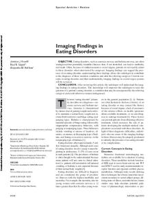
Fig 1. Case 4: 40-year-old man with multiple cerebral, cerebellar, and brain stem infarctions related to polyarteritis nodosa. A, Axial T2-weighted (2200/80/0.75) MR image shows multiple sites of hyperintense signal within the cerebellum (straight arrows) and pons (curved arrow). B, Axial T2-weighted (2200/80/0.75) MR image shows bilateral parietal (solid arrows) and left frontal (open arrows) regions of hyperintense signal within brain cortex, consistent with infarctions. Similar foci were also seen in the basal ganglia and internal capsules bilaterally. Cerebral angiography, performed a few days after the MR examination, was nomal.
Fig 2. Case 3: 58-year-old woman with hemorrhagic and nonhemorrhagic infarctions in the setting of cerebral arteritis related to polyarteritis nodosa. A, Sagittal T1-weighted (550/25/2) MR image shows a focus of hemorrhage within the inferior left frontal cortex (arrow) with an adjacent larger site of nonhemorrhagic infarction (arrowheads) primarily within the frontal cortex. B, Axial T1-weighted (500/25/1) MR image shows a focus of hemorrhage within the right frontal cortex (arrow). C, Right internal carotid catheter angiogram (cut-film angiography), lateral view, obtained 8 days after MR imaging. Multiple sites of narrowing and dilatation (arrows) are seen within medium-sized and small arteries.
bladder after cholecystectomy a few weeks earlier. Cerebral angiography, performed to define the extent of vascular involvement, was nondiagnostic. Because of the possibility of involvement of arteries too small for the resolution of angiography, corticosteroid therapy was initiated, resulting in marked improvement in cognitive function within 1 week. Three patients tested positive for the presence of hepatitis B surface antigen (Table). Four patients had systemic hypertension, which was initially poorly controlled in two patients but thought unlikely to be the cause of CNS signs and symptoms. Instead, CNS disease was thought most likely to be due to cardiogenic
embolism (case 1) and CNS arteritis (case 4). No patient had systemic or CNS infection at the time of neurologic symptoms or neuroradiologic examinations. All patients underwent echocardiography within a few weeks of neuroradiologic imaging. Three patients (cases 1, 2, and 4) had echocardiograms showing severe concentric left ventricular hypertrophy (resulting from hypertension) and global hypocontractility, with intracardiac thrombi seen in case 1. Three patients had a history of cocaine abuse (intravenous use in two and nasal inhalation in one), and all tested positive for hepatitis B surface antigen (Table). None tested positive for cocaine at admission.
1122
PROVENZALE
AJNR: 17, June 1996
Fig 3. Case 1: 25-year-old man with embolic infarction who had polyarteritis nodosa–related cardiomyopathy and intracardiac thrombus seen at echocardiography. A, Axial T2-weighted (2000/80/0.75) MR image obtained a few months after infarction of the hand shows a right parietal lobe wedge-shaped peripheral infarction consistent with an embolism. Cerebral atrophy is also present. B, Axial T2-weighted (2000/80/0.75) MR image shows hyperintense signal within the caudate heads (curved arrows) and putamina (straight arrows) bilaterally, presumed to be caused by ischemia.
Discussion In 1866, Kussmaul and Maier (14) described a patient with a widespread necrotizing arteritis involving small and medium-sized arteries. The name polyarteritis nodosa derives from the fact that their patient had palpable nodules along the course of the arteries caused by periarterial inflammatory exudates. Polyarteritis nodosa is one of three closely related systemic necrotizing vasculitides, the other two being allergic angiitis and granulomatosis (Churg-Strauss variant) and the polyangiitis overlap syndrome (which has features of both polyarteritis nodosa and the Churg-Strauss variant) (15). The disease affects every organ except the lungs and spleen (16). The cause of the arteritis is not known with certainty but is believed in many cases to be initiated by immune complex deposition (17). Approximately 30% of patients have hepatitis B surface antigenemia, and in those patients hepatitis B surface antigen, complement, and immunoglobulin have been demonstrated (16, 17). Symptom onset is usually in midlife although the disease can manifest as early as childhood (5, 18). Multiple organ systems are usually involved. The typical clinical presentation consists of nonspecific constitutional signs and symptoms (such as fever, weight loss, and generalized malaise) and inflammatory or ischemic findings involving multiple organ systems, including skin rash, joint pain, abdominal pain due to visceral infarction, renal failure, and polyneuropathy (1). Hypertension accompanying or signaling renal involvement is common.
New or worsening hypertension in a patient with features of a multisystem illness is a clue to search for this diagnosis. Cardiac manifestations are frequently seen and can include congestive heart failure, myocardial infarction, pericarditis, and, as in three of our patients, left ventricular hypertrophy resulting from hypertension (1). The diagnosis can be confirmed by nerve, muscle, or kidney biopsy findings of characteristic arteriopathy or by renal or mesenteric angiographic findings of characteristic multiple small (2 to 5 mm) aneurysms or arteritis, present in about 60% of cases (19, 20). There is evidence to suggest that clinically severe disease, including CNS involvement, may be more likely to develop in patients who have abnormal findings on renal and mesenteric angiograms (20). The 5-year survival rate in untreated patients is 13%, which increases to about 80% following treatment with both corticosteroids and cytotoxic agents (16). Polyarteritis nodosa is a rare disorder. In one study, which based the diagnosis on pathologic criteria, the prevalence was reported to be 6.3 per 100 000 population (21). Our study, which included only patients with neurologic features that were sufficiently severe to warrant neuroimaging examination, may underestimate the frequency of CNS involvement on neuroimaging studies in patients with polyarteritis nodosa, because patients with solely peripheral nervous system disease, cranial neuropathy not thought to be related to a brain abnormality, or CNS symptoms or signs that
AJNR: 17, June 1996
were not of sufficient severity to warrant an imaging study were not included in our patient population. For instance, all five of our patients had seizures and three had encephalopathy. It is possible that patients with polyarteritis nodosa who did not have seizures and who had only mild or transient encephalopathy did not undergo neuroradiologic examinations. The arteritis in polyarteritis nodosa has been characterized as hyalinelike necrosis of the media and internal elastic lamina of small and medium-sized arteries, followed by extension of an inflammatory reaction to the adjacent layers of the arterial wall and periarterial tissues (22). A secondary proliferation of the intima often results, causing varying degrees of luminal occlusion. Four stages of this inflammatory process have been described: an acute stage of necrosis of the intima or media; a subacute phase in which an inflammatory exudate of eosinophils, lymphocytes and polymorphonuclear leukocytes is seen; a chronic stage marked by intimal proliferation, fibrinoid necrosis, and the appearance of granulation tissue within and around the arterial wall; and a stage of healing that is often incomplete, leaving a scarred, fibrotic artery with a narrowed lumen (3, 16). Different stages of the inflammatory process are often seen concurrently. Segmental involvement of the artery is characteristic, with a predilection for bifurcations and branch points (22, 16). Inflammatory changes are commonly seen in perineural arteries, resulting in the peripheral neuropathy so common in polyarteritis nodosa, but they can also be seen in cerebral arteries. Small branches of the major cerebral arteries are most commonly affected. In some cases, marked loss of the muscular coatings of small (100 to 200 mm) intracranial arteries has been reported, with replacement by collagenous tissue, resulting in severe luminal narrowing (23). On occasion, however, arteries as large as the middle cerebral and anterior cerebral arteries can be involved (22). At any point in the inflammatory process, thrombosis leading to brain ischemia or infarction can result. Nervous system involvement in polyarteritis nodosa usually occurs in the form of mononeuropathy, polyneuropathy, or multiple peripheral nerves (mononeuritis multiplex). CNS disease, which is much less common, almost always involves the brain, although spinal cord involvement has occasionally been reported (24). The prevalence of CNS involvement varies among
POLYARTERITIS NODOSA
1123
reported series, and is in part dependent on the inclusion criteria used. In one large series in which cranial nerve and ocular fundus involvement were included as CNS manifestations, 12% of patients had CNS disease alone and another 34% had both CNS and peripheral nervous system involvement (5). CNS manifestations are reported to generally occur later than peripheral nervous system findings, and are usually seen along with physical signs (eg, fever, uremia), indicating a systemic disease process (5). In one series of 25 patients with systemic arteritis (including, but not restricted to, patients with polyarteritis nodosa), peripheral nervous system manifestations were usually part of the initial presentation or seen within a few months of the diagnosis, while CNS disease typically occurred 2 to 3 years after the diagnosis was established (6). The relatively late timing of CNS involvement has led investigators to hypothesize that manifestations of CNS disease are the result of accumulation of sequential vascular inflammatory changes occurring over many months or years, rather than solely the result of an acute arteritis (6). Reversible encephalopathy is the most common CNS manifestation, followed in frequency by focal neurologic deficits (8). Seizures are reported in up to 20% of patients (8). CNS involvement has rarely been reported to be the initial disease manifestation (8). Our patients appeared to differ from most of those previously reported in a few respects. First, CNS findings were an early disease manifestation in many of our patients. Second, seizures were the most common neurologic manifestation, present in all five patients. In other respects, however, the clinical characteristics of our patients were quite similar to those of previously reported series. Our patients all had the typical clinical features of an arteritis affecting multiple organ systems, including the CNS, causing ischemia or infarction and responsive to corticosteroids and immunosuppressive therapy. In the few reported cases of patients with polyarteritis nodosa who had undergone MR imaging, cortical and white matter hyperintense signal abnormalities on T2-weighted images, consistent in some cases with infarctions, have been noted (10 –12). In one case, large white matter lesions scattered throughout the centrum semiovale, which nearly completely resolved within a few months after initiation of
1124
PROVENZALE
corticosteroid and cyclophosphamide therapy, were found (11). The most common abnormalities seen in our patients were small lesions within the cerebral cortex and subcortical white matter. These lesions, which were seen on repeated examinations and associated with tissue loss, are consistent with infarctions. In general, the lesions seen in our patients were present in the periphery of major arterial distributions or, in some cases, in areas supplied by small end-arteries, such as the brain stem and basal ganglia. These sites are consistent with the known predilection of polyarteritis nodosa for involvement of small and mediumsized arteries. Such arterial involvement was shown at cerebral angiography in our case 3. However, in the single patient (case 4) with hyperintense signal abnormalities in the cerebellum and brain stem, which are rare sites of involvement in polyarteritis nodosa, no evidence of arteritis was seen at cerebral angiography. It is possible that the angiogram in our patient yielded false-negative results because of the involvement of small arteries that were below the limit of angiographic resolution. In one previous report of a polyarteritis nodosa–related brain stem infarction seen at MR imaging, cerebral angiography primarily showed involvement of such small arteries as the thalamoperforators (9). Postmortem examination revealed a necrotizing arteritis in small meningeal arteries supplying the brain stem. The possibility of arteritis in our case 4 is supported by the fact that marked improvement was noted in the patient’s neurologic status 1 week after initiation of corticosteroid therapy. With regard to the other two patients with small cortical and subcortical infarctions (cases 2 and 5), cerebral arteritis is also a possible diagnosis. One of these patients (case 2), in fact, had angiographic findings of arteritis on abdominal angiography. In both cases, however, cerebral arteritis cannot be definitively proved in the absence of cerebral angiography or biopsy of the brain or meninges. Intracranial hemorrhage, as was seen in case 3, is an uncommon complication of polyarteritis nodosa. In one large series, 4% of patients had gross intracranial hemorrhage at necropsy (5). In some of these patients, intraparenchymal hemorrhage occurred at typical sites for hypertensive hemorrhage, making the role of hypertension difficult to exclude on the basis of the clinical data provided by the authors (5). The
AJNR: 17, June 1996
hemorrhages seen in our case 3 were atypical in location for hypertensive hemorrhages and, furthermore, this patient was the only one in our series who did not have hypertension. In other reported series in which the role of hypertension has been reliably excluded, intraparenchymal hemorrhages (sometimes many centimeters in diameter) and intracranial and spinal subarachnoid hemorrhage have been found in association with the typical histologic findings of arteritis resulting from polyarteritis nodosa (5, 25– 28). Reports of the cerebral angiographic findings in polyarteritis nodosa are few and have shown either alternating segments of narrowing and widening of small and medium-sized intracranial arteries or occlusion of small arteries (9, 18, 29). The angiographic findings in polyarteritis nodosa are indistinguishable from those in other vasculitides affecting arteries in this size range. Aneurysms, which are a classic finding of polyarteritis nodosa in the renal and splanchnic circulation, are much less common in the intracranial circulation (19, 29). Intracranial arterial dissection has been reported in a patient with polyarteritis nodosa, but it is unclear whether this represented a spontaneous event or was related to an underlying arteriopathy (30). Fatal subarachnoid hemorrhage from a posterior cerebral artery aneurysm has been reported, but again it is unclear whether the aneurysm was specifically related to an arteritis (31). Cardiac disease, present in three of our patients, is common in polyarteritis nodosa. In one series of 17 patients with polyarteritis nodosa who were examined after death, a pathologic abnormality of the heart was found in 13 cases. The findings included left ventricular hypertrophy (10 cases), myocardial infarction or diffuse myocardial scarring (6 cases), and evidence of arteritis within large or small coronary arteries (7 cases) (26). In 1 of our patients, a severely dyskinetic and enlarged left ventricle was the setting for the development of intracardiac thrombus, which was the probable cause of cerebral infarction. Given the prevalence of cardiac disease in patients with polyarteritis nodosa, cardiogenic embolism must be considered high on the list of differential diagnoses in those with CNS findings when there are no features suggestive of active arteritis. It is notable that three of our patients had a history of cocaine abuse, raising the possibility that substance abuse either was the primary
AJNR: 17, June 1996
mechanism for, or contributed to, the neuroradiologic findings. We cannot exclude that possibility, but note that all three of these patients tested positive for hepatitis B surface antigen. The development of polyarteritis nodosa in patients who test positive for hepatitis B surface antigen is now a well-described phenomenon (32) and serves as evidence supporting the hypothesis that, at least in many cases, immune complex deposition is part of the pathogenesis of polyarteritis nodosa (16). Furthermore, intravenous drug abuse, present in our cases 1 and 2, is a common means of hepatitis B virus infection in patients with polyarteritis nodosa who test positive for hepatitis B surface antigen, accounting for 11% of patients in one large series (33). It is possible that the arteritis seen in some of our patients could have been a hypersensitivity arteritis resulting from cocaine use (ie, so-called cocaine-vasculitis). This entity appears to be a relatively rare phenomenon, given the widespread use of the drug. The few reports of cocaine vasculitis, however, have noted clinical and histologic findings confined to the CNS (rather than the systemic findings seen in our patients that are typical of polyarteritis nodosa) (34, 35). In addition, reports of cocaine vasculitis have described inflammatory changes only in small arteries (and in some cases, venules) (34, 35). Among the cocaine abusers in our series, however, one patient (case 4) had histologic findings of the medium-sized artery involvement typically seen in polyarteritis nodosa. In addition, two patients (cases 1 and 2) had abdominal angiograms showing the typical renal artery aneurysms of polyarteritis nodosa. Therefore, it is unlikely that the clinical and neuroradiologic findings seen in the three patients who abused cocaine were due to cocaine vasculitis. The findings of small, primarily cortical and subcortical infarctions in most of our patients, although nonspecific for polyarteritis nodosa, are consistent with an arteritis involving small and medium-sized arteries. The diagnosis of polyarteritis nodosa, if not already established by the time of neuroimaging, should be suspected in patients with these findings and evidence of a multisystem disease characterized by ischemia or infarction in various organs. Abdominal angiography and/or biopsy of accessible involved tissue (usually nerve, muscle, or kidney) should thereafter be performed to establish the diagnosis and to indicate appropriate
POLYARTERITIS NODOSA
1125
therapy. However, given the high rate of cardiac involvement in this disease, the possibility of a cardiac cause of CNS disease, most notably cardiogenic embolism, must also be borne in mind when examining patients with polyarteritis nodosa who have CNS dysfunction.
Acknowledgment We are grateful to Kathy Thompson for assistance in preparation of the manuscript.
References 1. Conn DL. Polyarteritis. Rheum Dis Clin North Am 1990;16:341– 362 2. Rose GA, Spencer H. Polyarteritis nodosa. Q J Med 1957;26: 43– 81 3. Arkin A. A clinical and pathologic study of polyarteritis nodosa. Am J Pathol 1930:6;401– 426 4. Leavitt RY, Fauci AS. Therapeutic approach to the vasculitic syndromes. Mt Sinai J Med 1986;53:440 – 448 5. Ford RG, Siekert RG. Central nervous system manifestations of periarteritis nodosa. Neurology 1965;15:114 –122 6. Moore PM, Fauci AS. Neurologic manifestations of systemic vasculitis: a retrospective and prospective study of the clinicopathologic features and responses to therapy in 25 patients. Am J Med 1981;71:517–524 7. Guillevin L, Houng Du LT, Godeau P, Jais P, Wechsler B. Clinical findings and prognosis of polyarteritis nodosa and Churg Strauss angiitis: a study in 165 patients. Br J Rheumatol 1988;27:258 – 264 8. Rosenberg MR, Parshley M, Gibson S, Wernick R. Central nervous system polyarteritis nodosa. West J Med 1990;153:553–556 9. Kasantikul V, Suwanwela N, Pongsabutr S. Magnetic resonance images of brain stem infarct in periarteritis nodosa. Surg Neurol 1991;36:133–136 10. Miller DH, Ormerod IEC, Gibson A, du Boulay EP, Rudge P, McDonald WI. MR brain scanning in patients with vasculitis: differentiation from multiple sclerosis. Neuroradiology 1987;29:226 –231 11. Koppensteiner R, Base W, Bognar H, Kiss A, Al Mubarak M, Tscholakoff D. Course of cerebral lesions in a patient with periarteritis nodosa studied by magnetic resonance imaging. Klin Woschenschr 1989;67:398 – 401 12. Wildhagen K, Stoppe G, Meyer GJ, Heintz P, Hundeshagen H, Deicher H. Bildgebende diagnostik der zentralnervosen Beteiligung bei der panarteriitis nodosa. Z Rheumatol 1989;48:323–325 13. Lightfoot RW Jr, Michel BA, Bloch DA, et al. The American College of Rheumatology 1990 criteria for the classification of polyarteritis nodosa. Arthritis Rheum 1990;33:1088 –1093 14. Kussmaul A, Maier R. Ueber eine bisher nicht beshriebene eigenthumliche Arterienerkrankung (Periarteritis nodosa), die mit Morbus Brightii und rapid fortschreitender allgemeiner Muskellahmung einhergeht. Dtsch Arch Klin Med 1866;1:484 –517 15. Haynes BF, Allen NB, Fauci AS. Diagnostic and therapeutic approach to the patient with vasculitis. Med Clin North Am 1986; 70:355–368 16. Moore PM, Cupps TR. Neurological complications of vasculitis. Ann Neurol 1983;14:155–167
1126
PROVENZALE
17. Michalak T. Immune complexes of hepatitis B surface antigen in the pathogenesis of periarteritis nodosa: a study of seven necropsy cases. Am J Pathol 1978;90:619 – 632 18. Engel DG, Gospe SM, Tracy KA, Ellis WG, Lie JT. Fatal infantile polyarteritis nodosa with predominant central nervous system involvement. Stroke 1995;26:699 –701 19. Travers RL, Allison DJ, Brettle RP, Hughes GRV. Polyarteritis nodosa: a clinical and angiographic analysis of 17 cases. Semin Arthritis Rheum 1979;8:184 –199 20. Ewald EA, Griffin D, McCune WJ. Correlation of angiographic abnormalities with disease manifestations and disease severity in polyarteritis nodosa. J Rheumatol 1987;14:952–956 21. Kurland LT, Hauser WA, Ferguson RH, et al. Epidemiologic features of diffuse connective tissue disorders in Rochester, Minnesota, 1951 through 1967, with special reference to systemic lupus erythematosus. Mayo Clin Proc 1969;44:649 – 663 22. Kernohan JW, Woltman HW. Periarteritis nodosa: a clinicopathologic study with special reference to the nervous system. Arch Neurol Psychiatry 1938;39:655– 686 23. Sheehan B, Harriman DG, Bradshaw JP. Polyarteritis nodosa with ophthalmic and neurological complications. Arch Ophthalmol 1958;60:537–547 24. Ojeda VJ. Polyarteritis nodosa affecting the spinal arteries. Aust N Z J Med 1983;13:287–289 25. Hiller F. Cerebral hemorrhage in hyperergic angiitis. J Neuropathol Exp Neurol 1953;12:24 – 40 26. Griffith GC, Vural IL. Polyarteritis nodosa: a correlation of clinical and postmortem findings in seventeen cases. Circulation 1951;3: 481– 491
AJNR: 17, June 1996
27. Henson RA, Croft PB. Spontaneous spinal subarachnoid hemorrhage. Q J Med 1956;25:53– 66 28. Iaconetta G, Benvenuti D, Lamaida E, Gallicchio B, Signorelli F, Maiuri F. Cerebral hemorrhagic complication in polyarteritis nodosa. Acta Neurol [Napoli] 1994;16:64 – 69 29. Ferris EJ, Levine HL. Cerebral arteritis: classification. Radiology 1973;109:327–341 30. Fujiwara S, Yokoyama N, Fujii K, Matsushima T, Matsubara T, Fukui M. Repeat angiography and magnetic resonance imaging (MRI) of dissecting aneurysms of the intracranial vertebral artery. Acta Neurochir 1993;121:123–129 31. Summers VK. The nervous manifestations of periarteritis nodosa. Lancet 1950;1:1148 –1149 32. Dally S, Guillevin B, Maidenberg M, Leclerc JP, Katz M, Gaultier M. Periarterite noueuse chez un toxicomane atteint d’hepatite B. Ann Med Interne 1979;130:649 – 652 33. Guillevin L, Lhote F, Jarrousse B, et al. Polyarteritis nodosa related to hepatitis B virus: a retrospective study of 66 patients. Ann Med Interne 1992;143(Suppl 1):63–74 34. Morrow PL, McQuillen JB. Cerebral vasculitis associated with cocaine abuse. J Forensic Sci 1993;38:732–738 35. Krendel DA, Ditter SM, Frankel MR, Ross WK. Biopsy-proven cerebral vasculitis associated with cocaine abuse. Neurology 1990;40:1092–1094