IJCRI 201 2;3(1 2):6–1 0.
Ikpeme et al.
www.ijcasereportsandimages.com
CASE SERIES
6
OPEN ACCESS
Neglected ruptures of the patellar tendon: Repair options in a resource poor setting Ikpeme A Ikpeme, Anthonia A Ikpeme, Innocent E Abang, Paul O Amah, Ahmed M Achaka
ABSTRACT
Introduction: Rupture of the patellar tendon is a disabling injury. Better results are reported in immediate repairs of fresh ruptures. In neglected ruptures, restoration of the structural and functional integrity of the extensor apparatus is difficult. This report highlights the use of an achilles osteotendinous autograft in the repair of an 8month old neglected rupture of the patellar tendon; and the Mandelbaum technique with open tendon harvest in the repair of a 10week old neglected rupture. Case Series: A 23yearold male presented with inability to extend the left knee; eight months after a road traffic accident. Physical and radiological findings were consistent with a rupture of the left patellar tendon. The patient was offered tendon reconstruction using an Achilles osteotendinous autograft from the contralateral lower limb. One year later, he was pleased with the outcome. A 29yearold army officer presented with pain, swelling and inability to extend his right knee since 10 weeks. Physical and Xray findings were in keeping with a rupture of the right patellar tendon. The patient was offered patellar tendon reconstruction using the Mandelbaum technique. Twentyeight weeks postoperatively,
Ikpeme A Ikpeme 1 , Anthonia A Ikpeme 2, Innocent E Abang 1 , Paul O Amah 1 , Ahmed M Achaka 3 Affiliations: 1 Orthopaedics Unit, Department of Surgery, University of Calabar Teaching Hospital, P.M.B 1 278, Calabar, Nigeria; 2Department of Radiology, University of Calabar Teaching Hospital, P.M.B 1 278, Calabar, Nigeria; 3 Nigerian Navy Hospital, Calabar, Nigeria. Corresponding Author: Dr. Ikpeme A Ikpeme, GPO Box 1 506, Calabar, Nigeria; Email:
[email protected] Received: 02 August 2011 Accepted: 11 April 201 2 Published: 01 December 201 2
the patient had a range of motion of 0–105o in the right knee and has since returned to a functional professional and recreational lifestyle. Conclusion: Neglected ruptures of patella commonly yield suboptimal results following repair. In resourcepoor settings, an autogenous achilles tendon technique as well as the Mandelbaum technique may prove beneficial in the treatment of this difficult condition. Keywords: Patellar tendon, Neglected rupture, Achilles osteotendinous autograft, Mandelbaum procedure, Resource poor setting
*********
Ikpeme IA, Ikpeme AA, Abang IE, Amah PO, Achaka AM. Neglected ruptures of the patellar tendon: Repair options in a resource poor setting. International Journal of Case Reports and Images 2012;3(12):6–10.
*********
doi:10.5348/ijcri201212228CS2
INTRODUCTION
Rupture of the patellar tendon is a disabling injury commonly seen in individuals under 40 years of age [1]. The injury is uncommon, can be easily missed in multi injured patients, in the obese and in knees with hemarthrosis [1]. These ruptures usually occur at the inferior pole of the patella and better results are reported in immediate repairs of fresh ruptures [1, 2]. In our community, late presentation to orthodox care is common and the traditional bone setter is often the first to be consulted after injuries [3–5]. Neglected patellar tendon ruptures are thus only seen when the patient is unwilling to live with the disability that follows unorthodox treatment [4]. Neglected ruptures of
IJCRI – International Journal of Case Reports and Images, Vol. 3 No. 1 2, December 201 2. ISSN – [0976-31 98]
IJCRI 201 2;3(1 2):6–1 0.
www.ijcasereportsandimages.com
the patellar tendon (defined as ruptures presenting after six weeks) are often difficult to repair. Restoration of the structural and functional integrity of the extensor mechanism is difficult because of the associated muscle contracture, scar tissue formation, proximal patella migration and fibrous adhesions between the patellar and the femur [2, 6]. Many different techniques have been reported in the management of these conditions in long standing cases. Endtoend anastomosis of the tendon ends is often not feasible [2, 7]. Quadriceps lengthening, quadriceplasty, traction (preoperative and intraoperative), and external fixation with pins and wires are some techniques used to relocate the patella to its anatomic position. Autogenous graft augmentation using the fascia lata or hamstring tendons, allograft reconstruction using the Achilles tendon or patellar tendon are the commonly used reconstruction techniques for neglected ruptures. Prolonged postoperative immobilization, loss of knee flexion, residual weakness of the quadriceps and construct failure are complications associated with repair of a neglected rupture [7].
Ikpeme et al.
7
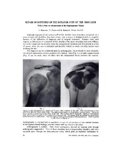
the patella and the medial and lateral tendinous expansions of the quadriceps tendon using non absorbable interrupted sutures. The osseous pedicle was anchored into an appropriately sized window cut into the tibial tuberosity by two 3.5 mm cancellous screws. Quadriceplastly was performed. Postoperatively, patellar traction was continued for two weeks, the left lower limb was encased in a long leg cast after removal of traction and patient was discharged. He was seen at four weeks post discharge, the long leg cast was removed and an exercise programme commenced. At 16th week postoperatively, the patient was pain free, had improved gait and an active range of motion of 5–950 in the left knee (Figures 1, 2). The ankle range of motion on the right side was normal and power of plantar flexion was grade 4+. He was subsequently lost to followup and reappeared in the clinic one year later. The knee range of motion at this time was 3–105o. He could run and walk normally and was very pleased with his progress.
CASE SERIES
Case 1: A 23yearold male presented to our out patient department with complaints of inability to extend his left knee. He had been involved in a road traffic accident eight months earlier and had spent the interval in a traditional bone setter’s practice. Examination showed wasting of the left quadriceps muscles, a visible and palpable defect over the left knee, patella alta, loss of active extension and a passive range of motion of 10–600. Xrays showed a proximally migrated patella with no fractures of the femur or tibia. The patient was admitted for preoperative traction for two weeks and subsequently offered quadriceplasty and tendon reconstruction. Neither preoperative traction nor intraoperative elongation, and quadriceplasty were successful in restoring the patella to its exact anatomic location. Lack of tendon harvesters precluded the use of the hamstring tendons. The fascia lata was adjudged unhealthy following chronic inflammation from the traditional bone setters scarification. As we did not have access to allograft Achilles tendon graft, a decision to use autogenous Achilles tendon graft was made. Intraoperative findings were a proximally migrated patella, a 10cm defect in the patellar tendon and marked fibrous adhesions involving the quadriceps femoris muscles. A 1cm central osteotendinous graft was harvested from the contralateral Achilles tendon. The defect at the donor site was closed with interrupted nonabsorbable sutures. The tendon graft was split into three slips, the central slip being thicker than the medial and lateral slips as described for the Achilles tendon allograft technique. A midline tunnel was drilled in the patella and the central slip passed through and anchored onto the quadriceps tendon from within outwards. The medial and lateral slips were anchored to either side of
Figure 1: Showing range of active extension in both the knees. (case 1)
Figure 2: Range of active flextion in the knees. (case 1)
IJCRI – International Journal of Case Reports and Images, Vol. 3 No. 1 2, December 201 2. ISSN – [0976-31 98]
IJCRI 201 2;3(1 2):6–1 0.
www.ijcasereportsandimages.com
Case 2: A 29yearold male army officer presented to the clinic with a 10week history of pain, swelling and inability to extend the right knee following a fall onto the right knee while playing recreative basketball. Prior to presentation, he had sought unorthodox treatment with massage and hot fermentations and had lost the partial extension of the affected knee which was present in the early post injury period. Examination revealed a swollen, mildly tender right knee, with a high riding patella and a distinct defect in the patellar tendon. The quadriceps femoris on the right side was wasted and patient had a range of active extension of 00 in the right knee. The range of right knee flexion was 1100 and Xrays showed patella alta. An impression of neglected and mismanaged rupture of the right patella tendon was made. The patient was admitted and offered patella tendon reconstruction using the Mandelbaum technique. The approach was an anterior midline approach. Intra operative findings were a transverse rupture of the right patellar tendon and healthy semitendinosus and gracilis tendons. Operative technique involved minimal quadriceplasty, mobilization of the patella and relocation to its anatomic position. Owing to a lack of tendon harvesters, the skin flaps were raised until the semitendinosus and gracilis tendons were identified on the medial side. The tendons were traced to their musculotendinous junctions, sectioned at the junctions and their distal ends detached from the tibia. The tendons were then sutured endtoend. Transverse holes were drilled across the patella and just below the tibial tubercle. The previously anastomosed semitendinosus and gracilis tendinous autografts were passed between the drill holes and used to reconstruct the extensor apparatus in a figureofeight configuration (Mandelbaum technique). A 0.5 guage malleable Kirschner wire was used to support the construct. The wound was closed in layers and a plaster of paris long leg cast was applied and worn for five weeks. The malleable Kirschner wire was removed six weeks postoperatively. Postoperative management included immediate, nonweight bearing, bilateral, axillary crutch aided mobilization and graded range of motion physiotherapy exercises from the sixth week. The patient made a complete recovery with right knee range of motion being 0–1050 at the last clinic visit 28th week after surgery. He has since returned to full military duties (including an international peace keeping tour of duty and a promotion) and has also returned to regular recreative basketball games.
DISCUSSION
Ruptures of the patella tendon usually follow long standing irritation. Chronic tendon degeneration due to repetitive micro trauma has been supported by histological evidence consistent with chronic inflammation and degeneration in ruptured tendon specimens [8]. Patella tendon rupture is usually
Ikpeme et al.
8
unilateral when it results from traumatic athletic injuries, road traffic accidents and local steroid injections. When it occurs in association with systemic disorders such as systemic lupus erythematosus, chronic renal failure or diabetes mellitus, the rupture can be bilateral [9, 10]. The condition can also occur in posterior dislocations of the knee. Suboptimal results have often been reported in the treatment of neglected injuries [1, 2, 6, 7]. In our setting, the combination of inappropriate unorthodox intervention and lack of basic requirements for tendon harvesting presented unique challenges. In our first patient, the fascia lata was also adjudged inappropriate for use due to evidence of chronic inflammation following scarifications by the traditional bone setter on the affected thigh. The combination of (possible) sub clinical soft tissue infection (from scarifications and herbal fomentations to drain “evilblood”) [3, 4] and poor nutrition can predispose to an unhealthy fascia lata. In our second patient, the challenges posed by inappropriate unorthodox interventions were obvious. In the immediate postinjury period and up to five weeks thereafter, the patient had weak extension of the right knee but lost that following massage and traditional ‘physiotherapy’. An apparently incomplete injury was converted to a complete rupture when the patient was presented to the clinic. The treatment of neglected ruptures of the patellar tendon aims to relocate the patella in its anatomic position and recreate the InsallSalvati ratio, ensuring mobility and correct tracking of the patella and restore the structural and functional integrity of the extensor apparatus [1, 2, 11]. This is difficult and the results are often suboptimal [2, 6, 11]. Patellar relocation can be achieved by preoperative and intraoperative traction, quadriceplasty and a VY elongation of the quadriceps tendon. The use of external fixators and the Ilizarov technique have also been reported. Tendon reconstruction involves the use of autogenous fascia lata or hamstring tendons; allograft patellar tendon or Achilles tendon in single or double graft techniques [2, 6, 7]. In our setting, there is no access to allograft tendons and autogenous hamstring or fascia lata grafts were not feasible in the first patient. An Achilles tendon autograft was thus the preferred option. Autogenous Achilles tendon grafts can potentially present such complications as residual donor site rupture, infection and loss of plantar flexion. These may discourage the routine use of this technique. However, in resourcepoor settings with no access to Achilles or patellar tendon allografts, no access to proper harvesting of an adequate length of hamstring tendons and where use of the fascia lata is precluded, our first case shows that an Achilles tendon autograft may be a feasible option. We believe the issues of donor site morbidity and infection can be addressed by ensuring harvest of only the central one cm of the tendon and meticulous attention to surgical technique. Graft take is improved by the inclusion of an osseous pedicle. In our first patient, the gait improved, range of motion in the left knee improved and plantar flexion power was grade
IJCRI – International Journal of Case Reports and Images, Vol. 3 No. 1 2, December 201 2. ISSN – [0976-31 98]
IJCRI 201 2;3(1 2):6–1 0.
Ikpeme et al.
www.ijcasereportsandimages.com
4+. We believe the outcome justifies our technique and may be useful in similar settings. In our second patient who presented relatively earlier, the issues of subclinical infection did not exist. In nonresource challenged settings, the Mandelbaum technique would have been offered with the less risk of donor site morbidity using a tendon harvester. Its absence left the managing team with the option of an open tendon harvest. Though our results support this as a viable option when indicated in resourcechallenged settings, the documented donor site morbidities such as infection must be borne in mind and appropriate steps taken to minimize the risk. Overall however, health systems and their managers must work towards the provision of an improved therapeutic armamentarium in our health sector. This will reduce the reported risks that attend orthodox intervention in the repair of this disabling injury. Health education to dissuade unorthodox intervention in orthopaedic maladies remains a credible intervention tool for improved outcomes. To the authors’ knowledge, an Achilles tendon autograft repair for neglected rupture of the patellar tendon has previously not been reported. We report our use of the technique of Achilles tendon autograft in the repair of an eightmonth old neglected rupture of the patellar tendon in a 23yearold male; and the use of the Mandelbaum procedure with open harvest of the hamstring tendons in the repair of a 10week old neglected rupture of the patella tendon in a 29 yearold male. Both patients had spent eight months and 10 weeks, respectively seeking unorthodox care. In nonresource challenged settings a tendon harvester makes tendon harvest easier with less risk of donor site morbidity.
data, Drafting the article, Final approval of the version to be published Innocent Abang – Substantial contributions to conception and design, Drafting the article, revising it critically for important intellectual content, Final approval of the version to be published Paul O Amah – Substantial contributions to conception and design, Drafting the article, revising it critically for important intellectual content, Final approval of the version to be published Ahmed M Achaka – Substantial contributions to conception and design, Drafting the article, revising it critically for important intellectual content, Final approval of the version to be published
Guarantor
The corresponding submission.
Neglected ruptures of the patellar tendon present unique treatment challenges especially in resource poor settings. An Achilles tendon autograft may prove beneficial in the treatment of this condition. The Mandelbaum technique using an open tendon harvest procedure is also a feasible option in these settings. Meticulous attention must be paid to the prevention of the possible complications that may occur following these techniques. Provision of appropriate intervention tools and health education to dissuade unorthodox interventions will help improve outcomes.
*********
Author Contributions
Ikpeme A Ikpeme – Substantial contributions to conception and design, acquisition of data, Drafting the article, Revising it critically for important intellectual content, Final approval of the version to be published Anthonia Ikpeme – Substantial contributions to conception and design, Analysis and interpretation of
author
is
the
guarantor
of
Conflict of Interest
Authors declare no conflict of interest.
Copyright
© Ikpeme A Ikpeme et al. 2012; This article is distributed under the terms of Creative Commons Attribution 3.0 License which permits unrestricted use, distribution and reproduction in any means provided the original authors and original publisher are properly credited. (Please see www.ijcasereportsandimages.com /copyrightpolicy.php for more information.)
REFERENCES 1.
2.
CONCLUSION
9
3. 4. 5.
6. 7. 8.
Matava MJ. Patellar Tendon Ruptures. J Am Acad Orthop Surg. 1996; 4:287–96. Bek D, Demiralp B, Komurcu M. Sehirlioglu A. Neglected patellar tendon rupture: a case of reconstruction without quadriceps lengthening. J Ortho Traumatology. 2008 Mar; 9 (1): 39–42. Ikpeme IA, Udosen AM, Asuquo ME, Ngim NE. Lighting Burns and Traditional Medical Treatment: A Case report. West Afr J Med 2007; 26(1): 53–4. Ikpeme IA, Udosen AM, OkerekeOkpa I. Patients perception of traditional bone setting in Calabar. Port Harcourt Med J 2007;1:104–8. Ikpeme IA, Udosen AM, Ngim NE, Amah P. Auto amputation of the foot following treatment of chronic leg ulcer by traditional bone setters. J. Orthopaedics 2008; 5(4i) e6. Available at: http://www.jortho.org/2008/5/4/e6/index.htm Kim HS, Park SR, Kang JS, Lee WH, Kim SH. Reconstruction of neglected patellar tendon rupture with Achilles tendon Allograft: A case report. J Korean Knee Soc. 2000 Jun; 12(1): 128–32. Levis PB, Rue JP, Bach BR. Chronic Patellar Tendon Rupture: Surgical Reconstruction technique using 2 Achilles Tendon Allografts. J Knee Surg. 2008; 21:130–5. Kannus P. Jozsa L. Histopathological changes preceding spontaneous rupture of a tendon. A controlled study of 891 patients. J Bone Joint Surg Am. Dec 1991; 73(10): 1507–25.
IJCRI – International Journal of Case Reports and Images, Vol. 3 No. 1 2, December 201 2. ISSN – [0976-31 98]
IJCRI 201 2;3(1 2):6–1 0.
www.ijcasereportsandimages.com 9.
Giblin P, Small A, Nichol R. Bilateral rupture of the Ligamentum patellae: two case reports and a review of the literature. Aust N Z J Surg. Apr 1982; 52(2): 145–8. 10. Rose PS, Francisca FJ. A traumatic bilateral patellar tendon rupture. A case report and review of the
Access full text article on other devices
Ikpeme et al.
10
literature. J Bone Joint Surg Am. Sept 2001; 83 A(9): 1382–6. 11. Heiden EA. Tendinopathies about the Knee. In: Chapman 250 MW(Ed). Chapman’s Orthopaedic Surgery. 3rd Edition. Chapter 88. Philadelphia. Lippincott Williams & Wilkins; 2001: 2339–45.
Access PDF of article on other devices
IJCRI – International Journal of Case Reports and Images, Vol. 3 No. 1 2, December 201 2. ISSN – [0976-31 98]