PROTOCOL
Multiplex amplification of ancient DNA Holger Römpler1, Paul H Dear2, Johannes Krause3, Matthias Meyer3, Nadin Rohland3, Torsten Schöneberg1, Helen Spriggs2, Mathias Stiller3 & Michael Hofreiter3 1Molecular Biochemistry, Institute of
Biochemistry, Medical Faculty, University of Leipzig, Johannisallee 30, 04103 Leipzig, Germany. 2MRC Laboratory of Molecular Biology, Hills Road, Cambridge CB2 2QH, UK. 3Department of Evolutionary Genetics, Max Planck Institute for Evolutionary Anthropology, Deutscher Platz 6, 04103 Leipzig, Germany. Correspondence should be addressed to M.H. (
[email protected]).
© 2006 Nature Publishing Group http://www.nature.com/natureprotocols
Published online 13 July 2006; doi:10.1038/nprot.2006.84
This method is designed to assemble long, continuous DNA sequences using minimal amounts of fragmented ancient DNA as template. This is achieved by a two-step approach. In the first step, multiple fragments are simultaneously amplified in a single multiplex reaction. Subsequently, each of the generated fragments is amplified individually using a single primer pair, in a standard simplex (monoplex) PCR. The ability to amplify multiple fragments simultaneously in the first step allows the generation of large amounts of sequence from rare template DNA, whereas the second nested step increases specificity and decreases amplification of contaminating DNA. In contrast to current protocols using many template-consuming simplex PCRs, the method described allows amplification of several kilobases of sequence in just one reaction. It thus combines optimal template usage with a high specificity and can be performed within a day.
INTRODUCTION Rationale Amplification of ancient mitochondrial, chloroplast and nuclear DNA is typically limited by small quantities of endogenous DNA, high levels of DNA degradation, contamination with exogenous DNA and the often restricted quantity of fossil material. Because of these problems, only limited information is usually obtained using standard simplex PCR. This protocol is specifically designed to bypass these limitations and to assemble long, continuous DNA sequences from degraded DNA templates, even if present in low copy numbers. The method is suitable for sequence data retrieval from disparate sources of DNA such as fossils, forensic specimens, noninvasively obtained samples and museum specimens. Thus, its applicability ranges from phylogenetic questions to population genetics, conservation genetics, forensic applications and functional genetics. The technique has been used to amplify both the first complete mitochondrial genome1 and the first complete nuclear gene (H.R., N.R., T.S. and M.H., unpublished data) from Pleistocene specimens. The two-step multiplex protocol was initially developed to analyze the sequences of single DNA molecules for genome mapping. When it is applied to ancient DNA, several modifications are required and additional precautions have to be taken. Working with ancient DNA Generally, and despite substantial progress in recent years, the retrieval of ancient DNA sequences from archeological and paleontological specimens is not routine. The problems in ancient DNA research arise from the fact that very little, usually extensively degraded and often no DNA survives in ancient tissues, whereas for certain species such as humans, bacteria or domestic animals, contemporary DNA is pervasive in the environment, both inside and outside the laboratory. To address these problems, a number of guidelines have been put forward in recent years. Although different studies require different levels of precautions2, there are certain standards for ancient DNA work3–5 to which all studies should adhere in order to obtain
720 | VOL.1 NO.2 | 2006 | NATURE PROTOCOLS
authentic sequences. However, it should be noted2 that even strict adherence to all guidelines may be insufficient to prevent incorrect results. Physically isolated work area. To avoid amplification of modern DNA contaminants, all work with ancient DNA preceding PCR cycling has to be carried out in a dedicated, isolated environment (in this protocol, Steps 3–7). The risk of cross-contamination with previously amplified PCR products is high, as copy numbers up to 1012 are generated for products amplified by PCR. Contamination problems can originate from any previous amplification product containing target sequences for the primers used. This problem becomes even more serious when products are cloned. Therefore, ancient DNA facilities should be located as far as possible from modern DNA laboratories. General considerations. To sustain the low-DNA status of such facilities, this laboratory should be irradiated with UV, the equipment should be treated with bleach (as far as possible) and personnel should use protective clothing and face shields. Personnel should not enter the low-DNA-status laboratory, after working in a normal molecular biology laboratory, without first changing clothes and showering. Whenever possible, consumables should be irradiated with UV light overnight before use. In general, disposable materials should be used. When chemicals are taken from stocks for preparing buffers, nothing should be returned to the stocks. Chemicals should be poured or transferred using disposable equipment. Aliquots should be prepared for water and all solutions. All tubes should be kept open for as short a time as possible. Control amplifications. To detect sporadic or lowcopy-number contamination, extraction and PCR controls must be performed in each experiment. Although carrier effects may limit their efficacy, a recent study questioned the existence of a strong carrier effect6. All results of contamination should be reported, and inclusion of positive controls from modern source DNA should generally be avoided, as they provide a contamination risk.
© 2006 Nature Publishing Group http://www.nature.com/natureprotocols
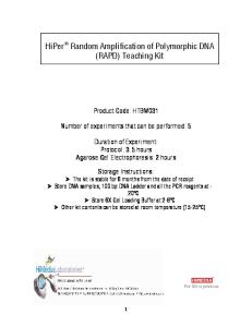
PROTOCOL Appropriate molecular behavior. For most ancient DNA amplifications, both quantity of PCR products and success rate should be inversely correlated with product size5. Large products (>500) are unusual, although under exceptional conditions amplification of products of length 1.6 kb7 to 2 kb8 have been reported. If single-copy nuclear or pathogen DNA is detected, mitochondrial DNA (mtDNA) should also be obtainable, as multicopy DNA sequences are more likely to survive in ancient specimens owing to their larger initial copy number. Generally, the maximum amplification length for nuclear DNA should be shorter than that for mtDNA. Biochemical preservation. Several studies have shown that there is no simple correlation between sample age and preservation of the DNA9,10. These studies revealed that not only the age of a fossil, but also burial temperature and environmental factors, are important for DNA preservation11,12. Indirect evidence for possible DNA survival, to support claims of retrieval of authentic ancient DNA from a specimen, can be provided by assessing the total amount, composition and relative extent of diagenetic change in amino acids and other organic molecules10,13. Copy number of the template. Often it is useful to assess the copy number of the DNA target using quantitative PCR. When the number of starting molecules is low (80 bp). In the strategy shown in d, completely nested primer pairs are used in the simplex PCRs. This two-sided nested variation can only be used for less fragmented DNA (>160 bp).
a
b © 2006 Nature Publishing Group http://www.nature.com/natureprotocols
c
d simplex PCRs are required for each multiplex PCR product to gain the complete sequence information of the initial multiplex amplification. It should be noted that the two simplex amplifications for each multiplex fragment, in this second strategy (Fig. 1c), do not represent independent amplifications according to ancient DNA guidelines, as they go back to the same initial multiplex product, which may have been derived from a single template molecule. Thus, a second multiplex PCR is necessary from which, in order to determine all sequence positions, two simplex amplifications again have to be done, resulting in a total of four simplex PCRs for every targeted multiplex fragment. For the amplification and assembly of continuous sequences, the first two strategies (Fig. 1b,c) are suitable if template DNA fragments are longer than approximately 80 bp. If the DNA is less fragmented, so that the template molecules are longer than approximately 160 bp, the third strategy (Fig. 1d) can be used. In this scenario, both simplex primers are
nested (contrary to the situation with the second strategy, in which only one primer is nested) and only one simplex amplification is done per multiplex fragment. This requires substantial overlap between the initial multiplex fragments, and thus this strategy is restricted to better-preserved samples. This approach can be modified to use only a single nested primer in the simplex amplification; this requires less overall overlap, but still substantially more than in the second strategy. Independent of the strategy chosen, ensure that, after subtraction of the simplex primer sequences, fragments still have an interset overlap, especially if nested primers are used (see Fig. 1a, ‘intraset overlap’). EQUIPMENT SETUP Work with ancient DNA requires that three labs be maintained, with absolutely no equipment and consumables exchanged between them. (i) Ancient DNA lab plus equipment and disposables: PCR setup and addition of template DNA to the first-step multiplex PCR should be carried out in this facility. (ii) Simplex setup lab or laminar-flow hood plus equipment and disposables: this lab should be used for opening of the finished multiplex PCRs, dilution and addition of the template to the simplex PCR reaction tubes. No PCR products from the simplex PCRs, modern genomic DNA, plasmids, colony PCRs or other sources of high-copy-number DNA must be handled in this laboratory. (iii) Standard molecular biology lab plus equipment and disposables: the lab is used for gel electrophoresis, product cloning and sequencing.
PROCEDURE Primer design for multiplex PCR 1| Design two sets of primer pairs for the initial multiplex step covering the sequence of interest, as outlined in the INTRODUCTION (‘Primer Setup’). Primer sets A and B are used in independent multiplex amplifications. For a flow diagram outlining the procedure, see Figure 2. ? TROUBLESHOOTING Primer design for simplex PCR 2| Choose the most suitable simplex amplification strategy for amplification of individual fragments, from three different approaches (Fig. 1b–d) as described in the INTRODUCTION. Design simplex PCR primers accordingly. ? TROUBLESHOOTING Multiplex PCR 3| Separately set up the multiplex primer sets A and B: Multiplex primer set
Volume (µl)
Concentration in multiplex primer set
All primers (100 µM)
2 each
1 µM each primer
Water
To 200
▲ CRITICAL STEP Steps 3–7 must be, and Steps 9–12 may be, carried out in the ancient DNA laboratory. ▲ CRITICAL STEP This dilution scheme is applicable for up to 50 primer pairs. If more than 50 pairs are used, either the primer concentration within the mix must be decreased or more concentrated stocks are needed. ? TROUBLESHOOTING NATURE PROTOCOLS | VOL.1 NO.2 | 2006 | 723
PROTOCOL 4| Prepare the multiplex master mix for each multiplex PCR: Multiplex master mix
Volume (µl)
Concentration in PCR
GeneAmp 10× PCR Buffer II
2
1×
BSA (10 mg ml–1)
2
1 mg ml–1
MgCl2 (25 mM)
3.2
4 mM
0.2
0.25 mM each
0.4
2U
dNTP (25 mM each) AmpliTaq Gold (5 U
µl–1)
© 2006 Nature Publishing Group http://www.nature.com/natureprotocols
Water
4.2
▲ CRITICAL STEP During the amplification process, the following controls must be performed (see Fig. 2): (i) an extraction control, treated identically to the extracts except that no sample is added, and (ii) two or more PCR water controls using water instead of sample (to detect sporadic or low-copy-number contamination). Setting up two or three identical multiplex PCRs is advisable when amplification of the target sequence can be expected, since, as in any ancient DNA study, each nucleotide position has to be determined from at least two independent primary PCRs. Working at this larger scale saves time in obtaining the necessary number of primary amplifications for an authentic ancient DNA reconstruction and prevents loss of ancient template DNA due to damage from freezing-thawing cycles and/or long-term storage. ? TROUBLESHOOTING 5| Combine the mixes prepared in Steps 3 and 4 separately for primer sets A and B for each multiplex PCR: Multiplex master mix, complete
Volume (µl)
Concentration in PCR
Multiplex primer set from Step 3
3
0.15 µM each primer
Multiplex master mix from Step 4
12
6| Distribute 15 µl of complete multiplex master mix for each primer set into PCR tubes (at least one extraction control, two PCR controls and one sample). 7| Add 5 µl of one of the following templates: DNA extract, blank extract or water. ▲ CRITICAL STEP Several methods have been published for extracting ancient DNA24–26, but these are not the subject of this protocol. However, ancient DNA standards have to be applied at this very first step, too.
Multiplex PCR (~4 h) Step 3 – 6: Set up multiplex primer set A master mix Step 7: Add samples and controls Primer set A
PCR water control 1
Temperature
Duration
Initial denaturation
94 °C
9 min
30 steps of: Denaturation
94 °C
20 s
Annealing
Primer dependent
30 s
Elongation
72 °C
30 s
Final elongation
72 °C
4 min
Storage
4 °C
Until use
▲ CRITICAL STEP Times and temperatures have to be adapted to the specific primers and to the targeted lengths. ? TROUBLESHOOTING 724 | VOL.1 NO.2 | 2006 | NATURE PROTOCOLS
Sample 1
Extraction blank
Sample 2
PCR water control 2
Step 8: Thermocycle multiplex PCRs
Simplex PCR (~6 h)
8| Put the multiplex tubes in a thermocycler outside the ancient DNA lab and amplify using the following durations and temperatures: Step
Handle set B identically to set A
Step 1 and 2: Design multiplex primer set A and corresponding simplex primers
Step 9 –12: Set up simplex primer master mixes Step 13 and 14: Add diluted multiplex PCR product
Simplex primer pair 1
PCR water control 1
Sample 1
Extraction blank
Sample 2
PCR water control 2
Simplex PCR water control
Simplex primer pair 2
PCR water control 1
Sample 1
Extraction blank
Sample 2
PCR water control 2
Simplex PCR water control
Remaining primer pairs
Step 15: Thermocycle simplex PCRs
Purification and sequence analysis of DNA (> 1 d)
Figure 2 | Flow diagram for the steps of the detailed protocol, including chronology, estimated duration and references to the corresponding protocol steps.
PROTOCOL Simplex PCR 9| Set up simplex primer pairs: Simplex primer pair
Volume (µl)
Concentration in mix
Forward primer (100 µM)
20
10 µM
Reverse primer (100 µM)
20
10 µM
Water
160
© 2006 Nature Publishing Group http://www.nature.com/natureprotocols
10| Prepare the simplex master mix for each simplex PCR: Simplex master mix
Volume (µl)
Concentration in PCR
GeneAmp 10× PCR Buffer II
2
1×
BSA (10 mg ml–1)
2
1 mg ml–1
MgCl2 (25 mM)
3.2
4 mM
0.2
0.25 mM each
0.05
0.25 U
dNTP (25 mM each) AmpliTaq Gold (5 U
µl–1)
Water
4.55
▲ CRITICAL STEP Simplex PCRs must be performed from all multiplex PCR reactions, including all controls. All controls must be performed with all primer pairs used in simplex PCRs. These controls include (i) a diluted extraction control from multiplex PCR to detect contamination during extraction process, (ii) diluted PCR water controls from multiplex PCR (testing each control separately) to uncover PCR contamination during multiplex or simplex setup, and (iii) a simplex control using water instead of template from multiplex PCR to identify PCR contamination during simplex setup. 11| Combine the mixes prepared in Steps 9 and 10 for each simplex PCR: Simplex master mix, complete
Volume (µl)
Concentration in PCR
Simplex primer pair from Step 9
3
1.5 µM each primer
Simplex master mix from Step 10
12
12| Pipette 15 µl of complete simplex master mix into PCR plates and store at 4 °C until continuing with Step 14. ▲ CRITICAL STEP Store no longer than a few hours. 13| Dilute a portion of each completed multiplex PCR with water in a ratio of 1:20 to 1:100 (we routinely use a dilution of 1:25). From each multiplex PCR, a single dilution that provides enough template for the subsequent simplex PCRs is sufficient. If the complete multiplex reaction is diluted, a 1:20 dilution results in sufficient template for 80 simplex amplifications and a 1:100 dilution suffices for 400 simplex reactions. If template for more than 400 simplex amplifications is required, a higher dilution factor than 1:100 is necessary. ? TROUBLESHOOTING ▲ CRITICAL STEP To avoid contamination and cross-contamination, Steps 13 and 14 are carried out in the simplex PCR laboratory or a dedicated laminar-flow hood with equipment reserved only for these two steps. 14| Set up the simplex PCRs by adding 5 µl diluted multiplex PCR from Step 13 to the corresponding wells of the plate prepared in Step 12.
NATURE PROTOCOLS | VOL.1 NO.2 | 2006 | 725
PROTOCOL 15| Place the tubes in a thermocycler and amplify using the cycling protocol described in Step 8.
© 2006 Nature Publishing Group http://www.nature.com/natureprotocols
Purification and sequencing 16| Final steps to obtain DNA sequences include PCR product purification (using either classical phenol/chloroform extraction or commercially available silica-based kits such as the QIAquick gel extraction or QIAquick PCR purification kits), Topo TA cloning and sequencing of the products. These standard procedures are not described, but all these steps should be carried out in a standard molecular biology lab with strict temporal and spatial separation from previous steps. ? TROUBLESHOOTING ● TIMING Steps 3–8: about 4 h (1 multiplex set of ~50 primer pairs and 8 vials) Steps 9–15: about 6 h (~50 simplex PCRs) We find it more convenient to set up both the multiplex PCRs (Steps 3–6) and the simplex PCRs (Steps 9–12) in the clean room before adding the extracts and controls to the multiplex PCRs (Step 7). While the simplex PCRs are being set up, the multiplex PCRs can be stored in the refrigerator, as can the simplex PCRs during Steps 7, 8 and 13. Performing the steps in this order allows the setup of all PCRs using the low-DNA status of the ancient DNA facilities before handling any DNA. After adding extracts and controls to the multiplex PCRs, take all multiplex and simplex tubes and plates for each primer set (from Steps 7 and 11) out of the ancient DNA laboratory. Put the simplex reactions in the refrigerator and place the multiplex tubes in a thermocycler (Step 8). ? TROUBLESHOOTING Troubleshooting is discussed in Table 1. TABLE 1 | Troubleshooting table. PROBLEM
MOST LIKELY CAUSE
Step 16: amplification product too short
Combining multiplex primer Separate primer pairs into two sets to prevent the amplification of short set A and B (Step 1 or 3) overlapping products. For example, combining set A and B (as in Fig. 1a) creates a primer pair 13F and 12R whose PCR product is much smaller than the intended product of 13F and 13R. This shorter product is preferentially amplified not only for kinetic reasons but also owing to the fact that shorter target DNA is present in higher template numbers due to ancient DNA fragmentation.
Step 16: no amplification product
Targeted PCR fragment too long (Step 1)
Redesign the multiplex primers, taking into account that the maximum fragment length amplified by any primer pair is limited by the degree of fragmentation of the ancient DNA template and has to be defined before multiplex primer design. Design a number of primer pairs that amplify nonoverlapping fragments of increasing length. Use these primers for initial multiplex PCRs from the extract to determine the likely target fragment length. Consider these results for final multiplex PCR primer design. Generally, better results are obtained when the amplification length is below the maximum amplification length observed for a sample.
Primers do not anneal (Step 1 or 2)
Primer design is based on the sequence of interest either from the same or closely related species. Sequence data with reasonably high sequence similarity are necessary to avoid failure of primer binding. Thus, it may be necessary to sequence suitable extant species prior to primer design. In the case of highly variable target sequences or the lack of sequence data from closely related species, it may be necessary to design degenerate primers.
Unsuitable PCR conditions (Step 4 or 8)
For some targets, recommended buffers or Taq polymerases might not work (e.g., because of a high GC content). Therefore, further optimization is needed. Try PCR additives such as glycerol or DMSO; optimize annealing temperature and elongation time.
726 | VOL.1 NO.2 | 2006 | NATURE PROTOCOLS
SOLUTION
PROTOCOL TABLE 1 | Troubleshooting table (continued).
© 2006 Nature Publishing Group http://www.nature.com/natureprotocols
Step 16: multiple products or smears
Degenerate primers are too nonspecific (Step 1 or 2)
Use less degenerate primers. The most degenerate primer pair we used so far within a multiplex primer set was composed of 320 different primers. However, to increase success rates, number and degeneracy of degenerate primers in a multiplex set should be kept as low as possible.
Primers are too nonspecific (Step 1 or 2)
Redesign primers and avoid known repeat regions, sequences that are similar between the target species, and potentially contaminating DNA such as human or bacterial DNA or sequences similar between paralogs of large gene families. Primers should be chosen by use of both a mispriming library (as provided with Primer 3.0) and comparison of potential primers to public sequence data using Blast Search27. If working with nuclear targets, consider paralogs to avoid chimeras.
Insufficient nesting (Step 2)
If strategy 1b was used, switch to another strategy and use at least one nested primer per simplex PCR. Try to nest simplex primers as much as possible by designing simplex primers that they have no overlap with the multiplex primers and by nesting both simplex primers of a fragment.
Step 16: product in PCR water control
Contamination in PCR reagents and/or during PCR setup (Steps 3–14)
Design primers in a way that amplification of human DNA is excluded. Sequence water control bands to ensure no cross contamination with previous PCR products has occurred. If this is the case, make sure that no member of the lab works in the clean room facilities the same day after having done work in the modern lab. Replace PCR reagents and run a small number of controls only to test for contamination before using extract again.
Step 16: product in extraction control
Contamination during extraction process
Design primers in a way that amplification of human DNA is excluded. Sequence contamination product to clarify its origin. If identical to target species, cross contamination either during extraction or with previous PCR products has occurred. In such case discard extracts and repeat extraction.
Step 16: product in simplex water control
Contamination in PCR reagents or during dilution (Step 13)
Repeat simplex PCRs. If contamination persists, perform a new multiplex PCR.
Step 16: Topo TA cloning failed
Unsuitable 5′ ends of one primer (Step 1 or 2)
Redesign primers so that they do not end with a 5′ thymine28.
ANTICIPATED RESULTS Step 1, primer design for multiplex PCR: to obtain the complete mitochondrial genome of a mammoth from a fossil bone, we designed multiplex primer pairs based on database sequences of African (GenBank ID: AJ224821) and Asian (ID: NC005129) elephants, mammoth (ID: DQ188829) and dugong (ID: AJ421723). This was done to obtain amplification products even if the sequence of an individual differed from the publicly available data. The resulting multiplex PCR products from set A and B range in size between 139 bp and 334 bp (including primer). Table 2 shows examples of three randomly chosen primer pairs. Complete primer sets A and B include 39 primer pairs each. The multiplex PCR setup (Steps 3–8) was carried out, with DNA extract from mammoth bone used as template, as described above including one extraction control, two PCR water controls and four samples (two with 1:5 and two with 1:10 diluted extract). Thermocycling was performed using the parameters stated above with an annealing temperature of 52 °C. TABLE 2 | Examples of three randomly chosen primer pairs. Primer name
Forward primer sequence 5′→3′
Primer name
Reverse primer sequence 5′→3′
Length of PCR product
B6-F
ATTTATTCCAGTACGAAAGGA
B6-R
TTACGAAGTTGTATRTAGCCTA
300 bp
A22-F
CTTAGGAGTCTACTTCACACTTCT
A22-R
GATCCTCATCAGTAAATDGATA
279 bp
A30-F
CCACTTTATTCACAGCYATCTG
A30-R
CAGTGAGTGCTARGCTGCC
294 bp
NATURE PROTOCOLS | VOL.1 NO.2 | 2006 | 727
PROTOCOL
© 2006 Nature Publishing Group http://www.nature.com/natureprotocols
TABLE 3 | Primer combinations chosen for simplex PCRs. Primer name
Forward primer sequence 5′→3′
Primer name
Reverse primer sequence 5′→3’
Length of PCR product
B6n-F*
ACAGAAAAAATGAGGCCA
B6-R
TTACGAAGTTGTATRTAGCCTA
280 bp
A22-F
CTTAGGAGTCTACTTCACACTTCT
A22n-R*
TGGATAAATAGAGGAATAGTCATAC
263 bp
A30-F
CCACTTTATTCACAGCYATCTG
A30n-R*
GGTATTGTTTTATAGAGTCCTCCTA
250 bp
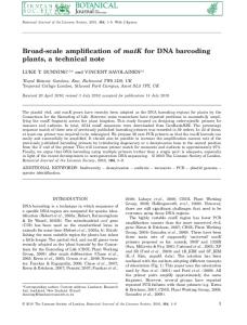
Step 2, primer design for simplex PCR: the third strategy described (see Fig. 1d) was employed for simplex PCR design, but using only one nested primer per primer pair. Nesting was done either completely (no overlap between the original and nested primer) or partially (some overlap between the original and nested primer). Table 3 shows the primer combinations chosen for simplex PCRs; nested primers are marked with asterisks (*). The setup of simplex PCRs (Steps 9–15) was done as stated above. Each multiplex reaction was diluted 1:25 and separately amplified with all simplex primer pairs. Additionally, a simplex PCR water control was included for all simplex primer pairs. Thermocycling was performed as described above at an annealing temperature of 53 °C. Step 16, Purification and sequencing: PCR products were visualized by electrophoresis on ethidium bromide– stained agarose gels as shown in Figure 3. The gel picture shows single bands of the expected size in the sample lanes and no amplification products in the control lanes. Moreover, no primer dimers or other secondary products are visible.
a
COMPETING INTERESTS STATEMENTS The authors declare that they have no competing financial interests.
2. 3. 4. 5. 6. 7. 8. 9. 10. 11. 12. 13.
Krause, J. et al. Multiplex amplification of the mammoth mitochondrial genome and the evolution of Elephantidae. Nature 439, 724–727 (2006). Gilbert, M.T.P. Bandelt, H.J., Hofreiter, M. & Barnes, I. Assessing ancient DNA studies. Trends Ecol. Evol. 20, 541–544 (2005). Cooper, A. & Poinar, H.N. Ancient DNA: do it right or not at all. Science 289, 1139 (2000). Hofreiter, M., Serre, D., Poinar, H.N., Kuch, M. & Pääbo, S. Ancient DNA. Nat. Rev. Genet. 2, 353–359 (2001). Pääbo, S. et al. Genetic analyses from ancient DNA. Annu. Rev. Genet. 38, 645–679 (2004). Malmstrom, H., Stora, J., Dalen, L., Holmlund, G. & Gotherstrom, A. Extensive human DNA contamination in extracts from ancient dog bones and teeth. Mol. Biol. Evol. 22, 2040–2047 (2005). Lambert, D.M. et al. Rates of evolution in ancient DNA from Adelie penguins. Science 295, 2270–2273 (2002). Rogaev, E.I. et al. Complete mitochondrial genome and phylogeny of Pleistocene mammoth Mammuthus primigenius. PLoS Biol. 4, e73 (2006). Höss, M., Jaruga, P., Zastawny, T.H., Dizdaroglu, M. & Pääbo, S. DNA damage and DNA sequence retrieval from ancient tissues. Nucleic Acids Res. 24, 1304–1307 (1996). Poinar, H.N., Höss, M., Bada, J.L. & Pääbo, S. Amino acid racemization and the preservation of ancient DNA. Science 272, 864–866 (1996). Smith, C.I. et al. Neanderthal DNA. Not just old but old and cold? Nature 410, 771–772 (2001). Smith, C.I., Chamberlain, A.T., Riley, M.S., Stringer, C. & Collins, M.J. The thermal history of human fossils and the likelihood of successful DNA amplification. J. Hum. Evol. 45, 203–217 (2003). Poinar, H.N. & Stankiewicz, B.A. Protein preservation and DNA retrieval
728 | VOL.1 NO.2 | 2006 | NATURE PROTOCOLS
c
Figure 3 | Anticipated results. Agarose gel electrophoresis of three different simplex PCR setups (a–c) and the NEB Low Molecular Weight DNA ladder (X). For each setup, lanes are loaded as follows: 1 and 5, PCR water controls (carried along from multiplex PCR); 2, extraction control; 3 and 4, a mammoth extract dilution of 1:10; 6 and 7, a dilution of 1:5; and 8, a 1:5 dilution of the PCR water control for the simplex PCR. Different primer pairs (see ANTICIPATED RESULTS) are presented as a for B6, b for A22 and c for A30.
ACKNOWLEDGMENTS We thank our lab members, especially C. Stäubert and I. Böselt, for comments that improved the manuscript. This work was funded by the Max Planck Society, the Deutsche Forschungsgemeinschaft and the Bundesministerium für Bildung und Forschung.
1.
b
from ancient tissues. Proc. Natl Acad. Sci. USA 96, 8426–8431 (1999). 14. Handt, O., Krings, M., Ward, R.H. & Pääbo, S. The retrieval of ancient human DNA sequences. Am. J. Hum. Genet. 59, 368–376 (1996). 15. Hofreiter, M., Jaenicke, V., Serre, D., Haeseler Av, A. & Pääbo, S. DNA sequences from multiple amplifications reveal artifacts induced by cytosine deamination in ancient DNA. Nucleic Acids Res. 29, 4793–4799 (2001). 16. Haak, W. et al. Ancient DNA from the first European farmers in 7500-yearold Neolithic sites. Science 310, 1016–1018 (2005). 17. Eichinger, L. et al. The genome of the social amoeba Dictyostelium discoideum. Nature 435, 43–57 (2005). 18. Greenwood, A., Capelli, C., Possnert, G. & Pääbo, S. Nuclear DNA sequences from late pleistocene megafauna. Mol. Biol. Evol. 16, 1466–1473 (1999). 19. Hummel, S., Schultes, T., Bramanti, B. & Herrmann, B. Ancient DNA profiling by megaplex amplications. Electrophoresis 20, 1717–1721 (1999). 20. Schultes, T., Hummel, S. & Herrmann, B. Amplification of Y-chromosomal STRs from ancient skeletal material. Hum. Genet. 104, 164–166 (1999). 21. Margulies, M. et al. Genome sequencing in microfabricated high-density picolitre reactors. Nature 437, 376–380 (2005). 22. Poinar, H.N. et al. Metagenomics to paleogenomics: Large-scale sequencing of mammoth DNA. Science 311, 392–394 (2006). 23. Kwok, S. et al. Effects of primer-template mismatches on the polymerase chain reaction: human immunodeficiency virus type 1 model studies. Nucleic Acids Res. 18, 999–1005 (1990). 24. Hofreiter, M. et al. Evidence for reproductive isolation between cave bear populations. Curr. Biol. 14, 40–43 (2004). 25. Kalmar, T., Bachrati, C.Z., Marcsik, A. & Rasko, I. A simple and efficient method for PCR amplifiable DNA extraction from ancient bones. Nucleic Acids Res. 28, E67 (2000). 26. Leonard, J.A., Wayne, R.K. & Cooper, A. Population genetics of ice age brown bears. Proc. Natl Acad. Sci. USA 97, 1651–1654 (2000). 27. Altschul, S.F. et al. Gapped BLAST and PSI-BLAST: a new generation of protein database search programs. Nucleic Acids Res. 25, 3389–3402 (1997). 28. Magnuson, V.L. et al. Substrate nucleotide-determined non-templated addition of adenine by Taq DNA polymerase: implications for PCR-based genotyping and cloning. Biotechniques 21, 700–709 (1996).