Multiparametric Imaging of Prostate Cancer Julia R. Fielding, M.D. Professor of Radiology, Obstetrics and Gynecology, and Urology Fried Lecture June 2...
Multiparametric Imaging of Prostate Cancer Julia R. Fielding, M.D. Professor of Radiology, Obstetrics and Gynecology, and Urology Fried Lecture June 20, 2014
Objectives • Review T2 weighted imaging of prostate cancer • Demonstrate the value of – 3 dimensional imaging – Diffusion imaging, perfusion imaging – Suggest current indications for prostate MRI
(a) Oblique sagittal and (b) axial schematic representations of the prostate zones and their relationship to the prostatic urethra (white arrow) and ejaculatory ducts (black arrow).
Basic MR imaging until 2008 • Endorectal coil + surface coil/1.5T magnet • Axial T2WI 5mm thickness with 1.0mm gap – peripheral zone white • Dark area cancer/prostatitis/blood • Decent identification of capsule • Focus on staging • Patients disliked endorectal coil
MRI in 2008 • Accuracy in predicting T3 cancer 68-85% • Not recommended as routine staging procedure in AUA guidelines – but things may be changing
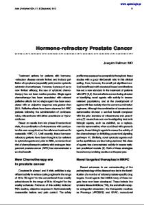
(a) Axial and (b) coronal T2-weighted MR mages (3150/105.4) in a 57-year-old patient with multifocal peripheral zone prostate cancer demonstrate similarity in T2 signal intensity of the normal central zone (arrows) and prostate cancer foci (dashed arrows).
Axial
Endorectal Coil Vargas H A et al. Radiology 2012;262:894-902
(a) Axial and (b) coronal T2-weighted MR mages (3150/105.4) in a 57-year-old patient with multifocal peripheral zone prostate cancer demonstrate similarity in T2 signal intensity of the normal central zone (arrows) and prostate cancer foci (dashed arrows).
MRI for initial staging, torso coil • The following 3 cases were done at UNC as part of a research protocol investigating MR spectroscopy using a torso coil and a 1.5T magnet • All subjects had low grade disease (PSA 0.5 cm, Gleason grade >7 • PPV 85% Low risk prostate CA: Accuracy of MPMR for detection. Kim HY et al. Radiology; May, 2014
Targeted ultrasound biopsy • How well does it work? • No clear answer yet, however several recent papers suggest that it has value • It is heavily marketed to physicians and patients
Recent literature 1. 499 patients, 241 low T2 SI lesions on MPMR 2. Biopsy with fused MR/US 3. Detection rate 80% Multiparametric MR and Subsequent MR/US fusion guided biposy inreases the detetion of anteriorly located prostate cancers. Volkin ,et al, in press
UCLA website/front page
Perfusion imaging – UCLA
Courtesy of Dr. Dan Margolis
Social media • Facebook • Linked in • Pinterest • Twitter • You tube
How does targeted biopsy work? • Radiologist supervises specialized MR protocol for prostate evaluation – includes T2/DWI/Perfusion • On review of MRI, suspicious lesions are marked in color • These lesions are fused with US for targeted biopsy
Bringing target information from MRI to realtime US Technology
Sensor
pre-procedural
intra-procedural
• Identify cancer in pre-acquired MRI • Track ultrasound probe and register with MRI • Show MRI-identified cancer on realtime ultrasound image Guide needle to target with realtime feedback Courtesy P. Choyke 36
Prostate Fusion-targeted biopsy workflow
Prior to procedure MRI Suite
T2w
MRS
DCE
MRI acquisition with ERC
Prostate segmentation and target identification Intra-procedure Realtime TRUS
Pre-procedure
Day of procedure Procedure
DWI
3D TRUS acquisition
MRI – 3D TRUS registration
Corresponding MRI
Targeted biopsy with realtime TRUS/MRI fusion
Anterior nodule
Why do prostate MRI? • Useful when: • Initial staging when physical exam and laboratory findings are at odds (small gland, high PSA) • Follow-up in patients with Gleason 6 disease • Negative biopsy with persistent elevation PSA - Biopsy guidance
UNC case • • • • •
Elevated PSA Multiple biopsies (outside location) MRI Target identified left mid gland Prostate cancer identified.